Immune memory vs. immune adaptation

This 2019 Dutch/German/Romanian perspective aimed for a better understanding of immune systems:

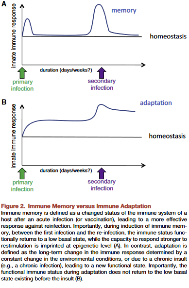
“Based on molecular, immunological, and evolutionary arguments, we propose that innate immune memory is a primitive form of immune memory present in all living organisms, while adaptive immune memory is an advanced form of immune memory representing an evolutionary innovation in vertebrates.

Innate immune responses have the capacity to be trained, and thereby exert a new type of immunological memory upon reinfection. The central feature of trained innate immune cells is their ability to mount a qualitatively and quantitatively different transcriptional response when challenged with microbes or danger signals. Evidence supports convergence of multiple regulatory layers for mediating innate immune memory, including changes in chromatin organization, DNA methylation, and probably non-coding RNAs such as microRNAs and/or long non-coding RNAs.

Two properties of adaptive immune response are mediated by two fundamentally different types of mechanisms:

  1. Higher magnitude and speed of the response is mediated by epigenetic programming.
  2. Specificity of the response is insured by gene recombination of TCR [T cell receptor] and BCR [B cell receptor] and clonal expansion of specific cell subpopulations upon antigen recognition.

To be effective, highly specific immune response requires huge diversity of receptors and antibodies, which is achieved by somatic rearrangement of gene segments. Recombination results in millions of TCR and antibody variants able to recognize and neutralize millions of various antigens.


This paper included speculations such as “Evidence supports..probably non-coding RNAs” quoted above, and the penultimate sentence:

“One can envision that vaccines that are capable of inducing both forms of immune memory at the same time would be more effective.”

100% factual evidence is preferred. Overall information can only be as accurate as the least accurate information.

This review highlighted a goal for humans to have both a functional innate immune system and a functional adaptive immune system. The lead author coauthored A dietary supplement that trains the innate immune system and a study referenced in Eat your oats.

https://www.sciencedirect.com/science/article/pii/S1931312818306334 “Innate and Adaptive Immune Memory: an Evolutionary Continuum in the Host’s Response to Pathogens” (not freely available)

Our brains are shaped by our early environments

This 2019 McGill paper reviewed human and animal studies on brain-shaping influences from the fetal period through childhood:

“In neonates, regions of the methylome that are highly variable across individuals are explained by the genotype alone in 25 percent of cases. The best explanation for 75 percent of variably methylated regions is the interaction of genotype with different in utero environments.

A meta-analysis including 45,821 individuals with attention-deficit/hyperactivity disorder and 9,207,363 controls suggests that conditions such as preeclampsia, Apgar score lower than 7 at 5 minutes, breech/transverse presentations, and prolapsed/nuchal cord – all of which involve some sort of poor oxygenation during delivery – are significantly associated with attention-deficit/hyperactivity disorder. The dopaminergic system seems to be one of the brain systems most affected by perinatal hypoxia-ischemia.

Exposure to childhood trauma activates the stress response systems and dysregulates serotonin transmission that can adversely impact brain development. Smaller cerebral, cerebellar, prefrontal cortex, and corpus callosum volumes were reported in maltreated young people as well as reduced hippocampal activity.

Environmental enrichment has a series of beneficial effects associated with neuroplasticity mechanisms, increasing hippocampal volume, and enhancing dorsal dentate gyrus-specific differences in gene expression. Environmental enrichment after prenatal stress decreases depressive-like behaviors and fear, and improves cognitive deficits.”


The reviewers presented strong evidence until the Possible Factors for Reversibility section, which ended with the assertion:

“All these positive environmental experiences mentioned in this section could counterbalance the detrimental effects of early life adversities, making individuals resilient to brain alterations and development of later psychopathology.”

The review’s penultimate sentence recognized that research is seldom done on direct treatments of causes:

“The cross-sectional nature of most epigenetic studies and the tissue specificity of the epigenetic changes are still challenges.”

Cross-sectional studies won’t provide definitive data on cause-and-effect relationships.

The question yet to be examined is: How can humans best address these early-life causes to ameliorate their lifelong effects?

https://onlinelibrary.wiley.com/doi/full/10.1111/dmcn.14182 “Early environmental influences on the development of children’s brain structure and function” (not freely available)

Epigenetic transgenerational inheritance mechanisms that lead to prostate disease

This 2019 Washington State University rodent study found:

“Ancestral exposure to toxicant vinclozolin induces an epigenetic transgenerational increase in susceptibility to prostate pathology in F3 [male great-grandchildren] generation rats. These results are in agreement with previous studies which found a transgenerational increase in rates of prostatic:

  • Epithelial atrophy;
  • Cystic hyperplasia; and
  • Prostatitis

in transgenerational F3 and F4 [male great-great-grandchildren] generations after exposure of F0 [great-great-grandmother] generation pregnant rats to vinclozolin. These effects were accompanied by transgenerational changes in mRNA expression in F3 generation ventral prostate epithelial cells.

A number of previous transgenerational studies have shown no ventral prostate histopathology or disease detected. Therefore, observations suggest ancestral exposure specificity in the ability to induce transgenerational inheritance of prostate disease.

There was also no increase in prostate histopathology in directly exposed F1 [male children] or F2 [male grandchildren] generation vinclozolin lineage rats compared to controls.

prostate pathology

The mechanism by which epigenetic transgenerational inheritance affects prostate epithelium involves control of gene expression by DNA methylation and lncRNAs. It will be necessary to determine exact gene targets of these epigenetic modifications to determine further mechanisms.

Future studies need to investigate if similar mechanisms are at work in human males who have adult-onset BPH or prostate cancer. Ancestral exposures to toxicants and epigenetic transgenerational inheritance may contribute to development of prostate disease in men today.”


This study’s above bolded sentence added to evidence that epigenetic effects may skip generations. A study by the same group, Epigenetic transgenerational inheritance of ovarian disease, found in females:

There was no increase in ovarian disease in direct fetal exposed F1 or germline exposed F2 generation vinclozolin or DDT lineage rats compared to controls.

A disturbance in the paradigm of child abuse referenced other studies that found generation-skipping effects.

Researchers are closer to discovering evidence for precise mechanisms of epigenetic transgenerational inheritance. It’s well past time that other researchers performing studies like Burying human transgenerational epigenetic evidence:

  • Turn things around;
  • Take their work seriously; and
  • Truly investigate human evidence for epigenetic transgenerational inheritance.

What are more important research and funding priorities than such human studies?

https://www.nature.com/articles/s41598-019-38741-1 “Environmental Toxicant Induced Epigenetic Transgenerational Inheritance of Prostate Pathology and Stromal-Epithelial Cell Epigenome and Transcriptome Alterations: Ancestral Origins of Prostate Disease”

Disproving the cholesterol paradigm

This 2018 review presented evidence that:

“For half a century, a high level of total cholesterol (TC) or low-density lipoprotein cholesterol (LDL-C) has been considered to be the major cause of atherosclerosis and cardiovascular disease (CVD), and statin treatment has been widely promoted for cardiovascular prevention. However, there is an increasing understanding that the mechanisms are more complicated and that statin treatment, in particular when used as primary prevention, is of doubtful benefit.

The authors of three large reviews recently published by statin advocates have attempted to validate the current dogma. This article delineates the serious errors in these three reviews as well as other obvious falsifications of the cholesterol hypothesis.

Our search for falsifications of the cholesterol hypothesis confirms that it is unable to satisfy any of the Bradford Hill criteria for causality and that the conclusions of the authors of the three reviews are based on:

  • Misleading statistics,
  • Exclusion of unsuccessful trials and by
  • Ignoring numerous contradictory observations.

The association between the absolute risk reduction of total mortality in 26 statin trials [squares] included in the study by Silverman et al. and in 11 ignored trials [triangles] and the year where the trial protocols were published. The vertical line indicates the year where the new trial regulations were introduced.

In 2004–2005, health authorities in Europe and the United States introduced New Clinical Trial Regulations, which specified that all trial data had to be made public. Since 2005, claims of benefit from statin trials have virtually disappeared.”


This paradigm was proven wrong eighty years ago! How much longer will its harmful consequences continue?

https://www.tandfonline.com/doi/full/10.1080/17512433.2018.1519391 “LDL-C does not cause cardiovascular disease: a comprehensive review of the current literature”

An hour of the epigenetic clock

Starting the fifth year of this blog with a 2018 presentation by the founder of the epigenetic clock method describing the state of the art up through July 2018. The webinar was given on the release day of The epigenetic clock now includes skin study.


Segments before the half-hour mark provide an introduction to the method and several details about the concurrently-released study. The Q&A section starts a little before the hour mark.

Stuck in the wrong paradigm

This 2019 article questioned the paradigm of determining substance carcinogenicity:

“In the absence of robust epidemiological data, the final arbiter of whether a chemical is considered to be a carcinogen or not has been based on the outcome of long-term rodent bioassays. This approach is incompatible with the current knowledge of the etiology of cancer. The current view of the etiology of cancer suggests that it is not useful to consider carcinogenicity as a single hazardous property with its own hazard category.

There is no bright line between carcinogens and non-carcinogens but rather there is a continuum with some chemicals having high potential, some having no potential, and others having potential at a point along the continuum. This continuum exists alongside other adverse effects. One problem is being stuck in the old practice of wishing to reproduce the binary “carcinogen/non-carcinogen” results of the long-term bioassay rather than move to a new paradigm in assessing the chemical’s position on the spectrum of carcinogenic potential.

The two-year bioassay has such high variability (because of the variability of the carcinogenic process it is trying to measure and the interplay between dose limiting toxicity and cell proliferation inducing toxicity) that the outcome of the assay for compounds with low to intermediate carcinogenic potential is little more than a lottery. After half a century, it has only been used to evaluate less than 5% of chemicals that are in use. It is not reproducible because of the probabalistic nature of the process it is evaluating combined with dose limiting toxicity, dose selection, and study design.”


Unscientific research paradigms will eventually collapse because they can’t withstand the scrutiny of the scientific method. Too bad the coauthors didn’t kill off this one while they were still in positions at the US Environmental Protection Agency, World Health Organization, etc.

https://www.sciencedirect.com/science/article/pii/S0273230019300248 “Chemical carcinogenicity revisited 2: Current knowledge of carcinogenesis shows that categorization as a carcinogen or non-carcinogen is not scientifically credible” (not freely available)

Epigenetic causes of sexual orientation and handedness?

This 2018 Austrian human study subject was various associations of prenatal testosterone levels to fetal development:

“The available evidence suggests, albeit not conclusively, that prenatal testosterone levels may be one cause for the association of sexual orientation with handedness. Associations among women were consistent with predictions of the Geschwind–Galaburda theory (GGT), whereas those among men were consistent with predictions of the callosal hypothesis. However, research on the associations between sexual orientation and handedness appears to be compromised by various methodological and interpretational problems which need to be overcome to arrive at a clearer picture.

The GGT posits that high prenatal testosterone levels cause a delay in the fetal development of the left cerebral hemisphere which results in a right-hemisphere dominance and hence in a tendency for left-handedness. According to the GGT, high prenatal testosterone levels entail not only a masculinization of the female fetus, but also a feminization of the male fetus (contrary to neurohormonal theory). Overall, the male fetus is subjected to higher levels of intrauterine testosterone than the female fetus. The GGT is thus consistent with the higher prevalence of left-handedness among men than among women.

The callosal hypothesis applies to men only and assumes, in line with neurohormonal theory, that low prenatal testosterone levels are associated with later homosexuality. According to the CH, high prenatal testosterone enhances processes of cerebral lateralization through mechanisms of axonal pruning, thereby resulting in stronger left-hemisphere dominance and a smaller corpus callosum. Consistent with this, women have a larger corpus callosum than men.”


The study’s Limitations section included the following:

  1. “Limitations of the current study pertain to the self-report nature of our data. Behavioral data may provide differing results from those obtained here.
  2. Assessment of sexual orientation relied on a single-item measure. Utilization of rating scales (e.g., the Kinsey Sexual Orientation Scale) or of multi-item scales, and assessing different components of sexual orientation, would have allowed for a more fine-grained analysis and for a cross-validation of sexual orientation ratings with sexual attraction.
  3. Albeit both our samples were large, the proportions of bisexual and homosexual individuals were, expectedly, only small, as were effects of lateral preferences. Thus, in analysis we could not differentiate bisexual from homosexual individuals. Bisexual and homosexual individuals may differ with regard to the distribution of lateral preferences.
  4. Some effect tests in this study have been underpowered. Independent replications with even larger samples are still needed.”

The largest unstated limitation was no fetal measurements. When a fetus’ epigenetic responses and adaptations aren’t considered, not only can the two competing hypotheses not be adequately compared, but causes for the studied phenotypic programming and other later-life effects will also be missed.

https://link.springer.com/article/10.1007/s10508-018-1346-9 “Associations of Bisexuality and Homosexuality with Handedness and Footedness: A Latent Variable Analysis Approach”

Burying human transgenerational epigenetic evidence

The poor substitutes for evidence in this 2018 US study guaranteed that human transgenerational epigenetically inherited effects wouldn’t be found in the generations that followed after prenatal diethylstilbestrol (DES) exposure:

“A synthetic, nonsteroidal estrogen, DES was administered to pregnant women under the mistaken belief it would reduce pregnancy complications and losses. From the late 1930s through the early 1970s, DES was given to nearly two million pregnant women in the US alone.

Use of DES in pregnancy was discontinued after a seminal report showed a strong association with vaginal clear cell adenocarcinoma in prenatally exposed women. A recent analysis of the US National Cancer Institute (NCI) DES Combined Cohort Follow-up Study showed elevated relative risks of twelve adverse health outcomes.

We do not have sufficient data concerning the indication for DES in the grandmother to determine whether adverse pregnancy outcomes in the third generation might resemble those of their grandmothers. Fourth generation effects of prenatal exposures in humans have not been reported.”

https://www.sciencedirect.com/science/article/pii/S0890623818304684 “Reproductive and Hormone-Related Outcomes in Women whose Mothers were Exposed in utero to Diethylstilbestrol (DES): A Report from the US National Cancer Institute DES Third Generation Study” (not freely available)


This study had many elements in common with its poor-quality reference [25] “Transgenerational effects of prenatal exposure to the 1944–45 Dutch famine” which is freely available at https://obgyn.onlinelibrary.wiley.com/doi/full/10.1111/1471-0528.12136.

That study’s Methods section showed:

  1. Its non-statistical data was almost all unverified self-reports by a self-selected sample of the F2 grandchildren, average age 37.
  2. No detailed physical measurements or samples were taken of the F2 grandchildren, or of their F1 parents, or of their F0 grandparents, all of which are required as baselines for any transgenerational epigenetic inheritance findings.
  3. No detailed physical measurements or samples were taken of the F3 great-grandchildren, which is the generation that may provide transgenerational evidence if the previous generations also have detailed physical baselines.

That study’s researchers drew enough participants (360) such that their statistics package allowed them to impute and assume into existence a LOT of data. But the scientific method constrained them to make factual statements of what the evidence actually showed. They admitted:

“In conclusion, we did not find a transgenerational effect of prenatal famine exposure on the health of grandchildren in this study.”


The current study similarly used the faulty methods 1-3 above to produce results such as:

“We do not have sufficient data concerning the indication for DES in the [F0] grandmother to determine whether adverse pregnancy outcomes in the [F2] third generation might resemble those of their grandmothers.

Fourth [F3] generation effects of prenatal exposures in humans have not been reported.

Zero studies of probably more than 10,000,000 F3 great-grandchildren of DES-exposed women just here in the US?

Who is against funding these studies? Who is afraid of what such studies may find?

One plausible hypothesis of these human studies would be of inherited effects that skipped generations! The rodent studies Epigenetic transgenerational inheritance mechanisms that lead to prostate disease and Epigenetic transgenerational inheritance of ovarian disease found inherited diseases that didn’t manifest until the F3 great-grand offspring:

The F3 generation can have disease while the F1 and F2 generations do not.

Ancestral exposure to toxicants is a risk factor that must be considered in the molecular etiology of ovarian disease.

For the current study:

  • What could be expected from a study design that didn’t include F3 women and men, which is the only generation that didn’t have direct DES exposure?
  • What a nonsensical study design to permit NON-evidence like educational level!

Human studies of possible intergenerational and transgenerational epigenetic inheritance are urgently needed. There will be abundant evidence to discover if researchers will take their fields seriously.

Fear of feeling?

Here’s a 2018 article from two researchers involved in the Dunedin (New Zealand) Longitudinal Study. They coauthored many studies, including People had the same personalities at age 26 that they had at age 3.

The paper’s grand hypothesis was:

“A single dimension is able to measure a person’s liability to mental disorder, comorbidity among disorders, persistence of disorders over time, and severity of symptoms.”

The coauthors partially based this on:

“Repeated diagnostic interviews carried out over 25 years, when the research participants were 11, 13, 15, 18, 21, 26, 32, and 38 years old, and include information about seven diagnostic groups: anxiety, depression, attention deficit hyperactivity disorder, conduct disorder, substance dependence, bipolar disorder, and schizophrenia.”


https://ajp.psychiatryonline.org/doi/full/10.1176/appi.ajp.2018.17121383 “All for One and One for All: Mental Disorders in One Dimension” (not freely available)


More about the coauthors:

Two psychologists followed 1000 New Zealanders for decades. Here’s what they found about how childhood shapes later life

“Dunedin and other studies show that most people have at least one episode of mental illness during their lifetime.”


What compels people to manufacture “universal” truths? Aren’t such beliefs poor substitutes for feeling? For understanding historical, factual, personal truths?

What if the price we pay for avoiding and pressuring down our feelings is: A wasted life?

What if the grand hypothesis worth proving is: For one’s life to have meaning, each individual has to regain their feelings?

Eat your oats!

Here’s some motivation to replenish your oats supply.

From a 2013 Canadian human review:

“Review of human studies investigating the post-prandial blood-glucose lowering ability of oat and barley food products” https://www.nature.com/articles/ejcn201325

“Change in glycaemic response (expressed as incremental area under the post-prandial blood-glucose curve) was greater for intact grains than for processed foods. For processed foods, glycaemic response was more strongly related to the β-glucan dose alone than to the ratio of β-glucan to the available carbohydrate.”

The review found that people don’t have to eat a lot of carbohydrates to get the glycemic-response benefits of β-glucan. Also, eating ~3 grams of β-glucan in whole oats and barley will deliver the same glycemic-response benefits as eating ~4 grams of β-glucan in processed oats and barley.

However, the glycemic index used in the review is a very flawed measure. What’s the point of indexing healthy choices like whole grains to unhealthy choices that healthy people aren’t going to make anyway?


The reviewer somewhat redeemed herself by participating in a 2018 review:

“Processing of oat: the impact on oat’s cholesterol lowering effect” https://www.ncbi.nlm.nih.gov/pmc/articles/PMC5885279/

“For a similar dose of β-glucan:

  1. Liquid oat-based foods seem to give more consistent, but moderate reductions in cholesterol than semi-solid or solid foods where the results are more variable;
  2. The quantity of β-glucan and the molecular weight at expected consumption levels (∼3 g day) play a role in cholesterol reduction; and
  3. Unrefined β-glucan-rich oat-based foods (where some of the plant tissue remains intact) often appear more efficient at lowering cholesterol than purified β-glucan added as an ingredient.”

The review’s sections 3. Degree of processing and functionality and 4. Synergistic action of oat constituents were informative:

“Both in vitro and in vivo studies clearly demonstrated the beneficial effect of oat on cholesterolemia, which is unlikely to be due exclusively to β-glucan, but rather to a combined and synergetic action of several oat compounds acting together to reduce blood cholesterol levels.”


Another use of β-glucan is to improve immune response. Here’s a 2016 Netherlands study where the researchers used β-glucan to get a dozen people well after making them sick with lipopolysaccharide as is often done in animal studies:

β-Glucan Reverses the Epigenetic State of LPS-Induced Immunological Tolerance” https://www.ncbi.nlm.nih.gov/pmc/articles/PMC5927328/

“The innate immune ‘training stimulus’ β-glucan can reverse macrophage tolerance ex vivo.”

I’ve curated other research on β-glucan’s immune-response benefits in:

Adverse epigenetic effects of prenatal and perinatal anesthesia

This 2018 Chinese animal review subject was prenatal and perinatal anesthesia’s adverse epigenetic effects on a fetus/neonate:

“Accumulating evidence from rodent and primate studies has demonstrated that in utero or neonatal exposure to commonly used inhaled and intravenous general anesthetics is associated with neural degeneration and subsequent neurocognitive impairments, manifested in learning and memory disabilities.

So far, conflicting data exist about the effect of anesthetic agents on neurodevelopment in humans and no definite conclusion has been given yet.”

The inhibitors in the above graphic counter anesthesia’s effects on the fetus/neonate, summarized as:

“Epigenetic targeting of DNA methyltransferases and/or histone deacetylases may have some therapeutic value.”


Do physicians consider possible epigenetic alterations of a newborn’s chromatin structure and gene expression when they administer anesthesia to mothers during childbirth?

https://www.ncbi.nlm.nih.gov/pmc/articles/PMC6079265/ “Epigenetic Alterations in Anesthesia-Induced Neurotoxicity in the Developing Brain”

A top-down view of biological goal-directed mechanisms

This 2016 US/Italy article was written from a perspective of regenerative bioengineering:

“Higher levels beyond molecular can have their own unique dynamics that offer better (e.g. more parsimonious and potent) explanatory power than models made at lower levels. Biological systems may be best amenable to models that include information structures (organ shape, size, topological arrangements and complex anatomical metrics) not defined at molecular or cellular level but nevertheless serving as the most causally potent ‘knobs’ regulating large-scale outcomes.

Top-down models can be as quantitative as familiar bottom-up systems biology examples, but they are formulated in terms of building blocks that cannot be defined at the level of gene expression and treat those elements as bona fide causal agents (which can be manipulated by interventions and optimization techniques). The near-impossibility of determining which low-level components must be tweaked in order to achieve a specific system-level outcome is a problem that plagues most complex systems.

The current paradigm in biology of exclusively tracking physical measurable and ignoring internal representation and information structures in patterning contexts quite resemble the ultimately unsuccessful behaviourist programme in psychology and neuroscience. For example, even if stem cell biologists knew how to make any desired cell type from an undifferentiated progenitor, the task of assembling them into a limb would be quite intractable.

Current state of the art in the field of developmental bioelectricity is that it is known, at the cellular level, how resting potentials are transduced into downstream gene cascades, as well as which transcriptional and epigenetic targets are sensitive to change in developmental bioelectrical signals. What is largely missing however is a quantitative understanding of how global dynamics of bioelectric circuits make decisions that orchestrate large numbers of individual cells, spread out over considerable anatomical distances, towards specific pattern outcomes.”


Regenerative research is gathering evidence for goal-directed memory and learning that doesn’t meet current definitions. For example:

salamander

“A tail grafted to the flank of a salamander slowly remodels to a limb, a structure more appropriate for its new location, illustrating shape homeostasis towards a normal amphibian body plan. Even tail tip cells (in red) slowly become fingers, showing that remodelling is not driven by only local information.”

These reviewers compared their findings to several existing research and real-world-operations domains. Other models may also benefit from concepts of:

“Quantitative, predictive, mechanistic understanding of goal-directed morphogenesis.”

https://royalsocietypublishing.org/doi/full/10.1098/rsif.2016.0555 “Top-down models in biology: explanation and control of complex living systems above the molecular level”


I came across this article as a result of its citation in The Body Electric blog post.

“Levin drops a hint that there are photo-sensitive drugs that can control ion gates that can be used to translate a projected geometric image into a pattern of membrane potentials. He argues that the patterns encode ‘blueprints’ rather than a ‘construction manual’ based on the fact that the program is adaptive in the face of physical barriers and disruptions.”

Epigenetic clock statistics and methods

This 2018 Chinese study was a series of statistical and methodological counter-arguments to a previous epigenetic clock study finding that:

“Only [CpG] sites mapping to the ELOVL2 promoter constitute cell and tissue-type independent aDMPs [age-associated differentially methylated positions].”

The study used external data sets and the newer epigenetic clock’s fibroblast data in its analyses to find:

“While we agree that specific sites mapping to ELOVL2 are special aDMPs in the sense that their effect sizes are particularly large across a number of different tissue-types, our analysis suggests that most aDMPs are valid across multiple different tissue types, suggesting that shared aDMPs are common.”

The details of each of the study’s counter-arguments were compelling. For example:

“We analyzed Illumina 850k data from an EWAS profiling blood, buccal and cervical samples from a common set of 263 women. Because blood is a complex mixture of many immune-cell subtypes, and buccal and cervical samples are highly contaminated by immune cells, we identified aDMPs in each tissue after adjustment for batch effects and cell-type heterogeneity.

Using either an FDR [false discovery rate] < 0.05 or Bonferroni adjusted P-value < 0.05 thresholds, the overlap of aDMPs between the 3 tissues was highly significant, mimicking the result obtained on blood cell subtypes. We observed a total of 2200 aDMPs in common between blood, buccal and cervix, an overlap which cannot be explained by random chance.”

The study’s Discussion section provided qualifications and limitations such as:

“It is important to point out that even if age-associated DNAm changes are widespread across the genome, downstream functional effects may be rare. While specific aDMPs may be shared between tissue-types, it is only in specific tissues or cell-types that any associated functional deregulation may be of biological and clinical significance.

https://www.aging-us.com/article/101666/text “Cell and tissue type independent age-associated DNA methylation changes are not rare but common”


The November 2018 issue of Aging also contained other articles of interest:

https://www.aging-us.com/article/101626/text “Accelerated DNA methylation age and the use of antihypertensive medication among older adults”

“DNAmAge and AA [age acceleration] may not be able to capture the preventive effects of AHMs [antihypertensive medications] that reduce cardiovascular risks and mortality.”

https://www.aging-us.com/article/101633/text “Azithromycin and Roxithromycin define a new family of senolytic drugs that target senescent human fibroblasts”

“Azithromycin preferentially targets senescent cells, removing approximately 97% of them with great efficiency. This represents a near 25-fold reduction in senescent cells.”

https://www.aging-us.com/article/101647/text “Disease or not, aging is easily treatable”

“Aging consists of progression from (pre)-pre-diseases (early aging) to diseases (late aging associated with functional decline). Aging is NOT a risk factor for these diseases, as aging consists of these diseases: aging and diseases are inseparable.”

Chronological age by itself is an outdated clinical measurement

This 2018 editorial in the New England Journal of Medicine concerned a clinical trial of an osteoporosis treatment:

“When measurement of bone density was first introduced 25 years ago, absolute bone mineral density (g per square centimeter) was considered as too onerous for clinicians to understand. Ultimately, these events led to a treatment gap in patients who had strong clinical risk factors for an osteoporotic fracture (particularly age) but had T scores in the osteopenic range.

The average age of the participants in the current trial was approximately 3.5 years older than that in the Fracture Intervention Trial. Owing to the interaction between age and bone mineral density, the results of the current trial should not be extrapolated to younger postmenopausal women (50 to 64 years of age) with osteopenia.

This trial reminds us that risk assessment and treatment decisions go well beyond bone mineral density and should focus particularly on age and a history of previous fractures.”

https://www.nejm.org/doi/pdf/10.1056/NEJMe1812434 “A Not-So-New Treatment for Old Bones”


This editorial provided some history of how a still-generally-accepted set of diagnostic measurements were selected for their relative convenience instead of chosen for their efficacy. Add chronological age to such ineffective measurements.

Let’s recognize better aging and diagnostic measurements, then incorporate them. How else will we advance past the above uninformative averaging and unhelpful recommendation based on chronological age?

The time has passed for physicians and clinicians to consider only chronological age when evaluating a patient’s clinical age. More effective human age measurements covering the entire person as well as their body’s components include:

F2.large

A slanted view of the epigenetic clock

The founder of the epigenetic clock technique was interviewed for MIT Technology Review:

“We need to find ways to keep people healthier longer,” he says. He hopes that refinements to his clock will soon make it precise enough to reflect changes in lifestyle and behavior.”


The journalist attempted to dumb the subject down “for the rest of us” with distortions such as the headline. The varying correlation of epigenetic age to chronological age was somewhat better reported in the story:

“The epigenetic clock is more accurate the younger a person is. It’s especially inaccurate for the very old.”

The journalist inappropriately used luck as a synonym for randomness/stochasticity:

“He estimates that about 40% of the ticking rate is determined by genetic inheritance, and the rest by lifestyle and luck.”

A third example of less-than-straightforward journalism started with:

“Such personalization raises questions about fairness. If your epigenetic clock is ticking faster through no fault of your own..”

Were MIT Technology Review readers unable to comprehend a straightforward story on the epigenetic clock? What was the purpose of slants and distortions in an introductory article?

https://www.technologyreview.com/s/612256/want-to-know-when-youre-going-to-die/ “Want to know when you’re going to die?”