No magic bullet, only magical thinking

Consider this a repost of Dr. Paul Clayton’s blog post The Drugs Don’t Work:

“The drug industry has enough funds to:

  • Rent politicians;
  • Subvert regulatory agencies;
  • Publish fake data in the most august peer-reviewed literature; and
  • Warp the output of medical schools everywhere.

Their products are a common cause of death. Every year, America’s aggressively modern approach to disease kills over 100,000 in-hospital patients, and twice that number of out-patients.

In 1900, a third of all deaths occurred in children under the age of 5. By 2000 this had fallen to 1.4%. The resulting 30-year increase in average life expectancy fed into the seductive and prevailing myth that we are all living longer; which is manifestly untrue. Improvements in sanitation were far more significant in pushing infections back than any medical developments.

There is currently no pharmaceutical cure for Alzheimer’s or Parkinsonism, nor can there be when these syndromes are in most cases driven by multiple metabolic distortions caused by today’s diet. The brain is so very complex, and it can go wrong in so many ways. The idea that we can find a magic bullet for either of these syndromes is ill-informed and philosophically mired in the past.

It is also dangerous. There is a significant sub-group of dementia sufferers whose conditions are driven and exacerbated by pharmaceuticals. Chronic use of a number of commonly prescribed drugs – and ironically, anti-Parkinson drugs – increases the risk of dementia by roughly 50%.

Big Pharma’s ability to subvert regulatory authorities is even more dangerous. The recent FDA approval of Biogen’s drug aducanumab is a scandal; not one member of the FDA Advisory Committee voted to approve this ineffective product, and three of them resigned in the aftermath of the FDA’s edict. This ‘anti-Alzheimer’s’ drug, which will earn Biogen $56,000 / patient / year, was licensed for financial reasons; it reduced amyloid plaque but was clinically ineffective.

So did the eagerly awaited gantenerumab and solanezumab. But they, too, failed to produce any significant clinical benefit.”


A knee-replacement patient enduring her daily workout

PXL_20210822_102713662

Prevent your brain from shrinking

My 800th curation is a 2021 human diet and lifestyle study:

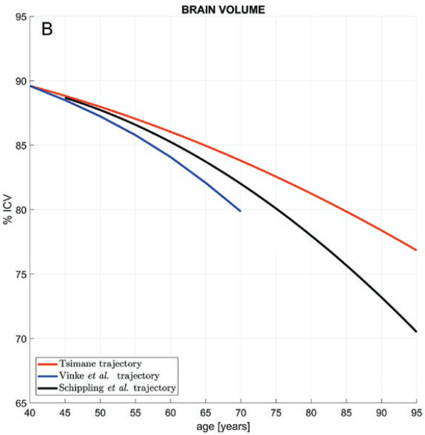
“Brain atrophy is correlated with risk of cognitive impairment, functional decline, and dementia. This study (a) examines the statistical association between brain volume (BV) and age for Tsimane, and (b) compares this association to that of 3 industrialized populations in the United States and Europe.

Tsimane forager-horticulturists of Bolivia have the lowest prevalence of coronary atherosclerosis of any studied population, and present few cardiovascular disease (CVD) risk factors. They have a high burden of infections and inflammation, reflected by biomarkers of chronic immune activation, including higher leukocytes counts, faster erythrocyte sedimentation rates, and higher levels of C-reactive protein, interleukin-6, and immunoglobulin-E than in Americans of all ages.

The Tsimane have endemic polyparasitism involving helminths and frequent gastrointestinal illness. Most morbidity and mortality in this population is due to infections.

brain volume

The Tsimane exhibit smaller age-related BV declines relative to industrialized populations, suggesting that their low CVD burden outweighs their high, infection-driven inflammatory risk. If:

  1. Cross-sectional data (which we believe are population-representative of Tsimane adults aged 40 and older) represent well the average life course of individuals; and
  2. The Tsimane are representative of the baseline case prior to urbanization;

these results suggest a ~70% increase in the rates of age-dependent BV decrease accompanying industrialized lifestyles.

Despite its limitations, this study suggests:

  • Brain atrophy may be slowed substantially by lifestyles associated with very low CVD risk; and
  • There is ample scope for interventions to improve brain health, even in the presence of chronically high systemic inflammation.

Lastly, the slow rate of age-dependent BV decrease in the Tsimane raises new questions about dementia, given the role of both infections and vascular factors in dementia risk.”

https://gurven.anth.ucsb.edu/sites/default/files/sitefiles/papers/irimiaetal2021.pdf “The indigenous South American Tsimane exhibit relatively modest decrease in brain volume with age despite high systemic inflammation”


I came across this study by its citation in Dr. Paul Clayton’s 2021 blog post We’ve got to get ourselves back to the garden.

ω-6 to ω-3 PUFA ratio

Three human-evidenced publications on omega-6 and omega-3 polyunsaturated fatty acids, with the first a 2021 blog post that cited 72 references:

“In the area of heart health, which is why most consumers swallow fish oil, the data is hopelessly conflicted:

  • One meta-analyses found that protective effects were dose-related, which is always persuasive;
  • In marked contrast, three recent powerful clinical trials found fish oil to have no effects on cardiovascular pathology in either primary or secondary prevention; and
  • Yet another meta-analysis found null results, except for a slight degree of protection in subjects who had gallantly taken fish oil supplements for over ten years.

Can these all be right? I think they can, based on secondary bioavailability.

Levels of omega 3s in the bloodstream are irrelevant, except in terms of their calorie content. That is not where they do their anti-inflammatory thing. They become precursors for resolvins, maresins, protectins, and anti-inflammatory eicosanoids only after they have been incorporated into the host’s cell membranes.

Getting them into cell membranes is secondary bioavailability (or bio-efficacy), and this is a much more complicated procedure. Seafood does it, but fish oil doesn’t.

Specifically, there is something in oily fish which enables secondary bioavailability, but which is missing in commercial fish oils. That something is a lipophillic polyphenol called phlorotannin.”

https://drpaulclayton.eu/blog/fish-oil-upgrade-to-snake-oil/ “Fish Oil? Upgrade to Snake Oil!”


A second paper was a 2021 review that focused on ratios of ω-6 to ω-3 PUFAs:

“Chronic diseases including obesity, type 2 diabetes, cardiovascular disease, cancer, and Alzheimer’s disease are rising exponentially in the modern world. Though these diseases are multifactorial in nature, their prevalence is mostly associated with an unbalanced increase in dietary n-6 PUFAs and decrease in n-3 PUFAs.

Mostly, these diseases escalate on the fact that inflammation in conjunction with obesity is the basis of every chronic disease.

Considering antagonistic effects of n-3 and n-6 PUFAs, both n-3 and n-6 SC-PUFAs and LC-PUFAs in their proportional ratio with each other, which is close to 4:1, play a significant role in regulating body homeostasis of inflammation and anti-inflammation, vasodilation and vasoconstriction, bronchoconstriction and bronchodilation, and platelet aggregation and antiaggregation.”

https://www.hindawi.com/journals/jl/2021/8848161/ “Overconsumption of Omega-6 Polyunsaturated Fatty Acids (PUFAs) versus Deficiency of Omega-3 PUFAs in Modern-Day Diets: The Disturbing Factor for Their ‘Balanced Antagonistic Metabolic Functions’ in the Human Body”


A third paper was a 2020 human adolescent study:

“Obese youth 9–19 y of age with nonalcoholic fatty liver disease were treated to see whether 12 wk of a low n–6:n–3 PUFA ratio (4:1) normocaloric diet mitigated fatty liver.

Independent of weight loss, a low n–6:n–3 PUFA diet ameliorated the metabolic phenotype of adolescents with fatty liver disease. This trial was registered at clinicaltrials.gov as NCT01556113.”

https://academic.oup.com/jn/article/150/9/2314/5870325 “A Low ω-6 to ω-3 PUFA Ratio (n–6:n–3 PUFA) Diet to Treat Fatty Liver Disease in Obese Youth”


My ω-6 to ω-3 PUFA 4 : 1 (1400 / 350) intake at breakfast and dinner via Balance Oil:

PXL_20210704_161714382

At lunch I eat an ounce of walnuts with a ω-6 to ω-3 PUFA 4.4 : 1 ratio:

walnuts 1 oz


PXL_20210710_093234225.NIGHT

A time to speak

“To every thing there is a season, and a time to every purpose under heaven:
A time to break down, and a time to build up;
A time to dance, and a time to mourn;
A time you may embrace, and a time to refrain from embracing;
A time to keep silent, and a time to speak.”


A review from 2017:

“Few, if any, other drugs can rival ivermectin for its beneficial impact on human health and welfare. Perhaps more than any other drug, ivermectin is a drug for the world’s poor. For most of this century, some 250 million people have been taking it.

The following are an indication of disease-fighting potential that has been identified for ivermectin thus far:

  • Antiviral – Ivermectin has been found to potently inhibit replication of yellow fever virus, with EC50 values in the sub-nanomolar range. It inhibits replication in several other flaviviruses, including dengue, Japanese encephalitis, and tick-borne encephalitis. Ivermectin interrupts virus replication. It demonstrates antiviral activity against several RNA viruses by blocking nuclear trafficking of viral proteins. It has been shown to have potent antiviral action against HIV-1.
  • Asthma – Ivermectin suppressed mucus hypersecretion by goblet cells, establishing that ivermectin can effectively curb inflammation, such that it may be useful in treating allergic asthma and other inflammatory airway diseases.
  • Bedbugs – Ivermectin is highly effective against bedbugs, capable of eradicating or preventing bedbug infestations.
  • Disease vector control – Ivermectin is highly effective in killing a broad range of insects. Comprehensive testing against 84 species of insects showed that avermectins were toxic to almost all insects tested. At sub-lethal doses, ivermectin inhibits feeding and disrupts mating behavior, oviposition, egg hatching, and development.
  • Malaria – Mosquitoes that transmit Plasmodium falciparum, the most dangerous malaria-causing parasite, can be killed by ivermectin present in the human bloodstream after a standard oral dose.
  • Myiasis – Myiasis is an infestation of fly larvae that grow inside the host. Oral myiasis has been successfully treated with ivermectin, which has also been effective as a non-invasive treatment for orbital myiasis, a rare and preventable ocular morbidity.
  • Schistosomiasis – Schistosoma species are the causative agent of schistosomiasis, a disease afflicting more than 200 million people worldwide. Ivermectin helps control one of the world’s major neglected tropical diseases.
  • Trichinosis – Globally, approximately 11 million individuals are infected with Trichinella roundworms. Ivermectin kills Trichinella spiralis, the species responsible for most of these infections.”

https://www.nature.com/articles/ja201711 “Ivermectin: enigmatic multifaceted ‘wonder’ drug continues to surprise and exceed expectations”


72 citations in CrossRef. Didn’t see citing 2020-2021 papers that noted any safety concerns when administered at proper doses.

Train your immune system every day, because:

“Rapid clearance following ivermectin dosing, results not from direct impact of the drug, but via suppression of a parasite’s ability to evade the host’s natural immune defense mechanisms.”

It’s safe, and it’s effective. Ivermectin’s main difficulty is that its patent expired in 1997.

PXL_20210714_093031845.NIGHT

Improving gut barriers

Three papers on gut barriers, with the first a 2020 review of four intestinal barrier layers:

“The epithelial cell layer and outer/inner mucin layer constitute the physical barrier. Intestinal alkaline phosphatase (IAP) produced by epithelial cells and antibacterial proteins secreted by Paneth cells represent the functional barrier.

Multiple layers of this barrier, from intestinal lumen to systemic circulation, include:

  1. Luminal intestinal alkaline phosphatase (IAP) that dephosphorylates bacterial endotoxin lipopolysaccharide (LPS) to detoxify it;
  2. Mucus layer that provides a physical barrier preventing interactions between gut bacteria and intestinal epithelial cells;
  3. Tight junctions between epithelial cells that limit paracellular transport of bacteria and/or bacterial products to systemic circulation; and
  4. Antibacterial proteins secreted by specialized intestinal epithelial cells or Paneth cells, and IgA [immunoglobulin A] secreted by immune cells present in lamina propria underlying the epithelial cell layer.

m_bvz039f0001

The presence of LPS in systemic circulation is identified as a causal or complicating factor in diverse diseases such as:

  • Diet-induced metabolic diseases;
  • Autism;
  • Alzheimer’s disease;
  • Parkinson’s disease;
  • Arthritis;
  • Obesity-induced osteoarthritis;
  • Asthma; and
  • Several autoimmune diseases.

Causal relationships between circulating LPS levels and development of multiple diseases underscore the importance of changes in intestinal barrier layers associated with disease development.

Correcting intestinal barrier dysfunction to modulate multiple diseases can be envisioned as a viable therapeutic option. Identifying precise defects by use of specific biomarkers would facilitate targeted interventions.”

https://academic.oup.com/jes/article/4/2/bvz039/5741771 “Intestinal Barrier Dysfunction, LPS Translocation, and Disease Development”


A second 2020 review focused on IAP:

“IAP plays a vital role in intestinal barrier function, affecting bicarbonate secretion, duodenal surface pH, nutrient resorption, local intestinal inflammation, and gut microbiota. Disturbances of IAP functions are associated with persistent inflammatory diseases associated with aging (i.e.,inflammageing), inflammatory bowel diseases, type 2 diabetes mellitus, obesity, metabolic syndrome, and chronic kidney disease (CKD).

Expression and activity of IAP are directly affected by food intake, i.e., quantity and type of macro- and micronutrients including vitamins and other bioactive nutrients, or by absence of food, as well as indirectly by composition of gut microbiota that in turn are highly dependent on food intake. Increased IAP gene expression and activity promoting detoxification of LPS may lead to improvement of both intestinal and systemic inflammation, reduced bacteria translocation, and maintaining gut barrier function.

IAP could be used as an inflammatory marker together with other markers, such as interleukins, to predict inflammation and diseases that are based on chronic inflammatory processes.”

https://doi.org/10.1007/s13167-020-00228-9 “Intestinal alkaline phosphatase modulation by food components: predictive, preventive, and personalized strategies for novel treatment options in chronic kidney disease” (not freely available)


A third paper was a 2021 rodent study by coauthors of the first paper:

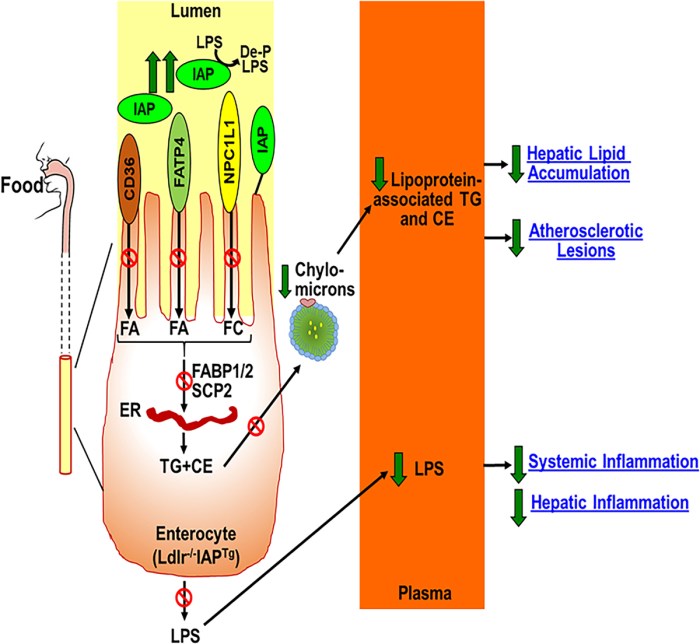
“We developed intestine-specific IAP transgenic mice (IAPTg) overexpressing human chimeric IAP to examine direct effects of increased IAP expression on barrier function and development of metabolic diseases. We evaluated effects of intestine-specific IAP overexpression in hyperlipidemic Ldlr−/− mice. The data presented demonstrated significant attenuation of Western-type diet (WD)-induced LPS translocation in Ldlr−/−IAPTg mice, with significant reduction in intestinal lipid absorption, hyperlipidemia, hepatic lipids, and development of atherosclerotic lesions.

circresaha.120.317144.fig09

IAP is produced by enterocytes, and catalyzes removal of 1 of the 2 phosphate groups from the toxic lipid A moiety of LPS. This produces monophosphoryl-LPS, and results in attenuation of the downstream TLR (Toll-like receptor)-4–dependent inflammatory cascade.

IAP also:

  • Dephosphorylates other proinflammatory molecules such as flagellin and ATP, resulting in their detoxification;
  • Regulates expression of key gap junction proteins (zonula occludens, claudin, and occludin) and their cellular localization, which directly modulates intestinal barrier function;
  • Promotes growth of various commensal bacteria in the gut by decreasing luminal concentrations of nucleotide triphosphates via dephosphorylation; and
  • Translocates from the apical surface of enterocytes during fat absorption. Increased serum IAP accompanies fat absorption, which is consistent with observed increased levels of circulating LPS in WD-fed mice, providing one more likely mechanism by which WD affects intestinal barrier function via IAP.

Nutrients and food components/supplements that increase IAP include galacto- or chito- oligosaccharides, glucomannan, and vitamin D3. These provide a novel opportunity to develop simple strategies for modulation of diet/nutrition to target metabolic diseases including diabetes, fatty liver disease, atherosclerosis, and heart disease.”

https://www.ahajournals.org/doi/10.1161/CIRCRESAHA.120.317144 “Over-Expression of Intestinal Alkaline Phosphatase Attenuates Atherosclerosis”


Previously curated IAP studies were:

PXL_20210710_093310161.NIGHT

Gut and brain health

This 2021 human review subject was interactions of gut health and disease with brain health and disease:

“Actions of microbial metabolites are key for appropriate gut-brain communication in humans. Among these metabolites, short-chain fatty acids (SCFAs), tryptophan, and bile acid metabolites / pathways show strong preclinical evidence for involvement in various aspects of brain function and behaviour.

Dietary fibres, proteins, and fats ingested by the host contain components which are metabolized by microbiota. SCFAs are produced from fermentation of fibres, and tryptophan-kynurenine (TRP-KYN) metabolites from dietary proteins. Primary bile acids derived from liver metabolism aid in lipid digestion, but can be deconjugated and bio-transformed into secondary bile acids.

1-s2.0-S0149763421001032-gr1

One of the greatest challenges with human microbiota studies is making inferences about composition of colonic microbiota from faeces. There are known differences between faecal and caecal microbiota composition in humans along with spatial variation across the gastrointestinal tract.

It is difficult to interpret microbiome-host associations without identifying the driving influence in such an interaction. Large cohort studies may require thousands of participants on order to reach 20 % explanatory power for a certain host-trait with specific microbiota-associated metrics (Shannon diversity, relative microbial abundance). Collection of metadata is important to allow for a better comparison between studies, and to identify differentially abundant microbes arising from confounding variables.”

https://www.sciencedirect.com/science/article/pii/S0149763421001032 “Mining Microbes for Mental Health: Determining the Role of Microbial Metabolic Pathways in Human Brain Health and Disease”


Don’t understand why these researchers handcuffed themselves by only using PubMed searches. For example, two papers were cited for:

“Conjugated and unconjugated bile acids, as well as taurine or glycine alone, are potential neuroactive ligands in humans.”

Compare scientific coverage of PubMed with Scopus:

  • 2017 paper: PubMed citations 39; Scopus citations 69.
  • 2019 paper: PubMed citations 69; Scopus citations 102.

Large numbers of papers intentionally missing from PubMed probably influenced this review’s findings, such as:

  1. “There are too few fibromyalgia and migraine microbiome-related studies to make definitive conclusions. However, one fibromyalgia study found altered microbial species associated with SCFA and tryptophan metabolism, as well as changes in serum levels of SCFAs. Similarly, the sole migraine-microbiota study reported an increased abundance of the kynurenine synthesis GBM (gut-brain module).
  2. Due to heterogeneity of stroke and vascular disease conditions, it is difficult to make substantial comparisons between studies. There is convincing evidence for involvement of specific microbial genera / species and a neurovascular condition in humans. However, taxa were linked to LPS biosynthesis rather than SCFA production.
  3. Several studies suggest lasting microbial changes in response to prenatal or postnatal stress, though these do not provide evidence for involvement of SCFA, tryptophan, or bile-acid modifying bacteria. Similar to stress, there are very few studies assessing impact of post-traumatic stress disorder on microbiota.”

These researchers took on a difficult task. Their study design could have been better.


PXL_20210628_095746132

Wildlife

PXL_20210710_100826663

Cow milk causes disease

This 2021 review followed up Epigenetic effects of cow’s milk and many papers since then:

“Epidemiological studies associate intake of cow milk with an increased risk of diseases, which are associated with overactivated mechanistic target of rapamycin complex 1 (mTORC1) signaling. Milk’s physiological function to maintain high mTORC1 signaling at the beginning of mammalian life turns into adverse health effects when this postnatal endocrine and epigenetic system is not discontinued as designated by physiological processing of the lactation genome.

Milk is a signaling interface between the maternal lactation genome and the infant’s cellular mTORC1 system that orchestrates growth, anabolism, metabolic, immunological, and neurological programming. Pasteurization combined with refrigeration exposed human milk consumers to bioactive milk exosome (MEX)-derived micro-ribonucleic acids (miRs), augmenting milk’s mTORC1 activity compared to boiled, ultra-heat-treated, or fermented milk.

milk-mediated mTORC1 signaling

Milk consumption activates five major pathways stimulating mTORC1 via:

  1. Growth factors, including growth hormone, insulin, and insulin-like growth factor 1;
  2. Amino acids, especially branched-chain amino acids;
  3. Milk fat-derived palmitic acid;
  4. Milk sugar lactose; and
  5. Epigenetic modifiers, especially MEX-derived miRs.

Understanding milk’s interaction with the central hub of metabolic regulation, mTORC1, will open new avenues for prevention of common diseases.”

https://www.mdpi.com/2218-273X/11/3/404/htm “Lifetime Impact of Cow’s Milk on Overactivation of mTORC1: From Fetal to Childhood Overgrowth, Acne, Diabetes, Cancers, and Neurodegeneration”


This reviewer is somewhat of a zealot. Still, he cited 555 references.

His genotype may tolerate lactose, but he didn’t argue for it:

“After breast feeding, mucosal expression of lactase, an intestinal enzyme hydrolyzing lactose into glucose and galactose, is downregulated in all mammals with the exception of Neolithic humans, who developed LCT [lactase gene] mutations allowing persistent lactase expression.

Lactose content of milk makes up around 2–8% by weight. Lactose hydrolysis provides glucose and galactose, which both activate mTORC1:

  • During glucose abundance and glycolysis, sufficient cellular energy is produced in the form of ATP, which suppresses AMPK activity. Aldolase operates as a sensor for glucose availability that directly links glucose shortage to activation of AMPK.
  • Galactose via induction of oxidative stress activates mTORC1. Galactose-induced overactivation of mTORC1 promotes senescence of neural stem cells and aging of mesenchymal stem cells.

Lactobacilli used in food and dairy fermentation increase NRF2 activation, resulting in NRF2-induced sestrin expression, which attenuates mTORC1 activation.”

Eat broccoli sprouts for your hearing

Two 2021 papers, both of which I found by each citing a 2009 Molecular mechanisms underlying cochlear degeneration in the tubby mouse and the therapeutic effect of sulforaphane (not freely available). First was a review:

“Hair cell damage and loss mediated by oxidative stress are important causes of hearing loss. Sensorineural hearing loss is the most common type of hearing loss, including noise induced hearing loss (NIHL), age-related hearing loss (ARHL), and ototoxic hearing loss.

Nrf2 reduces cell damage caused by oxidative stress, and maintains the dynamic balance of systematic redox by inducing and regulating expression of various antioxidant factors. This review summarizes correlation studies of Nrf2 in hearing loss, providing ideas for prevention and treatment of hearing loss with Nrf2 as the target.

fphar-12-620921-g002

There is positive feedback between p62-mediated autophagy and Nrf2. p62 promotes accumulation of Nrf2 and nuclear translocation. Concurrently, increased Nrf2 promotes p62 expression.

How Nrf2 regulates ROS changes in hair cells, and the upstream and downstream regulatory network of Nrf2 in hair cells, are still not fully understood. Studies on early prevention and treatment of hearing loss through the Keap1-Nrf2-ARE [antioxidant response element] signaling axis are still at the exploratory stage.”

https://www.frontiersin.org/articles/10.3389/fphar.2021.620921/full “The Role of Nrf2 in Hearing Loss”


Second paper was a rodent study:

“We examined oxidative stress and antioxidant response of the p62-Keap1-Nrf2 pathway in cochleae during age-related hearing loss (ARHL) and noise-induced hearing loss (NIHL). We elucidated the function of full-length and variant p62/Sqstm1 (referred to here as p62) in regulation of Nrf2 activation.

Cochlear damage was assessed by analyzing auditory brainstem response (ABR) and by counting hair cells (HCs). Malondialdehyde (MDA, a lipid peroxidation product) levels were measured in young and old mice to determine whether oxidative stress contributed to ARHL.

auditory brainstem response

  • (A) Audiometric threshold (dB) determined from click and pure tone evoked ABRs. Thresholds were each significantly different (P < 0.001) between young mice and old mice.
  • (B) HC loss percentage in basal cochlear turns. Significant differences (P < 0.001) were observed between young and old mice.
  • (C) MDA levels in the cochleae of old mice were significantly higher (P = 0.034) than those of young mice.

ROS accumulation is closely related to ARHL and NIHL. The inability of ROS accumulation to activate the Nrf2 antioxidant stress pathway under physiological conditions may be related to alternative splicing of p62 mRNA in cochleae.

However, the agonist of the Nrf2 pathway enhanced Nrf2 nuclear translocation. This suggests a mechanism in which the antioxidant pathway was difficult to be activated in the context of accumulation of ROS.”

https://www.researchsquare.com/article/rs-535219/v1 “New Target of Oxidative Stress Regulation in Cochleae: Alternative Splicing of the p62/Sqstm1 gene”


The study’s two-month-old mice were equivalent to a 20-year-old human. Its 13-to-14-month-old mice were equivalent to humans in their 60s to 70s.

I expected preconditioning to be mentioned in both papers. Maybe these researchers thought it was too obvious and didn’t need to be stated that:

  • Repeated use of a Nrf2 activator produces transient mild stress;
  • Which elicits a stronger response; and
  • Preconditions cells for future stress?

Sulforaphane in the Goldilocks zone and its cited papers exhaustively emphasized preconditioning’s importance. The main thing I’m trying to do with isothiocyanates is to send a weak pro-inflammatory signal to my endogenous ARE system to exercise natural defenses.

Twice-daily drills make me more proficient at responding to actual emergencies. Post-drill, my body recycles material to be ready to respond the next time.

I do the same thing once a day with yeast cell wall β-glucan 1,3/1,6 to train my innate immune system. Microphages in my gut are the first responders. Like the very reactive isothiocyanates, I don’t take anything with, or an hour before or after β-glucan 1,3/1,6.

Why tolerate “the antioxidant pathway was difficult to be activated in the context of accumulation of ROS” when a sulforaphane “agonist of the Nrf2 pathway enhanced Nrf2 nuclear translocation”? For all we know, diminished natural defenses and hearing loss may exist to turn old mammals into prey.

Continued in Part 2.

The amino acid ergothioneine

A trio of papers on ergothioneine starts with a 2019 human study. 3,236 people without cardiovascular disease and diabetes mellitus ages 57.4 ± 6.0 were measured for 112 metabolites, then followed-up after 20+ years:

“We identified that higher ergothioneine was an independent marker of lower risk of cardiometabolic disease and mortality, which potentially can be induced by a specific healthy dietary intake.

overall mortality and ergothioneine

Ergothioneine exists in many dietary sources and has especially high levels in mushrooms, tempeh, and garlic. Ergothioneine has previously been associated with a higher intake of vegetables, seafood and with a lower intake of solid fats and added sugar as well as associated with healthy food patterns.”

https://heart.bmj.com/content/106/9/691 “Ergothioneine is associated with reduced mortality and decreased risk of cardiovascular disease”


I came across this study by its citation in a 2021 review:

“The body has evolved to rely on highly abundant low molecular weight thiols such as glutathione to maintain redox homeostasis but also play other important roles including xenobiotic detoxification and signalling. Some of these thiols may also be derived from diet, such as the trimethyl-betaine derivative of histidine, ergothioneine (ET).

image description

ET can be found in most (if not all) tissues, with differential rates of accumulation, owing to differing expression of the transporter. High expression of the transporter, and hence high levels of ET, is observed in certain cells (e.g. blood cells, bone marrow, ocular tissues, brain) that are likely predisposed to oxidative stress, although other tissues can accumulate high levels of ET with sustained administration. This has been suggested to be an adaptive physiological response to elevate ET in the damaged tissue and thereby limit further injury.”

https://www.sciencedirect.com/science/article/pii/S2213231721000161 “Ergothioneine, recent developments”


The coauthors of this review were also coauthors of a 2018 review:

“Ergothioneine is avidly taken up from the diet by humans and other animals through a transporter, OCTN1. Ergothioneine is not rapidly metabolised, or excreted in urine, and has powerful antioxidant and cytoprotective properties.

ergothioneine in foods

Effects of dietary ET supplementation on oxidative damage in young healthy adults found a trend to a decrease in oxidative damage, as detected in plasma and urine using several established biomarkers of oxidative damage, but no major decreases. This could arguably be a useful property of ET: not interfering with important roles of ROS/RNS in healthy tissues, but coming into play when oxidative damage becomes excessive due to tissue injury, toxin exposure or disease, and ET is then accumulated.”

https://febs.onlinelibrary.wiley.com/doi/full/10.1002/1873-3468.13123 “Ergothioneine – a diet-derived antioxidant with therapeutic potential”


I’m upping a half-pound of mushrooms every day to 3/4 lb. (340 g). Don’t think I could eat more garlic than the current six cloves.

PXL_20210606_095517049

I came across this subject in today’s video:

Foods for your vision

This 2021 review by five ophthalmologists and two researchers characterized findings of food effects on human vision:

“The most challenging ocular disorders are uncorrected / under-corrected refractive errors, ocular surface dysfunction / dry eye disease, cataracts, glaucoma, diabetic retinopathy (DR), and age-related macular degeneration (AMD):

  • Severe visual impairment and blindness due to cataract or refractive error constitutes half of all global cases;
  • Glaucoma is the most common cause of irreversible blindness;
  • DR is the first cause of visual disability in working-age adults; and
  • AMD is the first cause of blindness in the elderly.

We identify directions for further research on:

  • The role of diet and nutrition in eyes and vision;
  • Potential antioxidant, anti-inflammatory, and neuroprotective effects of natural food (broccoli, saffron, tigernuts and walnuts);
  • The Mediterranean Diet; and
  • Nutraceutic supplements that may supply a promising and highly affordable scenario for patients at risk of vision loss.

We improve understanding of natural food nutritional hallmarks, benefits of the MedDiet, and appropriate oral supplements with vitamins, carotenoids and PUFAs for better eye and vision care.”

https://www.mdpi.com/2304-8158/10/6/1231/htm “Searching for the Antioxidant, Anti-Inflammatory, and Neuroprotective Potential of Natural Food and Nutritional Supplements for Ocular Health in the Mediterranean Population”


eyes

🙂

The next phase of reversing aging and immunosenescent trends

Dr. Greg Fahy earlier this week provided an update on the November 2020 TRIIM-X follow-on to the September 2019 TRIIM curated in Reversal of aging and immunosenescent trends. Emphasis was on reproducibility:

23:45 Dr. Steve Horvath reanalyzed TRIIM for the plasma portion of Levine’s PhenoAge epigenetic clock. Results were congruent with four other epigenetic clocks showing a 2.5 year reduction of biological age.

39:20 TRIIM-X preliminary results started with C-Reactive protein.

43:05 No backsliding in epigenetic age deceleration between TRIIM and TRIIM-X!

continued epigenetic age deceleration

55:07 Q & A session starts with how TRIIM-X controls for supplements. Answers for resveratrol and calorie restriction, emphasizing that CR doesn’t reverse aging.

1:10 TRIIM-X took photos of subjects’ hair at baseline!


Great update! The last 20 minutes emphasized a need for capital in aging research. TRIIM-X has another 1.5 years to go, and other aging research projects needing funding were mentioned.

Don’t know what happened to the unmentioned 3000 IU vitamin D and 50 mg zinc recommendations of TRIIM. So I asked. Dr. Fahy replied:

“They are still there! Just not mentioned!”

Thought briefly about enrolling in TRIIM-X, but there’s no way anyone but me gets to experiment with my body.

Vitamin K2 – What can it do?

A trio of papers on Vitamin K2, the first being a 2021 review that emphasized dual effects:

“Osteoporosis (OP) is the most common bone disease that affects elderly men and women. It is a metabolic skeletal disorder caused by an imbalance between bone formation and resorption, leading to a loss of bone mass and quality, skeletal structure deterioration, and an increased risk of fractures.

Vascular calcification is defined as ectopic deposition of mineral matrix in vessel wall. It occurs prevalently in aging and primary chronic conditions (hypertension, diabetes mellitus, and chronic kidney disease), representing an important risk factor for cardiovascular morbidity and mortality.

Studies have provided support for a close link between bone and vascular health. Findings suggest that bone loss in OP may promote and increase the risk of cardiovascular events and vascular atherosclerosis.

Vitamin K2 is involved in a phenomenon in which a low calcium deposition in bone tends to be associated with a parallel increase of calcium deposition in vessel wall as a consequence of impaired calcium metabolism. Most production of Vitamin K2 in humans takes place in intestines. However, the amount derived from intestinal bacteria is poorly absorbed, and is not able to reach concentrations required to exert physiological functions.

Vitamin K2‘s ability to reduce loss of bone mineral density and fracture risk, as well as to improve bone quality, has been described by several clinical studies, which have confirmed that osteocalcin (OC) γ-carboxylation is the main mechanism of action through which this natural compound is able to improve bone health. Clinical evidence suggests an analogous protective role of Vitamin K2 at the vascular level, emphasizing a strict association between:

  • Vitamin serum level;
  • Matrix gla protein (MGP) γ-carboxylation levels;
  • Reduction of vascular smooth muscle cells osteogenic trans-differentiation; and
  • Possible risk of cardiovascular events.”

https://www.mdpi.com/2072-6643/13/4/1222/htm “The Dual Role of Vitamin K2 in ‘Bone-Vascular Crosstalk’: Opposite Effects on Bone Loss and Vascular Calcification”


A second 2021 review emphasized aging:

“Vitamin K can:

  • Carboxylate OC (a protein capable of transporting and fixing calcium in bone);
  • Activate MGP (an inhibitor of vascular calcification and cardiovascular events); and
  • Carboxylate Gas6 protein (involved in brain physiology and a cognitive decline and neurodegenerative disease inhibitor).

By improving insulin sensitivity, Vitamin K lowers diabetes risk. It also exerts antiproliferative, proapoptotic, autophagic effects, and has been associated with a reduced risk of cancer.

The most common [Vitamin K2] subtypes in humans are the short-chain MK[menaquinone]-4, which is the only MK produced by systemic conversion of phylloquinone [Vitamin K1] to menaquinone, and MK-7 through MK-10, which are synthesized by bacteria. The main sources of Vitamin K2 are fermented foods, cheeses, eggs, and meats.”

https://www.mdpi.com/2076-3921/10/4/566/htm “The Role of Vitamin K in Humans: Implication in Aging and Age-Associated Diseases”


The third paper – somehow not cited by these two reviews – was a 2006 human study that performed four experiments:

“The synthetic short-chain vitamin K1 is commonly used in food supplements, but recently the natural long-chain MK-7 has also become available as an over-the-counter supplement. The purpose of this paper was to compare in healthy volunteers absorption and efficacy of K1 and MK-7.

Serum vitamin K species were used as a marker for absorption and OC carboxylation as a marker for activity. Both K1 and MK-7 were absorbed well, with peak serum concentrations at 4 hours after intake.

A major difference was:

  • Very long half-life time of MK-7, resulting in much more stable serum levels; and
  • Accumulation of MK-7 to higher levels (7- to 8-fold) during prolonged intake.

MK-7 induced more complete carboxylation of OC.

Vitamin K2 vs K1

Accumulation and efficacy of K vitamins during long-term daily administration. Participants received in a crossover design either K1 (○) or MK-7 (•) or placebo; in the latter case only K1 (▴) could be detected.

  • (A) Circulating levels of vitamin K; baseline levels for K1 were subtracted; no MK-7 could be detected at baseline.
  • (B) Ratio between circulating carboxylated and undercarboxylated osteocalcin (cOC/ucOC); at baseline the ratio was 1.74 for MK-7, 1.8 for K1, and 1.7 for the placebo group.

MK-7 accumulated during the first 2 weeks until it reached a plateau level of about 10 nM (6 μg/L), whereas K1 remained slightly above placebo values during the entire study period. Efficacy of both K vitamins for OC carboxylation was monitored using the ratio between circulating cOC and ucOC, and it turned out that within 3 days both vitamins had induced increased cOC.

But only by taking MK-7 did the effect continue to increase during the entire study period.

Taken together, these data demonstrate considerable differences between MK-7 and K1:

  • Higher and more stable serum levels are reached with MK-7; and
  • MK-7 has a higher efficacy in both hepatic and extrahepatic protein carboxylation.”

https://ashpublications.org/blood/article/109/8/3279/23729/Vitamin-K-containing-dietary-supplements “Vitamin K–containing dietary supplements: comparison of synthetic vitamin K1 and natto-derived menaquinone-7″


I’ve tried various things over the years to address hypertension. I stopped high blood pressure medications briefly to see if each intervention worked. They all haven’t, presumably because I didn’t address causes.

More recently, I broke my left big toe on furniture while walking around in the dark last month, and haven’t recovered. No pictures from walking on the beach at sunrise because it still isn’t possible. 😦

A link between these two health conditions could be Vitamin K2. I don’t eat fermented foods because of their high sodium, or dairy products, and haven’t supplemented Vitamin K2.

Next week I’ll start a 300 μg MK-7 daily dose. Current Vitamin D3 dose is 3800 IU, compared to the second paper of Part 2 of Vitamin K2 – What can it do? which is 400 μg MK-7 and 3200 Vitamin D3.

Astaxanthin bioavailability

By request, research on astaxanthin bioavailability. I used a “astaxanthin” “bioavailability” “quinone reductase” 2021 search term, and read citing papers.

“The bioaccessibility, bioavailability, and antioxidative activities of three astaxanthin geometric isomers were investigated using an in vitro digestion model.

  • 13Z-Astaxanthin showed higher bioaccessibility than 9Z- and all-E-astaxanthins during in vitro digestion, and
  • 9Z-astaxanthin exhibited higher transport efficiency than all-E- and 13Z-astaxanthins.

These might explain why 13Z- and 9Z-astaxanthins are found at higher concentrations in human plasma than all-E-astaxanthin.

9Z- and 13Z- astaxanthins exhibited a higher protective effect than all-E-astaxanthin against oxidative stress.”

https://pubs.acs.org/doi/10.1021/acs.jafc.7b04254 “Bioaccessibility, Cellular Uptake, and Transport of Astaxanthin Isomers and their Antioxidative Effects in Human Intestinal Epithelial Caco-2 Cells” (2017, not freely available)


jf1c00087_0005

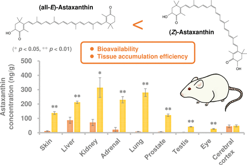
“Astaxanthin with a high proportion of Z-isomer (especially rich in 9Z- and 13Z-isomers) was prepared from (all-E)-astaxanthin by thermal treatment and solid–liquid separation. Z-isomer-rich astaxanthin diet resulted in higher levels of astaxanthin in blood and many tissues (in particular, skin, lung, prostate, and eye) compared to all-E-isomer-rich diet.

Z-isomer-rich diet enhanced the level of 13Z-isomer in blood and tissues rather than that of 9Z-isomer. (13Z)-astaxanthin would have higher bioavailability and tissue accumulation than other isomers.”

https://pubs.acs.org/doi/10.1021/acs.jafc.1c00087 “Z-Isomers of Astaxanthin Exhibit Greater Bioavailability and Tissue Accumulation Efficiency than the All-E-Isomer” (2021, not freely available)


“Astaxanthin is highly susceptible to light, oxygen, and heat stress degradation. In addition, poor water solubility and bioavailability limit its efficacy in vivo. Investigating novel astaxanthin delivery systems is necessary in order to solve these drawbacks.”

https://www.mdpi.com/1420-3049/24/14/2640/htm “The Neuroprotective Effects of Astaxanthin: Therapeutic Targets and Clinical Perspective” (2019)


“Astaxanthin Z-isomers potentially have greater bioavailability and biological activity than (all-E)-astaxanthin. However, stability of Z-isomers is lower than all-E-isomer, which is a serious problem affecting its practical use.

In this study, we investigated impacts of different suspension media (oils and fats) and additives on astaxanthin isomer stability.

  • Z-isomers of astaxanthin isomerized to all-E-isomer during storage.
  • When soybean and sunflower oils were used as the suspension medium, astaxanthin isomers were hardly degraded. However the total Z-isomer ratio decreased from ~80% to ~50% during 6-week storage at 30 °C.
  • (9Z)-astaxanthin showed higher stability than 13Z- and 15Z-isomers.”

https://www.sciencedirect.com/science/article/abs/pii/S0308814621003770 “Evaluation and improvement of storage stability of astaxanthin isomers in oils and fats” (2021, not freely available)


I looked for but didn’t find a graph similar to this one that comparatively plotted astaxanthin:

OMCL2019-2716870.006

I also didn’t find recent human studies.

It seems that a special delivery system is required for taking astaxanthin as a supplement. It would require investigating manufacturers’ claims about isomer content and stability.

Eating colorful seafood is another way to get astaxanthin. Don’t know about eating raw or dried algae.

Ride the waves of gene expression with betaine

This 2021 cell study investigated a dietary supplement’s role in preventing nerve disease:

“A loss of epigenetic control has been implicated in development of neurodegenerative diseases. Previous studies have implicated aberrant DNA and histone methylation in multiple sclerosis (MS) disease pathogenesis.

We have previously reported that methyl donor betaine is depleted in MS and is linked to changes in histone H3 trimethylation (H3K4me3) in neurons. We have also shown that betaine increases histone methyltransferase activity by activating chromatin bound betaine homocysteine S-methyltransferase (BHMT).

A hallmark of MS is the death of oligodendrocytes, the cells responsible for wrapping axons in myelin in the central nervous system and maintaining a healthy sheath. In demyelinating diseases like MS, oligodendrocyte progenitor cells (OPCs) fail to differentiate and make more myelin, resulting in sclerotic lesions.

Promoting differentiation of OPCs and generation of myelin is of great interest as a novel MS therapy. Waves of gene regulation (repression and activation) need to occur to promote myelination.

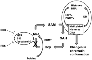
This BHMT-betaine methylation pathway ensures availability of S-adenosylmethionine (SAM) for a variety of DNA and histone methylation processes. OPC survival and differentiation are dependent upon DNA and histone methylation, and both processes require SAM.

journal.pone.0250486.g001

BHMT uses betaine to remethylate homocysteine to methionine. Betaine can be taken in through the diet or synthesized through the oxidation of choline in mitochondria.

We demonstrated that oligodendrocyte gene expression can be modulated by betaine supplementation through the BHMT-betaine methylation pathway. Our study suggests that dietary betaine supplementation may prove to be a therapeutic agent for MS and other demyelinating disorders.”

https://journals.plos.org/plosone/article?id=10.1371/journal.pone.0250486 “The BHMT-betaine methylation pathway epigenetically modulates oligodendrocyte maturation”


I started taking betaine 16 years ago. Didn’t know of these effects until reading this study.

Treating psychopathological symptoms will somehow resolve causes? had more on betaine (aka trimethyl glycine). Current dose is 1.5 grams twice daily.

Are rodent models of human neurodegenerative diseases realistic?

This 2020 stem cell review argued against rodent models of human neurodegenerative diseases:

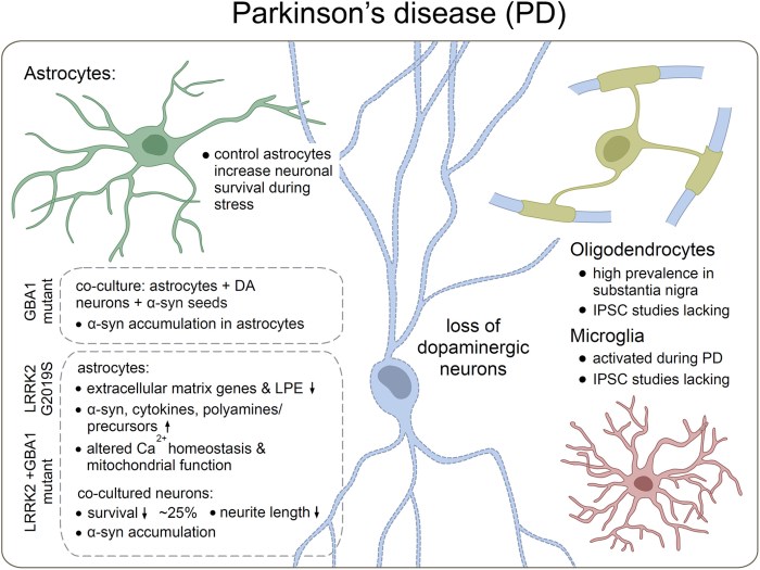
“Neuronal loss is not caused solely by intrinsic degenerative processes but rather via impaired interactions with surrounding glia and other brain cells. Dysfunctional astrocytes do not provide sufficient nutrients and antioxidants to neurons, while dysfunctional microglia cannot efficiently clear pathogens and cell debris from extracellular space, resulting in chronic inflammatory processes in the brain.

Human glia, especially astrocytes, differ significantly in morphology and function from their mouse counterparts. Recent advances in stem cell technology make it possible to reprogram human patients’ somatic cells to induced pluripotent stem cells (iPSC) and differentiate them further into patient‐specific glia and neurons, thus providing a source of human brain cells.

stem3309-fig-0002-m

Astrocytes do not efficiently utilize energy resources and cannot provide adequate metabolic support to neurons. A coculture of healthy human neurons with diseased astrocytes impaired neuronal calcium responses to glutamate and γ‐aminobutyric acid (GABA) as compared to coculture with healthy human astrocytes.

Treatment with sulforaphane:

  • Normalized basal level glycolysis;
  • Decreased basal level Aβ42 secretion; as well as
  • Ameliorated inflammatory response to pro‐inflammatory cytokines TNF-α and IL1-β in PSEN1 mutant iPSC astrocytes.

It is essential to make sure that what we see in the dish is the real patient‐specific phenotype. Transplantation of human brain organoids containing microglia into mice could provide a novel tool for drug screening in vivo.”

https://stemcellsjournals.onlinelibrary.wiley.com/doi/full/10.1002/stem.3309 “Metabolic and immune dysfunction of glia in neurodegenerative disorders: Focus on iPSC models”


This review’s thesis seems plausible. However, one problem with in vitro stem cell studies is that they often don’t have a control group.