Impact of processing and storage methods on nutritional values of a dozen fruits and vegetables

This 2023 study investigated a dozen fruits and vegetables processed with three methods and freezer times for impacts on their sixteen main nutrients. I’ll focus on sulforaphane:

“This paper compares how different processing methods (pasteurization vs. high hydrostatic pressure processing or pascalization) affect phytochemical concentrations of a complex mixture of fruits and vegetables, and investigates how these methods influence their stability during freezing and over time in frozen storage. Phytochemicals tested were vitamin C, quercetin-3-glucoside, delphinidin-3-glucoside, cyanidin-3-glucoside, peonidin-3-glucoside, catechin, epigallocatechin-3-gallate, epicatechin, epicatechin gallate, chlorogenic acid, sulforaphane, resveratrol, lycopene, lutein, alpha-carotene, and beta-carotene.

After freezing to −18 °C, one bottle from each condition was immediately removed from the freezer and thawed at 4 °C, which took about two days. Measurements at t = 0 for the fresh and frozen condition were technically made two days after processing.

antioxidants-12-01252-g001

antioxidants-12-01252-g010

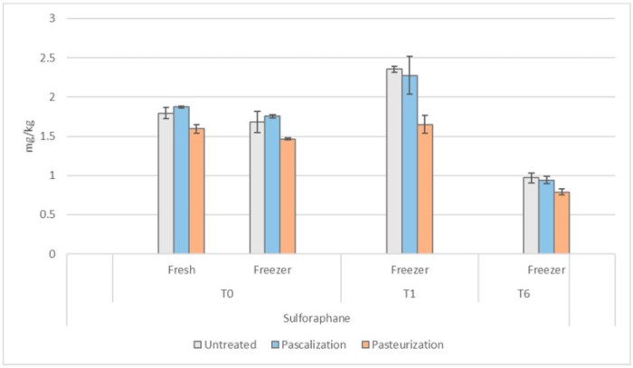
The effect of immediate freezing and thawing on broccoli, cauliflower, and Brussels sprouts sulforaphane levels was consistent despite the processing method (−6% for pascalized and untreated samples, and −8% for pasteurized) at t = 0. Pasteurized samples at t = 0 were 11% lower in sulforaphane than untreated in fresh samples and 13% lower in frozen.

At one month in the freezer, levels of sulforaphane increased in each processing method from t = 0:

  • Untreated by +18%;
  • Pascalized by +57%; and
  • Pasteurized by +94%.

At six months in the freezer, sulforaphane levels in all samples decreased below their t = 0 levels:

  • Untreated by -31%;
  • Pascalized by -35%; and
  • Pasteurized by -35%.

Optimal processing method seems to vary based on the phytochemical of interest. These impacts should be considered to produce foods aimed at preventing chronic disease development.'”

https://www.mdpi.com/2076-3921/12/6/1252 “Impact of Processing Method and Storage Time on Phytochemical Concentrations in an Antioxidant-Rich Food Mixture”


Untreated samples’ sulforaphane took a hit in this study from fresh levels to initial freezing at -18°C then thawing for two days at 4°C. Untreated levels recovered after a month to be more than their two-day levels, but lowered again after six months.

My refrigerator / freezer has one control for both compartments. Pretty sure the freezer can’t get to 0°F / -18°C without ruining refrigerator fruits and vegetables.

In any event, a (1 – .06) x 1.18 = +11% sulforaphane gain after a month isn’t worth my effort. We can increase sulforaphane more than 1100% by microwaving broccoli sprouts in a 1000W microwave on full power for 35 seconds to 60°C (140°F) per Week 6 of Changing an inflammatory phenotype with broccoli sprouts.

PXL_20230610_093652335

A biomarker for impaired cognitive function?

This 2023 rodent study investigated associations between a drug, a gut microbiota species, cognitive function, and proinflammatory cytokine interleukin-6:

“We show that gut microbiota is altered by metformin, which is necessary for protection against ageing-associated cognitive function declines in aged mice.

  • Mice treated with antibiotics did not exhibit metformin-mediated cognitive function protection.
  • Treatment with Akkermansia muciniphila improved cognitive function in aged mice.
  • A. muciniphila decreased proinflammatory-associated pathways, particularly that of proinflammatory cytokine interleukin (IL)-6, in both peripheral blood and hippocampal profiles, which was correlated with cognitive function improvement.
  • An IL-6 antibody protected cognitive function, and an IL-6 recombinant protein abolished the protective effect of A. muciniphila on cognitive function in aged mice.

40168_2023_1567_Figa_HTML

A. muciniphila, which is mediated in gut microbiota by metformin, modulates inflammation-related pathways in the host and improves cognitive function in aged mice by reducing proinflammatory cytokine IL-6 both systemically and in the hippocampus. This is direct evidence to validate that gut microbiota mediate the effect of metformin on cognitive improvement.”

https://microbiomejournal.biomedcentral.com/articles/10.1186/s40168-023-01567-1Akkermansia muciniphila, which is enriched in the gut microbiota by metformin, improves cognitive function in aged mice by reducing the proinflammatory cytokine interleukin-6″


IL-6 may be useful with other biomarkers of impaired cognitive function. It’s too coarse to track improved cognitive function past a certain point, though. Maybe the current IL-6 blood test can be refined as high-specificity CRP and regular CRP blood tests were done?

We don’t need to take this drug or be concerned about this gut bacteria species in order to lower inflammation. Click the IL-6 link above and see blog posts such as Part 2 of Rejuvenation therapy and sulforaphane for other methods.

PXL_20230630_092833465

Paradigms determine findings

This 2023 rodent study from Dr. Michael Skinner’s labs at Washington State University investigated epigenetic transgenerationally inherited differential DNA methylation regions (DMRs). I’ll focus on a paradigm shift that enabled some of this study’s findings:

“The current study was designed to assess if morula embryos escape the erasure of DDT-induced transgenerational sperm DMR methylation. Observations demonstrate:

  • 98% of transgenerational sperm DMR sites retain DNA methylation and are not erased, appearing similar to imprinted-like sites.
  • Maintenance of DNA methylation on a variety of imprinted sites in a comparison of sperm versus morula methylation levels using methylated DNA immunoprecipitation (MeDIP) followed by next-generation sequencing (MeDIP-Seq).
  • The majority of low-density CpG genomic sites had a significant increase in DNA methylation in the morula embryo compared to sperm.

The general erasure of DNA methylation during embryogenesis appears applicable to high-density DNA methylation sites (e.g. CpG islands) but neither to transgenerational DMR methylation sites nor to low-density CpG deserts, which constitute the vast majority of the genome’s DNA methylation sites.

dvad003f1

Bisulfite procedures have been extensively used followed by next-generation sequencing (BS-Seq) to assess genome-wide DNA methylation in early embryonic development. This has led to the concept that DNA methylation erasure occurs during early embryo development and primordial germ cell development.

A limitation with BS-Seq is that it is often biased toward detecting changes in higher-density CpG sites with >5 CpG/100 bp. A critical technical limitation to BS-Seq is that bioinformatics protocols used remove low-density (<3 CpG/100 bp) regions from the genome prior to analysis. In contrast, MeDIP-Seq analysis is biased to low-density CpG sites with <5 CpG/100 bp that constitute >90% of the genome.

Alteration of morula stem cell epigenetics will impact epigenomes and transcriptomes of all subsequently derived somatic cells. This is the molecular basis for epigenetic transgenerational inheritance phenotypes and pathologies.

Future studies need to re-evaluate the current dogma of a genome-wide erasure of DNA methylation, and consider a more dynamic regulation of early embryonic stem cell epigenetic development.”

https://academic.oup.com/eep/article/9/1/dvad003/7190131 “Transgenerational sperm DMRs escape DNA methylation erasure during embryonic development and epigenetic inheritance”


PXL_20230614_014127763

Comparing ten dietary fibers’ effects on obesity

This 2023 rodent study compared a high-fat high-sugar diet’s deleterious effects with adding ten bioactive dietary fibers vs. adding the weight-loss pill Orlistat vs. a normal diet:

“Different dietary fibers supplementation improved obesity in rats with diversely positive responses, improvement of dyslipidemia, serum hormone, serum metabolome, and gut microbiota disorders.

Model group ate 66.5% normal chow diet, 10% lard, 20% sucrose, 2.5% cholesterol and 1% sodium cholate. Normal group ate normal chow.

Treatment groups ate high-fat high-sugar diet plus 270 mg/kg each of either barley β-glucan, glucomannan, arabinoxylan, inulin, guar gum, xanthan gum, carrageenan, apple pectin, arabinogalactan, or xylan dietary fibers. Orlistat (Y) supplementation was equivalent to 120 mg taken 3 times per day for 70 kg humans.

dietary fiber effects

We found that supplementation with β-glucan, arabinoxylan, xanthan gum, guar gum, apple pectin, carrageenan, inulin, and xylan significantly reduced body weight and dyslipidemia, whereas glucomannan and arabinogalactan did not. Apple pectin, β-glucan and arabinoxylan improved the most biomarkers (15, 17 and 18 kinds) relevant to obesity.

Most dietary fibers improved physiological indicators which have a risk with obesity, including accumulation of body fat, dyslipidemia, glucose metabolic abnormality, oxidative stress, and adipocytokines secreted by adipose tissue, while β-glucan reversed almost all physiological indicators. Consequently, intake of β-glucan could be considered as therapy for obesity management induced by high fat diet.”

https://www.sciencedirect.com/science/article/abs/pii/S0268005X23001637 “Different dietary fibers unequally remodel gut microbiota and charge up anti-obesity effects” (not freely available). Thanks to Dr. Jiajia Wen for providing a copy.


A human equivalent to this study’s 270 mg/kg daily dietary fiber intake is (270 mg x .162) x 70 kg = 3062 mg. I eat > 4 grams of oat β-glucan daily, and much less than that of orange pectin. No human will eat > 10 grams of inulin every day without having severe gastrointestinal adverse effects.

I didn’t curate this study’s gut microbiota findings because it used fecal samples. Per Measuring gut microbiota, Part 1 and Part 2, fecal microbiota don’t adequately represent gut microbiota in either the entire gastrointestinal tract, or in any section of it.

Replicating other studies’ fecal microbiota findings doesn’t advance science when these don’t define subjects’ gastrointestinal tract situations. More work is needed to improve methods of investigating gut microbiota.

PXL_20230615_092427922

Transgenerational transmission of stress

This 2023 rodent study found that effects of stress during mid-late gestation were epigenetically transmitted to the first, second, and third female generations:

“We investigated effects of gestational chronic variable stress (CVS) in rats using restraint and social isolation stress in the parental F0 generation. Only the F0 pregnant dams were subjected to stress.

When a pregnant female experiences adversity, impacts of that stress affect exposed somatic tissues (F0 generation), the fetuses (F1 generation), and the fetuses’ germline (F2 generation). A true transgenerational inheritance arises when germline epimutations are transmitted to unexposed F3 offspring.

A subset of F1 rats was housed in an enriched environment (EE) to mitigate adverse effects of CVS. F2 offspring reared in EE had increased birth weights, but their uterine gene expression patterns remained comparable to those of stressed animals.

ijms-24-03734-g001

We provide evidence that psychological and psychosocial CVS alters inflammatory status and endocrine markers in uteri of adult dams through transgenerational programming of the female germline. EE therapy in prenatally stressed F1 offspring had no beneficial effects on uterine expression of inflammatory and endocrine markers for them or their future offspring.”

https://www.mdpi.com/1422-0067/24/4/3734 “Environmental Enrichment Promotes Transgenerational Programming of Uterine Inflammatory and Stress Markers Comparable to Gestational Chronic Variable Stress”


PXL_20230611_100728709

Taurine’s effects on healthspan and lifespan

A 2023 human / primate / rodent / worm study with 56 coauthors exhaustively investigated taurine effects:

“We measured the blood concentration of taurine during aging and investigated the effect of taurine supplementation on healthspan and lifespan in several species.

  • In C57Bl/6J wild-type (WT) mice, serum taurine concentrations declined from 132.3 ± 14.2 ng/ml at 4 weeks to 40.2 ± 7.1 ng/ml at 56 weeks.
  • In 15-year-old monkeys, serum taurine concentrations were 85% lower than in 5-year-old monkeys.
  • Taurine concentrations in elderly humans were decreased by more than 80% compared with concentration in serum of younger individuals.

Regardless of their sex, taurine-fed mice survived longer than control mice. The median lifespan increase was 10 to 12%, and life expectancy at 28 months increased by 18 to 25%.

Improved survival of taurine-fed mice was not a consequence of low survival of control animals or differences in diet. Taurine deficiency is a driver of aging in mice because its reversal increases lifespan.

lifespan extension starting taurine in middle age

We investigated the health of taurine-fed middle-aged mice and found an improved functioning of bone, muscle, pancreas, brain, fat, gut, and immune system, indicating an overall increase in healthspan. Taurine reduced cellular senescence, protected against telomerase deficiency, suppressed mitochondrial dysfunction, decreased DNA damage, and attenuated inflammation.

An association analysis of metabolite clinical risk factors in humans showed that lower taurine, hypotaurine, and N-acetyltaurine concentrations were associated with adverse health, such as increased abdominal obesity, hypertension, inflammation, and prevalence of type 2 diabetes. We found that a bout of exercise increased concentrations of taurine metabolites in blood, which might partially underlie antiaging effects of exercise.

Taurine abundance decreases during aging. A reversal of this decline through taurine supplementation increases healthspan and lifespan in mice and worms, and healthspan in monkeys.”

https://www.science.org/doi/10.1126/science.abn9257 “Taurine deficiency as a driver of aging”


One area curiously not investigated in this study was that taurine supplementation freed up cysteine to do things other than synthesize taurine, like synthesize glutathione, an idea in Treating psychopathological symptoms will somehow resolve causes? An introductory article brought up this point:

“One of the most studied mechanisms of action for taurine is an increase in antioxidant capacity. Although oxidative damage is not clearly linked to mammalian lifespan, it plays a role in many age-associated pathologies.

Taurine is a poor scavenger of reactive oxygen species, with the exception of hypochlorite, which it detoxifies to N-chlorotaurine. N-Chlorotaurine is anti-inflammatory and induces expression of antioxidant enzymes in mice and humans.

Taurine supplementation might also cause an increase in levels of its precursors, including the antioxidants hypotaurine and cysteine. An interesting corollary is that up-regulating endogenous taurine synthesis would have the opposite result—consuming hypotaurine and cysteine.”

https://www.science.org/doi/10.1126/science.adi3025 “Taurine linked with healthy aging”


A human equivalent taurine dose is (1 g x .081) x 70 kg = 5.67 grams. Dose tests from supplementary data were:

“Dose and frequency of taurine administration was selected based on a pilot study, which showed that when given once daily to middle-aged WT mice, this regimen increased peak blood taurine concentrations to baseline concentrations in young (4-week-old) mice.”

taurine dose

I’ve taken 2 grams every day for the past three years, and will now bump that up to 5 grams. My diet doesn’t regularly include any foods high in taurine.

I recommend reading the study rather than commentaries. Its publisher did a very good job of linking figures so that images can be viewed, then the reader returned to the right context.

Gatekeepers are out in full force on this study, and their viewpoints are probably what you’ll see first, to include unevidenced statements like “the study’s main authors cautioned the public not to self-dose with the supplement” and the above introductory article’s unreferenced “equivalent doses used in the study by Singh et al. would be very high in humans.” Pretty pathetic that such ‘authorities’ are even publicized after recent years of deliberately misleading the world about science and medicine.

This study and all commentaries called for clinical trials that are NOT going to happen:

  • Drug companies can’t make money from a research area that’s cheap, not patentable, and readily accessible.
  • Government sponsors are likewise not incentivized to act in the public’s interest per their recent behavior.

Take responsibility for your own one precious life. See Part 2 for a sample of citing papers.

PXL_20230601_181526429

Amphibian epigenetic clocks

This 2023 study of two frog species expanded one of the cited studies in Epigenetic clocks so far in 2022 to include post-embryonic epigenetic clock measurements:

“We generated DNA methylation data from African clawed frogs (Xenopus laevis) and Western clawed frogs (Xenopus tropicalis) and built multiple epigenetic clocks. Dual species clocks were developed that apply to both humans and frogs (human-clawed frog clocks), supporting that epigenetic aging processes are evolutionary conserved outside mammals.

The two species underlying our Xenopus clocks have markedly different maximum lifespans (30.3 for X. laevis and 16 for X. tropicalis), and average ages of sexual maturity (1 year for laevis and 0.375 for tropicalis). When building our Xenopus clocks, we addressed this fact in two ways:

  • In our pan-clock, we used a log-linear transformation of age that effectively normalizes ages with respect to age at sexual maturity.
  • In our relative pan-clock, we instead estimate relative age (chronological age divided by maximum lifespan), which normalizes ages with respect to maximum lifespan.

We also created dual-species clocks, referred to as human-clawed frog clocks, for estimates of chronological age and relative age. Relative age is the ratio of chronological age to maximum lifespan, and takes on values between 0 and 1. Maximum lifespan observed for humans was 122.5 years.

The relative age clock allows for alignment and biologically meaningful comparison between species with different lifespans.

relative age

Previous studies in humans showed that a hallmark of age-related CpGs is their association with target sites of Polycomb repressive complex 2 (PRC2), which gain methylation with age. This feature is fully recapitulated in Xenopus, and physiological significance of this association is an important open question.

PRC2 plays a prominent role during embryonic development and consequently, many aging-clock-associated genes relate to developmental processes. Given its evolutionary conservation from frogs to humans, methylation status of PRC2 targets supports some critical causal relationship to systemic aging.

Since the association with PRC2 with aging stems from analyses of adult postmitotic cells, and of different tissue origin rather than from embryonic cells, it is tempting to speculate that adult methylation status will get important input during embryonic development, the very phase when PRC2 target gene expression is prominent.

Genes associated with both positive and negative age-related CpGs relate to neural processes, although in somewhat opposite direction. While DNAm increase is linked to neural developmental genes, DNAm decrease links to synaptic transmission, roughly corresponding to processes of immature vs. mature neuronal cells, respectively. This leads to the counter-intuitive suggestion that studying Xenopus neural development may yield new insights into biological aging.”

https://link.springer.com/article/10.1007/s11357-023-00840-3 “DNA methylation clocks for clawed frogs reveal evolutionary conservation of epigenetic aging”


I’ve seen dual-species epigenetic clocks – introduced in A rejuvenation therapy and sulforaphane – referenced elsewhere, most recently in Selective Breeding for High Intrinsic Exercise Capacity Slows Pan-Tissue Epigenetic Aging in Rats. These clocks still aren’t in wide use by researchers, though. Don’t know what it will take to persuade researchers to use dual-species relative age clocks in their model organism studies so that they can justifiably invoke human applicability.

PXL_20230601_182718629

Hyaluronic acid bioavailability

A 2023 rodent study performed nearly a dozen experiments to investigate oral hyaluronic acid bioavailability:

“Hyaluronan (HA) is a simple repeating disaccharide polymer, consisting of glucuronic acid (GlcA) and N-acetylglucosamine (GlcNAc), which is found in all vertebrate tissues as an essential component of the extracellular matrix. In the human body, HA is most abundant in the knee joint, articular cartilage, and skin, where it acts as a lubricant, shock absorber, and moisturizer.

We used 13C-hyaluronan combined with LC–MS analysis to compare absorption and metabolism of oral hyaluronan in germ-free and conventional wild-type mice. The presence of Bacteroides spp. in the gut was crucial for hyaluronan absorption.

Specific microorganisms cleave hyaluronan into unsaturated oligosaccharides (<3 kDa) which are partially absorbed through the intestinal wall. The remaining hyaluronan fragments are metabolized into short-chain fatty acids. Unsaturated oligosaccharides and SCFAs are the only metabolites available to the host in vivo.

1-s2.0-S0144861723003454-ga1_lrg

Our main finding is that depolymerization of orally-administered HA by gut microorganisms is essential for ensuring its bioavailability, and is fully dependent on gut microbiota, since in GF animals high-molecular HA is not absorbed at all. The in vivo fate of HA is not related to the molecular weight of the administered HA (15–1600 kDa), and orally-administered HA does not serve as a nutrition for joints and skin.

Poor bioavailability (~0.2 %) of oral hyaluronan indicates that the mechanism of action is the result of systematic regulatory function of hyaluronan or its metabolites rather than direct effects of hyaluronan at distal sites of action.”

https://www.sciencedirect.com/science/article/abs/pii/S0144861723003454 “Molecular weight and gut microbiota determine the bioavailability of orally administered hyaluronic acid” (not freely available) Thanks to Dr. Matěj Šimek for providing a copy.


PXL_20230605_194719656

Measuring gut microbiota, Part 2

A 2023 porcine study expanded Part 1’s coverage to include stomach and small intestine microbiota:

“Identification of individual intestinal microbes affecting phenotypes and diseases depends on statistical analyses between these two main variables. Because the phenotypes or diseases are typically well-defined, success of statistical analyses on these studies depend on precise elucidation of gut microbiome composition.

This work with genetically homogenous sibling pigs grown in a cohoused condition to minimize experimental errors showed that composition of the gut microbiome constantly changed in response to local environmental changes of the GI tract. Pigs are omnivorous and have the most similar digestive mechanisms to humans.

The stomach and small intestine microbiomes – which are rich in nutrients – were very different from the large intestine and feces microbiomes in terms of both composition and diversity. Firmicutes, Proteobacteria, Actinobacteria, Cyanobacteria, and Fusobacteria phyla were relatively more dominant in the stomach and small intestine than the large intestine and feces. Bacteroidetes was more heavily dominated in the large intestine and feces.

Sampling locations within the GI tract were determined based on their anatomical feature: stomach, duodenum (small intestine_1), jejunum (small intestine_2 ~ small intestine_5), ileum (small intestine_6), cecum (large intestine_1), colon (large intestine_2 ~ large intestine_6), and rectum (large intestine_7).

diversity

The gut microbiome between locations within an individual was significantly different, while individual differences at the same locations of the GI tract were not as significant. Fecal microbiome was more closely related to the gut microbiome in large intestine than stomach or small intestine.

Intestinal bacteria in terms of both species number and their prevalence were dramatically increased as intestinal matter transited from the stomach to the large intestine. Cooccurrence network analysis showed the gradual adaptation of intestinal microbiota from stomach to large intestine:

  • At the same time, the highly dense and diverse bacteria in the large intestine were closely related to each other.
  • Fecal microbiome did not represent any microbiome at the 14 locations.

This work demonstrated that the fecal microbiome does not represent the overall composition of the gut microbiome. Despite significant roles of gut microbiome in various phenotypes and diseases of its host, causative microbes for such characteristics identified by one research fail to be reproduced in others.

Since fecal microbiome is a result of the gut microbiome rather than the representative microbiome of the GI tract of the host, there is a limitation in identifying causative intestinal microbes related to these phenotypes and diseases by studying fecal microbiome. It seems urgent to develop new methods for gut microbiome analysis.”

https://www.hindawi.com/journals/cmi/2023/6868417/ “Fecal Microbiome Does Not Represent Whole Gut Microbiome”


This study showed that pig stomach and small intestine microbiota had few associations with fecal microbiota samples. Part 1 showed that only 6% of large intestine microbiota genes producing a secondary metabolite were found in human fecal samples.

What’s the point of poop microbiota studies when those microbiota don’t fairly represent ANY preceding gut microbiota, either overall or in actionable stages?

I don’t endorse this study’s Conclusions section suggestions of “endoscopic methods” because it ignores iatrogenic injuries and deaths. I’ll continue to give my trillion+ microbiota partners what they need, and expect reciprocity.

PXL_20230529_163229866.MP

Measuring gut microbiota, Part 1

A 2023 paper combined results of two clinical trials focused on large intestine microbiota:

“Our current understanding of the gut microbiome places it at the center of multiple physiological processes, and establishes its relevance to many facets of health and disease. Microbiome databases are based upon stool samples or invasively-acquired colon samples obtained during procedures such as colonoscopy.

We present data from two prospective clinical studies describing significant differences between the stool microbiome and inner-colonic microbiome collected during FDA-cleared defecation-inducing, gravity-fed, and high-volume colonic lavage. We examined several microbiome characteristics, including microbial diversity, community differential abundance, and composition of biosynthetic gene clusters (BGCs).

BGCs are locally clustered groups of two or more genes that encode a biosynthetic pathway that produces a secondary metabolite:

  • 6% of identified BGCs were common to stool and pooled inner-colonic effluent samples, 25% were expressed only in stool, and 69% were unique to effluent samples.
  • When effluent-specific BGCs were divided according to colon areas, 25% were found in Effluent-1 (left descending colon), 21% in Effluent-2 (transverse colon), and 11% in Effluent-3 (right ascending colon).

gr4

Taxonomic and phylogenetic differences between inner-colonic effluent and stool samples increased gradually when approaching the proximal colon and small intestine:

  • Comparing the left colon to stool showed that 22 species were significantly enriched while only five species were significantly more abundant in stool.
  • A comparison between the transverse colon and stool revealed 76 species that were significantly more differentially abundant, while stool had 10 differentially abundant species.
  • The most significant differentially abundant species were found by comparing the right colon (closest to the small intestine) to stool, with 96 species differently enriched while stool had 20 species significantly enriched.

Individuals are far more distinct in their inner-colonic microbial community than in their stool samples. Microbiota are relatively similar across patients when examining stool samples, while expression of rare microbial strains is more specific to each individual.

Analyzing both stool and inner-colonic effluents can provide more information on the gut microbiome.”

https://www.cell.com/heliyon/fulltext/S2405-8440(23)00809-5 “The gut microbiome–Does stool represent right?”

Continued in Part 2.


PXL_20230529_162603190.MP

Nrf2 Week #8: Epithelium

A 2023 review of Nrf2 regulating repair of epithelial cells in the skin, eye, lung, liver, and kidney:

“Major functions of epithelial cells include secretion/excretion of material, absorption of nutrients, as well as filtration. Some epithelial cells also act as a barrier to, and sensor of, the external environment, and are actively involved in inflammatory processes.

The epithelium is equipped with efficient protective capabilities to handle diverse environmental challenges while maintaining its function, or in the case of injury, mounting an effective repair response. It coordinates a combination of proliferation, migration, cell spreading, and differentiation to restore the lost tissue and its functionality. Defects in any of these cellular processes can result in chronic tissue damage as seen, for example, in chronic skin ulcers.

m_bst-2022-0228c.02

We summarize evidence for a direct involvement of NRF2 in repair processes after injury has occurred and relevant NRF2 target genes whose function extend beyond cytoprotection. We report on tissues and organs for which such data are available, including skin, eye, lung, liver, and kidney. Roles of NRF2 in repair of additional epithelial tissues are likely, but remain to be determined.

A beneficial effect of NRF2 activation on epithelial repair was confirmed in multiple studies. However, prolonged activation negatively impacted repair of the lung, liver, and kidney under certain conditions.

Compounds or treatment regimens that allow a precise timing of the extent and duration of NRF2 activation are required for promoting tissue repair. Identification of further NRF2 target genes and their function could help predict for what tissues or injury situations NRF2 activation may offer the greatest benefit.”

https://portlandpress.com/biochemsoctrans/article/51/1/101/232562/Targeting-NRF2-to-promote-epithelial-repair “Targeting NRF2 to promote epithelial repair”

Nrf2 Week #7: Immunity

Two reviews of Nrf2 relationships with our two immune systems, starting with adaptive immunity:

“We highlight recent findings about the influence of Keap1 and Nrf2 in development and effector functions of adaptive immune cells, T lymphocytes and B lymphocytes. We summarize Nrf2 research potential and targetability for treating immune pathologies.

Immune cells have mechanisms in place to strike a perfect redox balance, and to modulate levels of ROS differentially during their naive, activated, and effector stages for tailored immune responses. Cells of the lymphoid lineage (T, B, and NK cells) and myeloid lineage (macrophages, granulocytes, dendritic cells, and myeloid-derived suppressor cells) are generated from self-renewing progenitors, hematopoietic stem cell (HSCs) in the bone marrow.

Nrf2 activation in HSCs skews hematopoietic differentiation toward the myeloid lineage at the cost of the lymphoid lineage cells. Nrf2 does not participate in late T cell development leading to generation of single-positive CD4 and CD8 T cells.

ih2200061f2

  • Nrf2 activation supports differentiation of the Th2 subset, regulatory T cells (Tregs), and the NKT2 subset while inhibiting differentiation of Th1, Th17, NKT1, and NKT17 subsets.
  • The absence of or low Nrf2 results in enhanced proinflammatory responses, characterized by differentiation of Th1, Th17, NKT1, and NKT17 subsets, and subdued generation of Th2, Treg, and NKT2 subsets.

Nrf2 activation levels also influence generation of humoral responses.

  • Low Nrf2 levels favor T cell–dependent production of IgG and IgM Abs by activated B cells.
  • High Nrf2 suppresses B cell responses such as differentiation of germinal center B cells and plasma cells.

Nrf2 negatively regulates T–cell mediated inflammatory responses and T-dependent B cell responses.

https://journals.aai.org/immunohorizons/article/7/4/288/263657/Beyond-Antioxidation-Keap1-Nrf2-in-the-Development “Beyond Antioxidation: Keap1–Nrf2 in the Development and Effector Functions of Adaptive Immune Cells”


And our innate immune system:

“Nrf2 regulates the immune response by interacting directly or indirectly with one or more of the major innate immune signaling components that maintain cellular homeostasis. Toll-like receptors (TLR) signaling can induce Nrf2 activation, and this is primarily found to be through autophagy-mediated degradation of Keap1.

TLR agonists may be considered as stimuli that induce Nrf2 to reduce stress and inflammation, linking the immune and antioxidant pathways. Conversely, Nrf2 activation may restrain TLR-mediated inflammatory response through induction of antioxidant proteins and inhibition of pro-inflammatory cytokines.

Following LPS stimulation, the NF-κB pathway is engaged to initiate a host of pro-inflammatory responses such as IL-6 and interleukin 1 beta (IL-1β) gene expression. Nrf2 induction inhibits LPS-mediated activation of pro-inflammatory cytokines in macrophages.

Inflammasome activation is an essential component of the innate immune response, and is critical for clearance of pathogens or damaged cells through pro-inflammatory cytokine secretion and/or cell-death induction. While Nrf2 activation is in general associated with an anti-inflammatory state, Nrf2 has also been reported to be required for optimal NLRP3 inflammasome activity.

The type-I interferon (IFN) system constitutes an essential part of innate immunity. Type-I IFNs are produced upon recognition of foreign or self-DNA or RNA, and are best-known for inducing an antiviral state through the induction of interferon-stimulated genes. While Nrf2 interferes with IRF3 activation, STING expression, and type-I IFN signaling, none of these crucial players in innate immunity have been demonstrated to be direct targets of Nrf2.

The antiviral effect of Nrf2 activation by 4-OI may use various pathways to limit viral replication that have not been identified yet. It is important to consider that Nrf2-activating metabolites may also act as immunomodulators in a Nrf2-independent manner.

Anti-inflammatory properties of Nrf2 are independent of redox control. Further mechanistic studies are needed to decipher the exact indirect and/or direct interactions between Nrf2 and innate immune players.”

https://www.sciencedirect.com/science/article/pii/S0952791522000942 “Regulation of innate immunity by Nrf2”

Nrf2 Week #6: Phytochemicals

This 2023 review explored Nrf2 relationships with plant chemicals:

“This review focuses on possible mechanisms of Nrf2 activation by natural phytochemicals in preventing or treating chronic diseases, and regulating oxidative stress. Excess oxidative stress is closely related to many kinds of chronic diseases, such as cardiovascular diseases, cancer, neurodegenerative diseases, diabetes, obesity, and other inflammatory diseases.

Mitochondrial dysfunction and hyperglycemia lead to the massive production of ROS, which triggers molecular damage, inflammation, ferroptosis, insulin resistance, and β-cell dysfunction.

antioxidants-12-00236-g001

Crosstalk between Keap1-Nrf2-ARE pathway and other signaling pathways endows it with high complexity and significance in the multi-function of phytochemicals. Limited human data makes an urgent need to open the new field of phytochemical-original supplement application in human chronic disease prevention.”

https://www.mdpi.com/2076-3921/12/2/236 “The Regulatory Effect of Phytochemicals on Chronic Diseases by Targeting Nrf2-ARE Signaling Pathway”


Top ten mentions, not including references:

  • 21 Sulforaphane
  • 16 Broccoli
  • 9 Curcumin
  • 5 Resveratrol
  • 5 Green tea catechins
  • 4 Luteolin
  • 3 Garlic
  • 3 Soy isoflavones
  • 3 Lycopene
  • 3 Quercetin

PXL_20230518_191008431

Nrf2 Week #5: Elements

Two 2023 papers, starting with a cell study of Nrf2 regulating sulfur:

“We demonstrated that NRF2 increased intracellular persulfides by upregulating cystine transporter xCT encoded by Slc7a11, a well-known NRF2 target gene. Persulfides have been shown to play an important role in mitochondrial function.

Supplementation with glutathione trisulfide (GSSSG), which is a form of persulfide, elevated mitochondrial membrane potential, increased oxygen consumption rate (OCR), and promoted ATP production.

glutathione trisulfide

The sulfur oxidation pathway is thought to protect cells from sulfide toxicity and to support electron transport efficiency. This study clarified that facilitating persulfide production and sulfur metabolism in mitochondria by increasing cysteine availability is one of the mechanisms for NRF2-dependent mitochondrial activation.”

https://www.sciencedirect.com/science/article/pii/S2213231723000253 “Contribution of NRF2 to sulfur metabolism and mitochondrial activity”


The second paper reviewed Nrf2 regulating iron:

“The central role of Nrf2 in dictating multiple facets of cellular stress response has defined the Nrf2 pathway as a general mediator of cell survival. Ferroptosis is an iron- and lipid peroxidation-dependent form of cell death. While Nrf2 was initially thought to have anti-ferroptotic function primarily through regulating antioxidant response, accumulating evidence has indicated that Nrf2 also exerts anti-ferroptotic effects via regulating key aspects of iron and lipid metabolism.

molce-46-3-165-f2

Iron exists in two redox states, ferrous (Fe2+) and ferric (Fe3+). While constant loss or gain of electrons to switch between two redox states makes iron useful for metabolic reactions, generation of free radicals due to an excess of the highly reactive Fe2+ form is toxic to cells. To prevent iron toxicity, free labile iron in the form of (Fe2+) is controlled by multiple systems at both systemic and cellular levels to maintain iron homeostasis.

Nrf2 regulates iron homeostasis by controlling both ferritin synthesis and degradation. Overall, Nrf2 regulation of iron homeostasis is a critical determinant of a cell’s sensitivity or resistance to ferroptosis, which is independent of its antioxidant function.”

https://www.molcells.org/journal/view.html?doi=10.14348/molcells.2023.0005 “Anti-Ferroptotic Effects of Nrf2: Beyond the Antioxidant Response”


PXL_20230520_183122738

Nrf2 Week #4: Aging

Two 2023 reviews of Nrf2 and aging, starting with Nrf2-mitochondria interactions:

“We discuss molecular mechanisms of interactions between Nrf2 and mitochondria that influence the rate of aging and lifespan. Nrf2 activity positively affects both mitochondrial dynamics and mitochondrial quality control.

Nrf2 influences mitochondrial function through regulation of nuclear genome-encoded mitochondrial proteins and changes in the balance of ROS or other metabolites. In turn, multiple regulatory proteins functionally associated with mitochondria affect Nrf2 activity and even form mutual regulatory loops with Nrf2. These loops enable fine-tuning of cellular redox balance and, possibly, of the cellular metabolism as a whole.

mtDNA-encoded signal peptides interact with nuclear regulatory systems, first of all, Nrf2, and are possibly involved in regulation of the aging rate. Interactions between regulatory cascades that link programs ensuring maintenance of cellular homeostasis and cellular responses to oxidative stress are a significant part of both aging and anti-aging programs.

Understanding these interactions will be of great help in searching for molecular targets to counteract aging-associated diseases and aging itself. Future research on Nrf2 signaling and ability of various substances that activate the Nrf2 pathway to prevent age-associated chronic diseases will provide further insight into the role of Nrf2 activation as a possible longevity-promoting intervention.”

https://link.springer.com/article/10.1134/S0006297922120057 “Transcription Factor Nrf2 and Mitochondria – Friends or Foes in the Regulation of Aging Rate” (not freely available) Thanks to Dr. Gregory A. Shilovsky for providing a copy.


The second review evaluated whether Nrf2 is a master regulator of aging:

“This paper briefly presents main mechanisms of mammalian aging and roles of inflammation and oxidative stress in this process. Mechanisms of Nrf2 activity regulation, its involvement in aging and development of the senescence-associated secretory phenotype are also discussed.

The age-related decrease in Nrf2 activity is of universal interspecies character:

  • Rodents with high Nrf2 activity have a longer lifespan than rodents with low activity.
  • Genetic knockout of Nrf2 usually leads to the increased senescent phenotype in a variety of animal organs and tissues, and also reduces lifespan of female mice.
  • There are also opposite examples, where Nrf2 knockout in aging mice reduced iron ions deposition in the brain, lowered the level of oxidative damage in the striatum, and also alleviated age-related motor dysfunction.

10541_2022_2401_Fig2

It would be incorrect to consider the effect of Nrf2 transcription factor at the organism level as exclusively antioxidant, anti-inflammatory, and, ultimately, anti-aging. Nrf2 controls many genes, products of which have complex, pleiotropic effects on the body:

  • No experiments that use Nrf2 chemical inducers as anti-aging drugs have been performed so far.
  • Nrf2 is not involved in life extension caused by caloric restriction.
  • Epigenetic clocks do not reveal transcription factors activity of which changes with aging.

Aging is accompanied by changes in gene expression profiles, which are tissue- and species-specific. These changes only to a small extent include genes controlled by Nrf2. At the moment, it cannot be concluded that Nrf2 is the master regulator of the aging process.”

https://link.springer.com/article/10.1134/S0006297922120045 “Does Nrf2 Play a Role of a Master Regulator of Mammalian Aging?”


PXL_20230520_182643299