People will forgive you for being wrong, but they will never forgive you for being right – especially if events prove you right while proving them wrong. Thomas Sowell
“Sperm miRs [microRNAs, a small non-coding RNA that has a role in gene expression] function to reduce maternal mRNA[messenger RNA, a large RNA that carries codes for protein production] stores in early zygotes, ultimately reprogramming gene expression in the offspring hypothalamus and recapitulating the offspring stress dysregulation phenotype.”
These researchers caused stress-induced changes at an early stage of embryonic development with microRNA injections. Resultant adverse effects weren’t observed until subjects were adults!
Most news coverage focused on it being a male’s stress, not a female’s, that affected a developing embryo. Either or both sexes can epigenetically disadvantage a fetus – okay.
Demonstrating how a damaging influence can begin immediately after conception, but symptoms didn’t present until adulthood made this study newsworthy.
Although the term “transgenerational” was used in the study’s title, abstract, and elsewhere, studied epigenetic effects were intergenerational rather than transgenerational. Per A review of epigenetic transgenerational inheritance of reproductive disease, for the term to apply, researchers need to provide evidence in at least the next 2 male or non-gestating female generations and/or 3 gestating female generations of:
“Altered epigenetic information between generations in the absence of continued environmental exposure.”
“Bale suspects that when a male experiences stress it may trigger the release of miRs contained in exosomes from epithelial cells that line the epididymis, the storage and maturation site for sperm between the testes and the vas deferens. These miRs may be incorporated into maturing sperm and influence development at fertilization.”
Not all stress-related gene expression in pituitary and adrenal glands differed.
This 2015 Alabama combined animal and human review noted:
“Memories can last a lifetime, yet the proteins that enable synaptic plasticity, allowing for the establishment and maintenance of the memory trace, are subject to perpetual turnover.
DNA methylation may likely serve as the principle cellular information storage device capable of stably and perpetually regulating cellular phenotype.”
The authors developed a framework for understanding disparate findings of DNA methylation and demethylation concerning memory.
The dependencies expressed in the framework among the numerous factors – with their relative strengths, timings, and durations – reminded me of this video:
1) If such an error-prone framework accurately reflected the evolved architecture of our memory, we wouldn’t have the variety and number and intensity of memories that we have.
2) The framework neither accounted for prenatal memory processes nor differentiated emotional memories, although some of the referenced studies’ findings were applicable.
3) DNA methylation and demethylation aren’t the entirety of memory formation explanations. For example, they don’t explain state-dependent memories that can be instantiated, reactivated, and amnesia induced without involving “the proteins that enable synaptic plasticity” described in the authors’ framework. For completeness, the authors could have assessed the relative contributions of other memory processes, or at least enumerated them.
4) DNA methylation and demethylation explanations don’t cover all epigenetic biochemical processes. There are also placental interactions, histone/protein interactions, microRNA interactions, etc. For completeness, the authors could have placed the review’s topic within appropriate contexts of other epigenetic processes that influence memory.
This review of DNA methylation and demethylation roles in memory formation opened up a few slats in the blind covering one window. There’s more to be done to fully open that blind, and more window blinds to be opened before the workings of our memory are illuminated.
This 2015 New York combined animal and human review of epigenetic studies noted:
“While genetic factors are important in the etiology of most mental disorders, the relatively high rates of discordance among identical twins, particularly for depression and other stress-related syndromes, clearly indicate the importance of additional mechanisms.
Environmental factors such as stress are known to play a role in the onset of these illnesses.
Exposure to such environmental insults induces stable changes in gene expression, neural circuit function, and ultimately behavior, and these maladaptations appear distinct between developmental versus adult exposures.
Increasing evidence indicates that these sustained abnormalities are maintained by epigenetic modifications in specific brain regions.”
Placing the “maladaptations” and “sustained abnormalities” phrases into their contexts:
A fetusbiologically adapted to their environment – however toxic it was – in order to best survive.
These adaptations for survival were subsequently viewed as Disrupted Neurodevelopment and “maladaptations” from the perspectives of normal development and environments.
The “sustained abnormalities” caused within the earlier environments “are maintained by epigenetic modifications.”An improved environment wasn’t impetus enough to change developmental “maladaptations.”
Per the below link, it’s been a month since this review was published. Why has there been ZERO news coverage of it?
One reason may be that the Friedman Brain Institute, Icahn School of Medicine at Mount Sinai, didn’t issue a press release or otherwise publicize it. Another reason may be the groups that are opposed to its findings:
Parents who provided harmful environments for their children, beginning at conception;
People who feel threatened when scientific causal evidence resonates with what happened in their own lives, and in response, limit their empathetic understanding of others’ problems;
Social workers, psychologists, and others in industries whose paychecks depend on efforts that aren’t directed towards ameliorating the causes for these later-life effects;
Psychiatrists and medical personnel whose livelihoods depend on pharmaceutical and other treatments that only alleviate symptoms;
Researchers whose funding depends on producing non-etiologic findings.
Despite resistance to this review’s findings, a large number of people would benefit from publicizing evidence for:
“These sustained abnormalities are maintained by epigenetic modifications in specific brain regions.”
What does it take to empathetically understand, to make a part of oneself, to grok an ACE score?
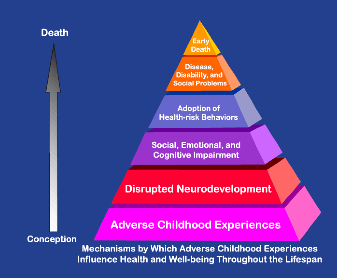
The ACE effort was initiated in 1985 in an era before epigenetics was well-studied. Its artifacts included the ACE pyramid:
The historical ACE lifespan continuum on the left began at conception. The pyramid on the right promoted a limited view of ACE that assigned childhood as the pyramid’s base.
Current official depictions of the ACE pyramid assign an expanded view of ACE as the pyramid’s base. The viewer’s attention is directed to “Scientific Gaps” between pyramid layers, but the largest gap remains: the continuum starts at conception but the pyramid still starts at childhood. The narrative claims:
“To provide scientific information that would be useful for developing new and more effective prevention programs.”
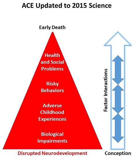
The official ACE pyramid doesn’t accurately reflect current science documented in, for example, Epigenetic effects of early life stress exposure. By downplaying Disrupted Neurodevelopment that may begin at conception, governing agencies implicitly endorse approaches that fail to address prenatal causes for later-life adverse effects.
If the ACE diagram was drawn thirty years later in 2015 to incorporate evidence for epigenetics, Disrupted Neurodevelopment wouldn’t be a consequent layer to an ACE base. The potential start of Disrupted Neurodevelopment would coincide with conception:
What’s an example of current ACE-related scientific evidence that wasn’t present three decades ago and also isn’t represented in the official ACE pyramid? Prenatal Disrupted Neurodevelopment may be considered today as a possible consequence of a “Yes” answer to half of the original ACE questions:
Were your parents were too drunk or high to take care of you or take you to the doctor?
Were your parents ever separated or divorced?
Was your mother often or very often pushed, grabbed, slapped, or had something thrown at her?
Did you live with anyone who was a problem drinker or alcoholic or who used street drugs?
Was a household member depressed or mentally ill?
These threats and other stresses cause a fetus to biologicallyadapt. When such adaptations occur during prenatal development, they may:
Emphasizing Disrupted Neurodevelopment that may begin at conception would encourage:
Research that’s directed toward producing causal evidence for adaptations that largely occur during the early periods of an individual’s lifespan; and
Research on how these adaptations consistently influence our later-life ideas, biology, and behavior.
The above recommendations for research are neither the current focus of ACE research nor the direction of related efforts to assist affected individuals. Relevant studies that I’ve curated on this blog often only produced symptomatic evidence:
If a study couched its findings in non-etiologic phrases such as “is associated with” or “is linked to” or “may relate to,” it didn’t address ACE originating causes.
“New and more effective prevention programs” seldom address Disrupted Neurodevelopment and Biological Impairments with efforts to reduce the source of the damage.
If a program’s presentation showed multivariate analyses with ACE score probabilities and percentages, it didn’t address originating causes.
Here’s a YouTube search of ACE + adverse. Evaluate the current focus of ACE efforts by people employed in the social sciences and services. What did you notice?
How many presentations emphasized prenatal Disrupted Neurodevelopment, a period during which problems may be prevented by addressing causes? Did you instead see that these were outnumbered by many more presentations that emphasized Health and Social Problems symptom interventions?
So, what does it take to empathetically understand, to make a part of oneself, to grok a person’s ACE score?
Regarding empathy – it’s best to avoid the advice of studies such as:
People who are helped may not recognize it at first, but over time, they’ll sense whether the helper’s empathy is genuine.
Regarding understanding – I feel that people first need to ameliorate the origins of their own problems. Then they may be able to help others therapeutically address causes for ACE symptoms.
Need proof? Think of someone you’ve met whose thoughts and feelings and behavior were caught up in and motivated by their own problems:
Did you feel they could empathetically understand others?
Wasn’t the welfare of the people who may have been helped truly incidental and secondary to someone who was acting out their own problems?
The last sentence in the Significance section of this 2015 Emory/Harvard rodent study was:
“These data highlight the potential to exploit sensory system plasticity as a means of ameliorating negative emotional memories that may be tied to peripheral sensory systems.”
The “ameliorating negative emotional memories” part of this statement was incongruent with what the study actually found, as summarized by the Abstract’s last sentence:
“These data suggest that learning-induced freezing behavior, structural alterations, and enhanced neural sensory representation can be reversed in adult mice following extinction training.”
Based on the previous research, the subjects’ “negative emotional memories” possibly weren’t affected at all by the current study’s extinction experiments!
The researchers provided neither direct evidence for “ameliorating negative emotional memories” nor studied areas of the subjects’ brains that contained or processed emotional memories, such as the hippocampus, amygdala, and prefrontal cortex. But – after all – Harvard.
What purposes did it serve for the researchers to make a Significance statement about “ameliorating negative emotional memories” when this wasn’t supported by the study’s findings? What part did the reviewer play in approving this statement?
Where was the study’s evidence to support the headline and statements in the news release such as:
“New Study Indicates That Sense of Smell Could Play Major Role in New Approaches to Treating PTSD
It’s possible for fear behaviors associated with emotional learning to be reversed through exposure-based talk therapy.”
Could this rodent study’s olfactory system findings be properly extrapolated to human talk therapy?
This 2015 Australian plant summary study made several points:
“Non-transmission of epigenetic marks through meiosis may be regarded as an epigenetic modification in itself. We should understand the implications for plant evolution in the context of both selection for and selection against transgenerational epigenetic memory.
Both epigenetic inheritance and resetting are mechanistically directed and targeted. Stress-induced epigenetic modifications may buffer against DNA sequence-based evolution to maintain plasticity, or may form part of plasticity’s adaptive potential.
In some cases the signature of the stress experience remains in the epigenome after relief from the stress, providing a “memory.” If this memory conditions the response to stress during subsequent development, the organism is said to be epigenetically primed. If the memory of the stress experienced by a parent conditions the response of its progeny, this epigenetic priming may be transgenerational.
Epigenetic and genetic variation co-evolve. Epigenetic plasticity does not completely buffer evolvability and reduce the correlation between fitness and genotype, slowing selection.”
One of the summarized studies found that a transgenerational epigenetic change eventually silenced itself after the 40th copy!
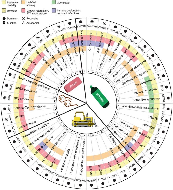
This 2015 human summary study was of 44 genetic disorders that disrupt the maintenance of epigenetic modifications:
“..making them likely to have significant downstream epigenetic consequences. Interestingly, these patients often demonstrate neurological dysfunction, suggesting that precise epigenetic regulation may be critical for neuronal homeostasis. However, at the same time, it is important to keep in mind that many of these proteins have additional non-epigenetic roles.
Mutations in many of these components have now been linked to a number of well-known causes of intellectual disability. Intellectual disability is generally defined as deficits of intellectual function and adaptivebehavior that occur during the developmental period.
Given the opposing activity of many of the components of the epigenetic machinery, the pathogenic sequence in these disorders involves an imbalance of chromatin states. Keeping a subset of genes under “pressure” from two opposing systems may allow the cellular system to rapidly respond to environmental stimuli.
These disorders, on average, have unusual phenotypic breadth. Similarly, there is a shift in distribution toward a higher number of organ systems affected.
In addition to developmental phenotypes (multiple congenital anomalies), in some cases there appear to be ongoing defects that remain consequential in post-natal life. An example of the latter is the hippocampal memory defects seen in many of the mouse models.
This raises the question whether cells undergoing neurogenesis and synaptogenesis are particularly sensitive to subtle defects of the epigenetic machinery and downstream epigenetic abnormalities. A major remaining question is whether neurogenesis defects and/or abnormalities of synaptic plasticity are a unifying pathophysiological process.”
The researchers represented the 44 genetic disorders on a wheel graph:
I look forward to further research that includes non-genetic disruptors of epigenetic modifications.
“With reference to methylation profiles of different tissues, we developed a general approach for studying the major tissue contributors to the circulating DNA pool. This development has opened up numerous research avenues and diagnostic applications.
Our study takes advantage of the recent availability of reference methylomes of a number of tissues. It is likely that such reference databases would be continually updated to include more sample types and from more individuals.”
Up to 41% of plasma DNA in pregnant women was from the placenta. However, I didn’t understand why the non-pregnant women in the control group had measurable placental DNA of up to 2.9%. Maybe it was leftover from a prior pregnancy?
This 2015 study was of the collective behavior of sheep, to:
“Alternate slow group dispersion in space with rapid aggregation phenomena induced by a sudden behavioral shift at the individual level.
The behavioral shift occurs without any perceived threat in the neighborhood, resulting in a spontaneous transition at the collective level that can be interpreted as a consequence of random individual decisions.”
The researchers’ assertion that the “sudden behavioral shift” was due to “random individual decisions” had a lot to do with facilitating their model.
If the researchers had taken individual neurobiological measurements, they may have found that these were non-random actions, deterministically driven by instincts or needs, instead of “random individual decisions.”
“Memories can be established and maintained without de novo protein synthesis and that experimental amnesia may not result from a disruption of memory consolidation/reconsolidation.
Posttraining/postreactivation treatments induce an internal state, which becomes encoded with the memory, and should be present at the time of testing to ensure a successful retrieval.
This integration concept includes most of the previous explanations of memory recovery after retrograde amnesia and critically challenges the traditional memory consolidation/reconsolidation hypothesis, providing a more dynamic and flexible view of memory.”
“A different drug, lithium chloride, produces the same pattern of effects – it blocks ‘reconsolidation’, but this can be reversed by a second dose at the time of recall. However, lithium chloride is not an amnestic [a drug that blocks memory formation] – it doesn’t block protein synthesis. Rather, it causes nausea.
The implication of the lithium experiment is that any drug that causes an ‘internal state change’, even if it’s just nausea, can trigger state-dependent memory and behave just like an ‘amnestic’.”
“Fear-inducing memories can be state dependent, meaning that they can best be retrieved if the brain states at encoding and retrieval are similar.”
Memories triggered while in a brain state reentered through an emotion or a physical reaction are experienced by Primal Therapy patients and observed by therapists every day. However, as mentioned in What scientific evidence can be offered for Primal Therapy’s capability to benefit people’s lives? there’s a difficulty in developing human evidence for such state-dependent emotional memories.
Standard procedures would use human subjects and control groups in a way that retrieved memories according to the researchers’ schedule and experimental parameters. In order for the retrieval of an emotional memory to be therapeutic, though, the methods of an experiential therapy such as Dr. Arthur Janov’s Primal Therapy leave the timing of entering a triggering brain state up to the patient.
When a brain state protects a human emotional memory from being accessed, it probably wouldn’t be therapeutic to:
Force a return to that brain state, and thereby
Remove the memory’s protection, then
Retrieve and re-experience the memory
just for the sake of research.
The evidence for retrieving and re-experiencing a state-dependent memory lies mainly within the individual’s experiences.
A challenge is to find innovative ways to document human evidence for state-dependent emotional memories while ensuring a therapeutic process.
http://www.jneurosci.org/content/35/33/11623 “Integration of New Information with Active Memory Accounts for Retrograde Amnesia: A Challenge to the Consolidation/Reconsolidation Hypothesis?”
“Maternal ADN [adiponectin, a hormone produced by fat cells, that regulates fat and glucose metabolism] supplementation reversed the adverse effects of maternal obesity on placental function and fetal growth.
Babies of mothers with obesity and/or gestational diabetes mellitus (GDM) are often large at birth and have increased adiposity, which predisposes them to the development of metabolic disease later in life.
Maternal ADN infusion in obese dams from E14.5 to E18.5 [the last 4 days of pregnancy, a period that accounts for 70% of the total fetal growth] normalized maternal insulin sensitivity, placental insulin/mTORC1 and PPARα signaling, nutrient transport, and fetal growth without affecting maternal fat mass.”
As the study may apply to humans:
“This hormone or a similar agent could feasibly do the same thing for humans that it did for mice,” Jansson said.
Jansson said more work needs to be done to track the long-term effects of the hormone treatment on the mice.”
The study focused on epigenetic effects of the mothers’ environment on fetuses, and didn’t assess possible genetic contributions.
http://www.pnas.org/content/112/41/12858.full “Adiponectin supplementation in pregnant mice prevents the adverse effects of maternal obesity on placental function and fetal growth”
“15-mo-old infants exposed to stress thereafter kept performing a previously effective action, even after the action suddenly became ineffective.
Infants in a no-stress control group flexibly adjusted their behavior by disengaging from the newly ineffective action in favor of exploring an alternative action.
This finding demonstrates that stress impairs infants’ ability to adjust their behavior to changing circumstances.”
The primary measurement of stress levels was cortisol. Stressful conditions were:
A stranger sat down next to them;
A dancing robot played loud music and moved around;
The infant’s caregivers left the room for up to four minutes.
News coverage stated that the study’s design was an adaptation of experiments that produced the same findings in adults. But would adult humans be stressed by being left alone for four minutes?
“Maternal separation in rodents is a useful model of early-life stress that results in enduring physiological and behavioral changes that persist into adulthood.”
A study limitation was that it involved just 26 infants.
This 2015 Northwestern University rodent study found:
“Fear-inducing memories can be state dependent, meaning that they can best be retrieved if the brain states at encoding and retrieval are similar.
Memories formed in a particular mood, arousal or drug-induced state can best be retrieved when the brain is back in that state.
‘It’s difficult for therapists to help these patients,’ Radulovic said, ‘because the patients themselves can’t remember their traumaticexperiences that are the root cause of their symptoms.’
The best way to access the memories in this system is to return the brain to the same state of consciousness as when the memory was encoded.”
The study demonstrated one method of activating neurobiological pathways with a drug to remove a hippocampal memory’s protection, which played a part in enabling subjects to relive their remembered experiences. This rodent study’s methods weren’t designed to therapeutically access similarly protected memories with humans.
“There are two kinds of GABA[gamma-Aminobutyric acid] receptors. One kind, synaptic GABA receptors, works in tandem with glutamate receptors to balance the excitation of the brain in response to external events such as stress.
The other population, extra-synaptic GABA receptors, are independent agents.
If a traumatic event occurs when these extra-synaptic GABA receptors are activated, the memory of this event cannot be accessed unless these receptors are activated once again.
‘It’s an entirely different system even at the genetic and molecular level than the one that encodes normal memories,’ said lead study author Vladimir Jovasevic, who worked on the study when he was a postdoctoral fellow in Radulovic’s lab.
This different system is regulated by a small microRNA, miR-33, and may be the brain’s protective mechanism when an experience is overwhelmingly stressful.
The findings imply that in response to traumatic stress, some individuals, instead of activating the glutamate system to store memories, activate the extra-synaptic GABA system and form inaccessible traumatic memories.”
I’d point out that “can’t remember” and “inaccessible traumatic memories” phrases used above were in reference to what’s usually called “memory” i.e., a recall initiated by the cerebrum.
The study’s findings should inform memory-study researchers if they care to understand how emotional memories can be formed and re-experienced.
The study provided evidence for fundamentals of Dr. Arthur Janov’s Primal Therapy, such as:
Experiences associated with pain can be remembered below our conscious awareness.
The retrieval and re-experiencing of emotional memories can engage our lower-level brain areas without our higher-level brain areas’ participation.
The obvious nature of this study’s straightforward experimental methods made me wonder why other researchers hadn’t used the same methods decades ago.
Use of this study’s methodology could have resulted in dozens of informative follow-on study variations by now, and subsequently found whether subjects’ physiological, behavioral, and epigenetic measurements differed from control group subjects, as in:
“miR-33 is downregulated in response to gaboxadol [the drug used to change subjects’ brain state] and modulates its effects on state-dependent fear.”
An interview with Jeff Link, the editor of Dr. Arthur Janov’s 2011 book “Life Before Birth: The Hidden Script that Rules Our Lives” with Ken Rose:
“Even further confirmation for some of the views of Janov, that maybe weren’t widely accepted for a time, it’s new research now being done into memory and what a lot of scientist are seeing, a lot of different studies is that memory reactivates the same neuroimpulses that were initially firing off when the event happened.
So a traumatic event when you remember it, the act of remembering it is actually creating a neuromirror of what went on initially.
In a lot of ways that is what Primal Therapy is attempting to do; is to go back to that place and reconnect, or as it’s sometimes referred to, reconsolidate the brain state so that real healing can take place.”
“Gene expression profiling in postmortem human brain and studies using animal models have implicated the fibroblast growth factor (FGF) family in affect regulation and suggest a potential role in the pathophysiology of major depressive disorder (MDD).
We show that FGF9 expression is up-regulated in the hippocampus of individuals with MDD, and that FGF9 expression is inversely related to the expression of FGF2.”
The researchers went down the evolutionary scale from human findings to replicate many of the findings with rodents:
“We found that chronic social defeat stress, an animal model recapitulating some aspects of MDD, leads to a significant increase in hippocampal FGF9 expression.
Collectively, these results suggest that high levels of hippocampal FGF9 play an important role in the development or expression of mood and anxiety disorders.”