Transgenerational effects of early environmental insults on aging and disease

The first paper of Transgenerational epigenetic inheritance week was a 2017 Canadian/Netherlands review that’s organized as follows:

“First, we address mechanisms of developmental and transgenerational programming of disease and inheritance. Second, we discuss experimental and clinical findings linking early environmental determinants to adverse aging trajectories in association with possible parental contributions and sex-specific effects. Third, we outline the main mechanisms of age-related functional decline and suggest potential interventions to reverse negative effects of transgenerational programming.”

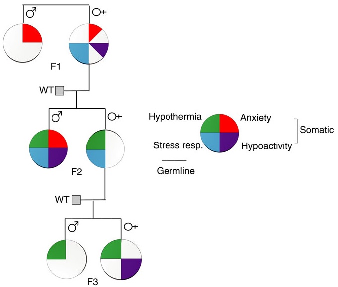
A transgenerational phenotype was defined as an epigenetic modification that was maintained at least either to F2 grandchildren in the paternal lineage, or to F3 great-grandchildren in the maternal lineage.

The reviewers noted that mechanisms of transgenerational programming are complex and multivariate.  Severity, timing, and type of exposure; lineage of transmission; germ cell exposure; and gender of an organism were the main factors that may determine consequences. Mechanisms reviewed were:

  1. Parental exposure to an adverse environment;
  2. Altered maternal behavior and care of offspring; and
  3. Experience-dependent modifications of the epigenome.

There was a long list of diseases and impaired functionalities that were consequences of ancestral experiences and exposures. Most studies were of animals, but a few were human, such as those done on effects of extended power outages during a Quebec ice storm of January 1998.


One intervention that was effective in reversing a transgenerational phenotype induced by deficient rodent maternal care was to place pups with a caring foster female soon after birth. It’s probably unacceptable in human societies to preemptively recognize all poor-care human mothers and remove the infant to caring foster mothers. But researchers could probably find enough instances to develop studies of the effectiveness of such placements in reversing a transgenerational phenotype.

The review didn’t have suggestions for reversing human transgenerational phenotypes, just “potential interventions to reverse negative effects of transgenerational programming.” Interventions suggested for humans – exercise, enriched lifestyle, cognitive training, dietary regimens, and expressive art and writing therapies – only reduced impacts of transgenerational epigenetic effects.

Tricky wording of “reverse negative effects of transgenerational programming” showed that research paradigms weren’t aimed at resolving causes. The review was insufficient for the same reasons mentioned in How one person’s paradigms regarding stress and epigenetics impedes relevant research, prompting my same comment:

Aren’t people interested in human treatments of originating causes so that their various symptoms don’t keep bubbling up? Why wouldn’t research paradigms be aligned accordingly?

When reversals of human phenotypes aren’t researched, problems may compound by being transmitted to the next generations.

http://www.sciencedirect.com/science/article/pii/S014976341630714X “Transgenerational effects of early environmental insults on aging and disease incidence” (not freely available)

Does living near a forest keep your amygdala healthier?

A thought-provoking post from A Paper a Day Keeps the Scientist Okay entitled “Living Near a Forest Keeps Your Amygdala Healthier” referenced a 2017 German human study which found:

“..a relationship between place of residence and brain health: those city dwellers living close to a forest were more likely to show indications of a physiologically healthy amygdala structure and were therefore presumably better able to cope with stress.”

The researchers accomplished the imperative of meeting the study’s stated objective:

“We set out to identify and characterize the geographical elements of a city that are associated with these brain structures following a suggestion by Kennedy and Adolph that studies should begin to derive recommendations for urban planning and architecture.

The results of our study may suggest that forests in and around the cities are a valuable resource that should be promoted. However future longitudinal studies are needed to investigate the causal directionality of the effect in order to disentangle whether more forest in ones habitat facilitates brain structural integrity or potentially those people with better brain structural integrity choose to live closer to forests. Moreover we need to investigate whether living close to the forest is associated with an absence of risk factors such as noise, air pollution or stress and thereby has beneficial effects or whether the forest itself constitutes a salutary factor that promotes well-being.”

https://www.nature.com/articles/s41598-017-12046-7 “In search of features that constitute an “enriched environment” in humans: Associations between geographical properties and brain structure”


A major limitation of this study’s methodology was intentional non-use of an available data source. Referring to Do we need to study the brain to understand the mind? posted earlier this week:

“Self-report is still the gold standard for assessing emotional experience and the contents of thought. Isn’t it easier just to ask?”

These researchers put the forest before the trees, and designed a study that didn’t ask subjects important questions such as why they lived where they lived. The researchers inferred sketchy fMRI-geography associations because they didn’t solicit relevant primary information via individual self-reports.


I don’t live in Berlin, and I’m not part of the selected cohort, but I otherwise generally meet this study’s subject parameters. Something in my past causes me to actively select housing that isn’t in a noisy environment. If I were asked why I lived where I lived, my answer would have included:

  • A deciding factor in why I sold my second house was traffic noise in wintertime;
  • A deciding factor in why I bought my fourth house was its location in the housing development’s center, away from street noise; and
  • A deciding factor in why I live where I now live is the house’s orientation away from both direct and reflective traffic noise sources.

Processing my hypothetical fMRI data with my self-reported historical housing choices may or may not have found:

“Geographical features in the proximal participants’ habitat are associated with brain integrity.”

Using better-quality information of self-reports, though, it’s unlikely that an association this study would have found to be significant – a chance fact that I live within one kilometer of a forest – would have been deemed significant.

Do we need to study the brain to understand the mind?

A coauthor of the studies referenced in:

offered an opinion piece in A Paper a Day Keeps the Scientist Okay entitled “Do We Need To Study The Brain To Understand The Mind?”

“The emerging consensus appears to be that implementation is important. Interestingly, the inverse question is also being asked by neurobiologists—do we need consider the mind to understand the brain?—and answered largely and increasingly in the affirmative.

Is pain different from negative emotions such as sadness and anger, or are they variants on a common theme? Pain appears to be distinct from negative emotion, but commonalities suggest ways in which they may share underlying processes such as heightened attention.

One of the biggest pitfalls is the temptation to observe brain activity and make inferences about the psychological state—for example, to infer:

  • Episodic memory retrieval from hippocampal activity,
  • Fear from amygdala activity, or
  • Visual processing from activity in the ‘visual cortex.’

These inferences ignore the scope of processes which may activate each of these areas and involve a fallacy in reasoning: “if memory then hippocampus” is not the same thing as “if hippocampus then memory.”

The fact that few brain areas, including the ‘visual cortex,’ are dedicated to one process means that self-report is still the gold standard for assessing emotional experience and the contents of thought. This is a serious challenge for those who would like, for example, to assess your brand preferences or your political affiliation from a brain scan. (And isn’t it easier just to ask?)”

Epigenetic effects of early life stress exposure

This 2017 Netherlands review subject was the lasting epigenetic effects of early-life stress:

“Exposure to stress during critical periods in development can have severe long-term consequences.

One of the key stress response systems mediating these long-term effects of stress is the hypothalamic-pituitary-adrenal (HPA) axis.

Early life stress (ELS) exposure has been reported to have numerous consequences on HPA-axis function in adulthood.

ELS is able to “imprint” or “program” an organism’s neuroendocrine, neural and behavioral responses to stress. Research focuses along two complementary lines:

  1. ELS during critical stages in brain maturation may disrupt specific developmental processes (by altered neurotransmitter exposure, gene transcription, or neuronal differentiation), leading to aberrant neural circuit function throughout life.
  2. ELS may induce modifications of the epigenome which lastingly affect brain function.

These epigenetic modifications are inducible, stable, and yet reversible, constituting an important emerging mechanism by which transient environmental stimuli can induce persistent changes in gene expression and ultimately behavior.”


In early life, the lower brain and limbic system brain structures are more developed and dominant, whereas the cerebrum is less developed (use the above rodent graphic as a rough guide). Stress and pain generally have a greater impact on a fetus than an infant, and a greater impact on an infant than an adult.

The reviewers cited 50+ studies from years 2000-2015 in the “Early Life Stress Effects in a “Matching” Stressful Adult Environment” section to argue for the match / mismatch theory:

“Encountering ELS prepares an organism for similar (“matching”) adversities during adulthood, while a mismatching environment results in an increased susceptibility to psychopathology, indicating that ELS can exert either beneficial or disadvantageous effects depending on the environmental context.

Initial evidence for HPA-axis hypo-reactivity is observed for early social deprivation, potentially reflecting the abnormal HPA-axis function as observed in post-traumatic stress disorder.

Experiencing additional (chronic) stress in adulthood seems to normalize these alterations in HPA-axis function, supporting the match / mismatch theory.”

Evidence for this theory was contrasted with the allostatic load theory presented in How one person’s paradigms regarding stress and epigenetics impedes relevant research.


The review mainly cited evidence from rodent studies that mismatched reactions in adulthood may be consequences of early-life events. These events:

“Imprint or program an organism’s neuroendocrine, neural and behavioral responses..leading to aberrant neural circuit function throughout life..which lastingly affect brain function.”

Taking this research to a personal level:

  • Have you had feelings that you were unsafe, although your environment was objectively safe?
  • Have you felt uneasy when people are nice to you?
  • Have you felt anxious when someone pays attention to you, even after you’ve acted to gain their attention?

Mismatched human feelings are one form of mismatched reactions. These may be consequences of early-life experiences, and indicators of personal truths.

If researchers can let go of their biases and Advance science by including emotion in research, they may find that human subjects’ feelings produce better evidence for what actually happened during the subjects’ early lives than do standard scientific methods of:

Incorporating feeling evidence may bring researchers and each individual closer to discovering the major insults that knocked their development processes out of normally robust pathways and/or induced “persistent changes in gene expression and ultimately behavior.”

https://www.frontiersin.org/articles/10.3389/fncel.2017.00087/full “Modulation of the Hypothalamic-Pituitary-Adrenal Axis by Early Life Stress Exposure”


I came across this review as a result of it being cited in http://www.sciencedirect.com/science/article/pii/S1084952117302884 “Long-term effects of early environment on the brain: Lesson from rodent models” (not freely available)

Epigenetic effects of THC differ between female adolescents and adults

This 2017 Italian rodent study found:

“THC [delta9-tetrahydrocannabinol, the psychoactive compound of cannabis] exposure affects histone modifications in the brain of female rats in a region- and age-specific manner. Specifically, THC acts on different targets depending on the considered brain area and, remarkably, the adolescent brain is generally more sensitive to THC than the adult brain.

Adolescent exposure to THC, or to synthetic cannabinoids, induced sex-dependent brain and behavioral alterations at adulthood. In female rats, the phenotype was more complex, as both depressive-like and psychotic-like signs were present.

Development of the depressive/psychotic-like phenotype is restricted to adolescent THC exposure. Not only the behavioral phenotype developed after adolescent, and not adult, exposure, but also changes in both histone modifications and gene expression were more widespread and intense after adolescent treatment, further confirming a specific adolescent susceptibility.

The primary effect in the adolescent brain was represented by changes leading to transcriptional repression, whereas the one observed after adult treatment led to transcriptional activation. Moreover, only in the adolescent brain, the primary effect was followed by a homeostatic response to counterbalance the THC-induced repressive effect, except in the amygdala.”

The authors’ interpretation of the brain area results was:

“The amygdala is more responsive in adult than adolescent animals. Since it has been established that the amygdala is activated during exposure to aversive stimuli, functioning as a “behavioral brake”, different response between adult and adolescent animals could represent the biological bases of the adolescent propensity for risk-taking and novelty-seeking behaviors. Also in adolescent humans, neuroimaging studies have shown a weaker involvement of the amygdala, and a greater contribution of the NAc [nucleus accumbens], in response to negative and positive stimuli compared to adults.”

http://www.mdpi.com/1422-0067/18/10/2094 “Chronic Δ9-THC Exposure Differently Affects Histone Modifications in the Adolescent and Adult Rat Brain”

A gaping hole in a review of nutritional psychiatry

This December 2016 Australian review published in September 2017 concerned:

“..the nutritional psychiatry field..the neurobiological mechanisms likely modulated by diet, the use of dietary and nutraceutical interventions in mental disorders, and recommendations for further research.”


The reviewers inexplicably omitted acetyl-L-carnitine, which I first covered in A common dietary supplement that has rapid and lasting antidepressant effects. A PubMed search on “acetyl carnitine” showed over a dozen studies from the past twelve months that were relevant to the review’s subject areas. Here’s a sample, beginning with follow-on research published in June 2016 of the study I linked above:

Reply to Arduini et al.: Acetyl-l-carnitine and the brain: Epigenetics, energetics, and stress

Dietary supplementation with acetyl-l-carnitine counteracts age-related alterations of mitochondrial biogenesis, dynamics and antioxidant defenses in brain of old rats

Neuroprotective effects of acetyl-l-carnitine on lipopolysaccharide-induced neuroinflammation in mice: Involvement of brain-derived neurotrophic factor

ALCAR promote adult hippocampal neurogenesis by regulating cell-survival and cell death-related signals in rat model of Parkinson’s disease like-phenotypes

Analgesia induced by the epigenetic drug, L-acetylcarnitine, outlasts the end of treatment in mouse models of chronic inflammatory and neuropathic pain

The cited references in these recent studies were older, of course, and in the time scope of the review. There’s no excuse for this review’s omission of acetyl-L-carnitine.

https://www.cambridge.org/core/journals/proceedings-of-the-nutrition-society/article/nutritional-psychiatry-the-present-state-of-the-evidence/88924C819D21E3139FBC48D4D9DF0C08 “Nutritional psychiatry: the present state of the evidence” (not freely available)

How one person’s paradigms regarding stress and epigenetics impedes relevant research

This 2017 review laid out the tired, old, restrictive guidelines by which current US research on the epigenetic effects of stress is funded. The reviewer rehashed paradigms circumscribed by his authoritative position in guiding funding, and called for more government funding to support and extend his reach.

The reviewer won’t change his beliefs regarding individual differences and allostatic load pictured above since he helped to start those memes. US researchers with study hypotheses that would develop evidence beyond such memes may have difficulties finding funding except outside of his sphere of influence.


Here’s one example of the reviewer’s restrictive views taken from the Conclusion section:

Adverse experiences and environments cause problems over the life course in which there is no such thing as “reversibility” (i.e., “rolling the clock back”) but rather a change in trajectory [10] in keeping with the original definition of epigenetics [132] as the emergence of characteristics not previously evident or even predictable from an earlier developmental stage. By the same token, we mean “redirection” instead of “reversibility”—in that changes in the social and physical environment on both a societal and a personal level can alter a negative trajectory in a more positive direction.”

What would happen if US researchers proposed tests of his “there is no such thing as reversibility” axiom? To secure funding, the prospective studies’ experiments would be steered toward altering “a negative trajectory in a more positive direction” instead.

An example of this influence may be found in the press release of Familiar stress opens up an epigenetic window of neural plasticity where the lead researcher stated a goal of:

“Not to ‘roll back the clock’ but rather to change the trajectory of such brain plasticity toward more positive directions.”

I found nothing in citation [10] (of which the reviewer is a coauthor) where the rodent study researchers even attempted to directly reverse the epigenetic changes! The researchers under his guidance simply asserted:

“A history of stress exposure can permanently alter gene expression patterns in the hippocampus and the behavioral response to a novel stressor”

without making any therapeutic efforts to test the permanence assumption!

Nevermind that researchers outside the reviewer’s sphere of influence have done exactly that, reverse both gene expression patterns and behavioral responses!!

In any event, citation [10] didn’t support an “there is no such thing as reversibility” axiom.

The reviewer also implied that humans respond just like lab rats and can be treated as such. Notice that the above graphic conflated rodent and human behaviors. Further examples of this inappropriate rodent / human merger of behaviors are in the Conclusion section.


What may be a more promising research approach to human treatments of the epigenetic effects of stress? As pointed out in The current paradigm of child abuse limits pre-childhood causal research:

“If the current paradigm encouraged research into treatment of causes, there would probably already be plenty of evidence to demonstrate that directly reducing the source of the damage would also reverse damaging effects. There would have been enough studies done so that the generalized question of reversibility wouldn’t be asked.

Aren’t people interested in human treatments of originating causes so that their various symptoms don’t keep bubbling up? Why wouldn’t research paradigms be aligned accordingly?”

http://journals.sagepub.com/doi/full/10.1177/2470547017692328 “Neurobiological and Systemic Effects of Chronic Stress”

Epigenetics account for two-thirds of Alzheimer’s disease

The genetics percentage from a 2017 summary of Alzheimer’s disease research caught my eye:

“Although numerous single nucleotide polymorphisms (SNPs) have now been robustly associated with AD via genome-wide association studies and subsequent meta-analyses, collectively these common SNPs are believed to only account for 33% of attributable risk and the mechanism behind their action remains largely unknown.”

This citation aligned with other studies’ findings per Using twins to estimate the extent of epigenetic effects that on cellular levels, our experiences account for two-thirds of who we are.


The promise of this category of epigenetics research?

“One of the most exciting aspects of identifying disease-associated epigenomic dysfunction is that these mechanisms are potentially reversible.”

Let’s make research on reversing epigenetic changes a priority for funding, and get studies underway here in 2017!

https://www.epigenomicsnet.com/users/27784-katie-lunnon/posts/14634-robust-evidence-for-dna-methylomic-variation-in-alzheimer-s-disease “Robust evidence for DNA methylomic variation in Alzheimer’s disease” (Registration required)

The hypothalamus couples with the brainstem to cause migraines

This 2016 German human study with one subject found:

“The hypothalamus to be the primary generator of migraine attacks which, due to specific interactions with specific areas in the higher and lower brainstem, could alter the activity levels of the key regions of migraine pathophysiology.”

The subject underwent daily fMRI scans, and procedures to evoke brain activity. She didn’t take any medications, and suffered three migraine attacks during the 31-day experimental period.

Neuroskeptic commented:

“The dorsal pons has previously been found to be hyperactive during migraine. It’s been dubbed the brain’s ‘migraine generator.’ Schulte and May’s data suggest that this is not entirely true – rather, it looks like the hypothalamus may be the true generator of migraine, while the brainstem could be a downstream mediator of the disorder.

A hypothalamic origin of migraines would help to explain some of the symptoms of the disorder, such as changes in appetite, that often accompany the headaches.”


The above graphic looks to me like the result of feedback mechanisms that either didn’t exist or inadequately handled the triggering event. Other examples of the hypothalamus lacking feedback or being involved in a deviated feedback loop include:

There are many unanswered questions with a one-person study, of course. Addressing the cause of this painful condition would find out when, where, and how a person’s hypothalamus became modified to express migraine tendencies.

I’d guess that migraine tendencies may appear as early as the first trimester of pregnancy, given that a highly functional hypothalamus is needed for survival and development in our earliest lives. Gaining as much familial and historical information as possible from the person would be necessary steps in therapies that address migraine causes.

http://blogs.discovermagazine.com/neuroskeptic/2016/05/22/pinpointing-origins-of-migraine/ “Pinpointing the Origins of Migraine in the Brain”

https://academic.oup.com/brain/article/139/7/1987/2464241 “The migraine generator revisited: continuous scanning of the migraine cycle over 30 days and three spontaneous attacks”


As mentioned in How to cure the ultimate causes of migraines? comments are turned off for this post due to it somehow becoming a magnet for spammers. Readers can comment on that post instead.

A limited study of parental transmission of anxiety/stress-reactive traits

BehavioralTraitsThis 2016 New York rodent study found:

“Parental behavioural traits can be transmitted by non-genetic mechanisms to the offspring.

We show that four anxiety/stress-reactive traits are transmitted via independent iterative-somatic and gametic epigenetic mechanisms across multiple generations.

As the individual traits/pathways each have their own generation-dependent penetrance and gender specificity, the resulting cumulative phenotype is pleiotropic. In the context of genetic diseases, it is typically assumed that this phenomenon arises from individual differences in vulnerability to the various effects of the causative gene. However, the work presented here reveals that pleiotropy can be produced by the variable distribution and segregated transmission of behavioural traits.”


A primary focus was how anxiety was transmitted from parents to offspring:

“The iterative propagation of the male-specific anxiety-like behaviour is most compatible with a model in which proinflammatory state is propagated from H [serotonin1A receptor heterozygote] F0 to F1 [children] females and in which the proinflammatory state is acquired by F1 males from their H mothers, and then by F2 [grandchildren] males from their F1 mothers.

We propose that increased levels of gestational MIP-1β [macrophage inflammatory protein 1β] in H and F1 mothers, together with additional proinflammatory cytokines and bioactive proteins, are required to produce immune system activation in their newborn offspring, which in turn promotes the development of the anxiety-like phenotype in males.

In particular, increase in the number of monocytes and their transmigration to the brain parenchyma in F1 and F2 males could be central to the development of anxiety.”


The researchers studied transmission of behavioral traits and epigenetic changes. Due to my quick take on the study title – “Behavioural traits propagate across generations..” – I had expectations of this study that weren’t born out. What could the researchers have done versus what they did?

The study design removed prenatal and postnatal parental behavioral transmission of behavioral traits and epigenetic changes as each generation’s embryos were implanted into foster wild-type (WT) mothers.

The study design substituted the foster mothers’ prenatal and postnatal parental environments for the biological parents’ environments. So we didn’t find out, for example:

  • To what extents the overly stress-reactive F1 female children’s prenatal environments and postnatal behaviors induced behaviors and/or epigenetic changes in their children; and
  • Whether the F2 grandchildren’s parental behaviors subsequently induced behaviors and/or epigenetic changes in the F3 great-grandchildren.

How did the study meet the overall goal of rodent studies: to help humans?

    1. Only a minority of humans experienced an early-life environment that included primary caregivers other than our biological parents.
    2. Very, very few of us experienced a prenatal environment other than our biological mothers.
    3. The study’s thorough removal of parental behavior was an outstanding methodology to confirm by falsifiability whether parental behavior was both an intergenerational and transgenerational epigenetic inheritance mechanism.
    4. Maybe the researchers filled in some gaps in previous rodent studies, such as determining what is or isn’t a “true transgenerational mechanism.”

As an example of a rodent study that more closely approximated human conditions, the behavior of a mother whose DNA was epigenetically changed by stress induced the same epigenetic changes to her child’s DNA when her child was stressed per One way that mothers cause fear and emotional trauma in their infants:

“Our results provide clues to understanding transmission of specific fears across generations and its dependence upon maternal induction of pups’ stress response paired with the cue to induce amygdala-dependent learning plasticity.”


How did parental behavioral transmission of behavioral traits and epigenetic changes become a subject not worth investigating? These traits and effects can be seen everyday in real-life human interactions, and in every human’s physiology.

But when investigating human correlates with behavioral epigenetic changes of rodents in the laboratory, parental behavioral transmission of behavioral traits is often treated the way this study treated it: as a confounder.

I doubt that people who have reached some degree of honesty about their early lives and concomitant empathy for others would agree with this prioritization. The papers of Transgenerational epigenetic inheritance week show the spectrum of opportunities to advance science that were intentionally missed.

http://www.nature.com/ncomms/2016/160513/ncomms11492/full/ncomms11492.html “Behavioural traits propagate across generations via segregated iterative-somatic and gametic epigenetic mechanisms”

Why drugs aren’t ultimately therapeutic

This 2016 Oregon review’s concept was the inadequacy of drug-based therapies, explored with the specific subject of epilepsy:

“Currently used antiepileptic drugs:

  • [aren’t] effective in over 30% of patients
  • [don’t] affect the comorbidities of epilepsy
  • [don’t] prevent the development and progression of epilepsy (epileptogenesis).

Prevention of epilepsy and its progression [requires] novel conceptual advances.”

The overall concept that current drug-based therapies poorly address evolutionary biological realities was illustrated by a pyramid, with the comment that:

“If the basis of the pyramid depicted in Figure 1 is overlooked, it becomes obvious that a traditional pharmacological top-down treatment approach has limitations.”

Why drug ultimately aren't therapeutic


I would have liked the reviewer to further address the “therapeutic reconstruction of the epigenome” point he made in the Abstract:

“New findings based on biochemical manipulation of the DNA methylome suggest that:

  1. Epigenetic mechanisms play a functional role in epileptogenesis; and
  2. Therapeutic reconstruction of the epigenome is an effective antiepileptogenic therapy.”

As it was, the reviewer lapsed into the prevalent belief that the causes of and cures for human diseases will always be found on the molecular level – for example, the base of the above pyramid – and never in human experiences. This preconception leads to discounting human elements – notably absent in the above pyramid – that generate epigenetic changes.

A consequence of ignoring experiential causes of diseases is that the potential of experiential therapies to effect “therapeutic reconstruction of the epigenome” isn’t investigated.

http://journal.frontiersin.org/article/10.3389/fnmol.2016.00026/full “The Biochemistry and Epigenetics of Epilepsy: Focus on Adenosine and Glycine”

Using salivary microRNA to diagnose autism

This 2016 New York human study found:

“Measurement of salivary miRNA in this pilot study of subjects with mild ASD [autism spectrum disorder] demonstrated differential expression of 14 miRNAs that are:

  • expressed in the developing brain,
  • impact mRNAs related to brain development, and
  • correlate with neurodevelopmental measures of adaptive behavior.”

Some problems with current diagnostic methods for autism are:

“The first sign of ASD commonly recognized by pediatricians is a deficit in communication and language that does not manifest until 18–24 months of age.

The mean age of diagnosis for children with ASD is 3 years, and approximately half of these are false-positives.

Despite a substantial genetic component, no single gene variant accounts for >1 % of ASD incidence.

Nearly 2000 individual genes have been implicated in ASD, but none are specific to the disorder.”

Study limitations included:

“Aside from the sample size and cross-sectional nature of this pilot study, another limitation is the age of ASD and control subjects it describes (4–14 years) which are not representative of the target population in which ASD biomarkers would ideally be utilized (0–2 years). However, selecting a homogenous group of subjects with mild ASD (as measured by ADOS) that was well-established and diagnosed by a developmental specialist requires subjects with long-standing diagnoses.”


Understanding later-life consequences of disrupted neurodevelopment is critical for tracing symptoms back to their causes, as noted in Grokking an Adverse Childhood Experiences (ACE) score. I wonder how long it will take for researchers in other fields to stop wasting resources and do what this study did: focus on epigenetic biomarkers that have developmental origins.

http://bmcpediatr.biomedcentral.com/articles/10.1186/s12887-016-0586-x “Salivary miRNA profiles identify children with autism spectrum disorder, correlate with adaptive behavior, and implicate ASD candidate genes involved in neurodevelopment”

Contending with epigenetic consequences of violence to women

This 2016 UK review subject was the interplay of genomic imprinting and intergenerational epigenetic information transfer:

“A range of evolutionary adaptations associated with placentation transfers disproportionate control of this process to the matriline, a period unique in mammalian development in that there are three matrilineal genomes interacting in the same organism at the same time (maternal, foetal, and postmeiotic oocytes).

Genomic imprinting is absent in egg laying mammals and only around 6 imprinted genes have been detected in a range of marsupial species; this is in contrast to eutherian mammals where around 150 imprinted genes have been described.

The interactions between the maternal and developing foetal hypothalamus and placenta can provide a template by which a mother can transmit potentially adaptive information concerning potential future environmental conditions to the developing brain.

In circumstances either where the early environment provides inaccurate cues to the environmental conditions prevailing when adult due to rapid environmental change or when disruptions to normal neural development occur, the mismatch between the environmental predictions made during early development and subsequent reality may mean that an organism may have a poorly adapted phenotype to its adult environment. An appreciation of these underlying evolutionary salient processes may provide a novel perspective on the [causal] mechanisms of a range of health problems.

The concept of a brain that is not pathological in the classical sense but it is simply mismatched to its environment has been most extensively studied in the context of ancestral and early developmental nutrition. However, this concept can be extended to provide insights into the development of a range of alternative neural phenotypes.”

The review’s final sentence was:

“Examination of the adaptive potential of a range of neural and cognitive deficits in the context of evolutionary derived foetocentric brain and placental development, epigenetics and environmental adaptation may provide novel insights into the development and potential treatment of a range of health, neurological, and cognitive disorders.”

One of the reviewers was cited in Epigenetic DNA methylation and demethylation with the developing fetus, which the review cited along with Epigenetic changes in the developing brain change behavior.


Researchers who avoid hypotheses that can’t be proven wrong could certainly test the subject matter of this review if they investigated their subjects’ histories.

For example, let’s say a patient/subject had symptoms where the “150 imprinted genes” were implicated. What are the chances a clinician or researcher would be informed by this review’s material and investigate the mother’s and grandmother’s histories?

For clinicians or researchers who view histories as irrelevant busywork: How many tens of millions of people alive today have mothers who were fetuses when their grandmothers were adversely affected by violence? Wouldn’t it be appropriate to assess possible historical contributions of:

“The mismatch between the environmental predictions made during early development and subsequent reality”

to the patient’s/subject’s current symptoms?

http://www.hindawi.com/journals/np/2016/6827135/ “Placental, Matrilineal, and Epigenetic Mechanisms Promoting Environmentally Adaptive Development of the Mammalian Brain”

A one-sided review of stress

The subject of this 2016 Italian/New York review was the stress response:

“The stress response, involving the activation of the hypothalamic-pituitary-adrenocortical [HPA] axis and the consequent release of corticosteroid hormones, is indeed aimed at promoting metabolic, functional, and behavioral adaptations. However, behavioral stress is also associated with fast and long-lasting neurochemical, structural, and behavioral changes, leading to long-term remodeling of glutamate transmission, and increased susceptibility to neuropsychiatric disorders.

Of note, early-life events, both in utero and during the early postnatal life, trigger reprogramming of the stress response, which is often associated with loss of stress resilience and ensuing neurobehavioral (mal)adaptations.”


The reviewers’ intentional dismissal of the role of GABA in favor of the role of glutamate was a key point:

“The changes in neuronal excitability and synaptic plasticity induced by stress are the result of an imbalance of excitatory (glutamatergic) and inhibitory (GABAergic) transmission, leading to long-lasting (mal)adaptive functional modifications. Although both glutamate and GABA transmission are critically associated with stress-induced alteration of neuronal excitability, the present review will focus on the modulation of glutamate release and transmission induced by stress and glucocorticoids.”

No particular reason was given for this bias. I inferred from the review’s final sentence that the review’s sponsors and funding prompted this decision:

“In-depth studies of changes in glutamate transmission and dendrite remodeling induced by stress in early and late life will help to elucidate the biological underpinnings of the (mal)adaptive strategies the brain adopts to cope with environmental challenges in one’s life.”

The bias led to ignoring evidence for areas the reviewers posed as needing further research. An example of relevant research the reviewers failed to consider was the 2015 Northwestern University study I curated in A study that provided evidence for basic principles of Primal Therapy that found:

“In response to traumatic stress, some individuals, instead of activating the glutamate system to store memories, activate the extra-synaptic GABA system and form inaccessible traumatic memories.”

http://www.ncbi.nlm.nih.gov/pmc/articles/PMC4812483/ “Stress Response and Perinatal Reprogramming: Unraveling (Mal)adaptive Strategies”

The cerebellum ages more slowly than other body and brain areas

This 2015 UCLA human study used the epigenetic clock methodology to find:

“All brain regions have similar DNAm ages in subjects younger than 80, but brain region becomes an increasingly significant determinant of age acceleration in older subjects. The cerebellum has a lower epigenetic age than other brain regions in older subjects.

To study age acceleration effects in non-brain tissues as well, we profiled a total of 30 tissues of a 112 year old woman. The cerebellum exhibited the lowest (negative) age acceleration effect compared to the remaining 29 other regions. In contrast, bone, bone marrow, and blood exhibit relatively older DNAm ages.”

Limitations included:

  • “While the epigenetic age of blood has been shown to relate to biological age, the same cannot yet be said about brain tissue.
  • Cellular heterogeneity may confound these results since the cerebellum involves distinct cell types.
  • This cross-sectional analysis does not lend itself for dissecting cause and effect relationships.”

The study didn’t determine why the cerebellum was relatively younger. Some hypotheses were:

  • “Our findings suggest that cerebellar DNA is epigenetically more stable and requires less ‘maintenance work.’
  • The cerebellum has a lower metabolic rate than cortex.
  • It has far fewer mitochondrial DNA (mtDNA) deletions than cortex especially in older subjects, and it accumulates less oxidative damage to both mtDNA and nuclear DNA than does cortex.”

http://impactaging.com/papers/v7/n5/full/100742.html “The cerebellum ages slowly according to the epigenetic clock”