Epigenetic regulation of natural killer cells

This 2016 German review focused on how epigenetic processes affected the natural killer cell part of the immune system:

“Natural killer (NK) cells recognize and eliminate tumor- and virus-infected cells, parasites as well as certain types of bacteria. NK cell activity is related to a complex interaction of activating and inhibiting receptors on the NK cell surface.

During the development of HPCs [hemopoietic progenitor cells] to mature NK cells, the DNA demethylation of KIR [killer cell immunoglobin-like receptors] genes leads to KIR expression. But DNA methylation does not just determine which KIR gene is expressed, it also determines which allele expresses the KIR gene. KIR genes are also regulated by microRNA.

KIR genes exhibit highly similar histone acetylation signatures, which are typically found in expressed genes. This fact puts the KIR genes into a state of readiness for transcription which is depending on the DNA methylation as critical epigenetic modification in the regulation of KIR gene expression.

Epigenetic modifications have been reported to be involved in the expression of NKG2D, which is one the most important activating NK cell receptor.”

The reviewers included a section on NK cell activity and external stimuli. They summarized:

“The significance of the described findings is limited by study designs. Although human NK cells were frequently used, in most cases treatment took place in ex vivo experiments.”

The reviewers also provided a good three-paragraph explanation of general epigenetic mechanisms.

http://www.mdpi.com/1422-0067/17/3/326/htm “Natural Killer Cells—An Epigenetic Perspective of Development and Regulation”

Beneficial epigenetic effects of mild stress with social support during puberty

This 2016 Pennsylvania rodent study found:

“Stress in the context of social support experienced over the pubertal window can promote epigenetic reprogramming in the brain to increase resilience to age-related cognitive decline in females.

These findings are actually consistent with previous studies showing that some amount of adversity, or adversity under more favorable circumstances such as social support or a protective gene polymorphism, provides a measure of ‘grit’ in coping with later life challenges.

Our findings provide a unique perspective on this relationship, as they highlight the important link between experience during the pubertal window and cognitive health during aging.”

These researchers made efforts to further investigate causes of unexpected results, such as:

“Peripubertal stress alone did not significantly alter Barnes maze performance in aging compared to aged Controls. Mice that had experienced stress with concurrent social support (CVS + SI) actually performed better than Control aged mice, specifically in learning the reversal task faster.

Peripubertal stress had no effect on corticosterone levels in response to an acute restraint stress or in sensorimotor gating and baseline startle reactivity.”

Their investigations led to epigenetic findings:

“Consistent with our behavioral findings, stress in the context of social interaction resulted in long-term reprogramming of gene expression in the PFC [prefrontal cortex]. While there were no differentially expressed genes between Control and CVS females, there were 88 genes that were significantly different between Control and CVS + SI groups. Of genes that were downregulated, a large portion (23 genes; 35%) were microRNAs.

We found that the PFC transcriptome of CVS + SI aged females was significantly enriched for predicted targets of the 23 microRNAs that were downregulated in the PFC in these mice. This suggests that microRNAs represent a mode of regulation capable of enacting far-reaching programmatic effects, and are a critical epigenetic gene expression regulatory mechanism.”

Applicability to humans was suggested by associations such as:

“A single microRNA can target more than a hundred different mRNA targets, and more than 45,000 conserved microRNA binding sites have been annotated in the 3′ UTR of 60% of human genes.”


A few limitations were noted:

“Given that mice at this age (1 year) are commonly compared to ‘late middle aged’ humans, later aging time points may yield differences in this group. Alternatively, it is possible that there was an effect of peripubertal stress that was not long-lasting due to the mild nature of our chronic stress model.

To include early neglect as a part of the stressor experience, CVS females were weaned one week earlier (PN21) than Control and CVS + SI mice. Addition of stress of this earlier weaning likely poses a significant contribution to programming of the PFC.”

One of the study coauthors was also a coauthor of:

https://www.ncbi.nlm.nih.gov/pmc/articles/PMC4870871/ “Peripubertal stress with social support promotes resilience in the face of aging”

A review that inadvertently showed how memory paradigms prevented relevant research

This 2016 Swiss review of enduring memories demonstrated what happens when scientists’ reputations and paychecks interfered with them recognizing new research and evidence in their area but outside their paradigm: “A framework containing the basic assumptions, ways of thinking, and methodology that are commonly accepted by members of a scientific community.”

A. Most of the cited references were from decades ago that established these paradigms of enduring memories. Fine, but the research these paradigms excluded was also significant.

B. All of the newer references were continuations of established paradigms. For example, a 2014 study led by one of the reviewers found:

“Successful reconsolidation-updating paradigms for recent memories fail to attenuate remote (i.e., month-old) ones.

Recalling remote memories fails to induce histone acetylation-mediated plasticity.”

The researchers elected to pursue a workaround of the memory reconsolidation paradigm when the need for a new paradigm of enduring memories directly confronted them!

C. None of the reviewers’ calls for further investigations challenged existing paradigms. For example, when the reviewers suggested research into epigenetic regulation of enduring memories, they somehow found it best to return to 1984, a time when dedicated epigenetics research had barely begun:

“Whether memories might indeed be ‘coded in particular stretches of chromosomal DNA’ as originally proposed by Crick [in 1984] and if so what the enzymatic machinery behind such changes might be remain unclear. In this regard, cell population-specific studies are highly warranted.”


Two examples of relevant research the review failed to consider:

1. A study that provided evidence for basic principles of Primal Therapy went outside existing paradigms to research state-dependent memories:

“If a traumatic event occurs when these extra-synaptic GABA receptors are activated, the memory of this event cannot be accessed unless these receptors are activated once again.

It’s an entirely different system even at the genetic and molecular level than the one that encodes normal memories.”

What impressed me about that study was the obvious nature of its straightforward experimental methods. Why hadn’t other researchers used the same methods decades ago? Doing so could have resulted in dozens of informative follow-on study variations by now, which is my point in Item A. above.

2. A relevant but ignored 2015 French study What can cause memories that are accessible only when returning to the original brain state? which supported state-dependent memories:

“Posttraining/postreactivation treatments induce an internal state, which becomes encoded with the memory, and should be present at the time of testing to ensure a successful retrieval.”


The review also showed the extent to which historical memory paradigms depend on the subjects’ emotional memories. When it comes to human studies, though, designs almost always avoid studying emotional memories.

It’s clearly past time to Advance science by including emotion in research.

http://www.hindawi.com/journals/np/2016/3425908/ “Structural, Synaptic, and Epigenetic Dynamics of Enduring Memories”

Using an epigenetic clock with older adults

This 2016 German human study found:

“Epigenetic age acceleration is correlated with clinically relevant aging-related phenotypes through pathways unrelated to cellular senescence as assessed by telomere length.

The current work employed the frailty index, a multi-dimensional approach that combines [34] parameters of multiple physiological systems and functional capacities. The present findings were based on [1,820] study participants aged 50 to 75 years.

Innovative approaches like Mendelian randomization will be needed to elucidate whether epigenetic age acceleration indeed plays a causal role for the development of clinical phenotypes.”

The study had an informative “Age acceleration and telomere length are not correlated” section with references. It was another step toward establishing the Horvath epigenetic clock for widespread usage.

http://clinicalepigeneticsjournal.biomedcentral.com/articles/10.1186/s13148-016-0186-5 “Frailty is associated with the epigenetic clock but not with telomere length in a German cohort”

What’s the origin of the problem of being fat?

This 2016 UK human study attempted to replicate the DNA methylation and adiposity associations found by studies on a long-term longitudinal UK cohort:

“We tested for replication of associations between previously identified CpG sites at HIF3A [the hypoxia inducible factor 3 alpha subunit gene] and adiposity in ∼1,000 mother-offspring pairs from the Avon Longitudinal Study of Parents and Children.”

The researchers had sufficient data to test the unidirectional and causal findings of previous studies:

“Availability of methylation and adiposity measures at multiple time points, as well as genetic data, allowed us to assess the temporal associations between adiposity and methylation and to make inferences regarding causality and directionality.”

The analyses didn’t replicate the previous studies’ findings, and a new finding was indicated:

“Our results were discordant with those expected if HIF3A methylation has a causal effect on BMI [body mass index, derived from height and weight] and provided more evidence for causality in the reverse direction i.e. an effect of BMI on HIF3A methylation.

These results are based on robust evidence from longitudinal analyses and were also partially supported by Mendelian randomization analysis, although this latter analysis was underpowered to detect a causal effect of BMI on HIF3A methylation.

Our results also highlight an apparent long-lasting inter-generational influence of maternal BMI on offspring methylation at this locus, which may confound associations between own [offspring] adiposity and HIF3A methylation.”


A person’s parents contributed all of their genetic material and the prenatal environment, and usually almost all of their postnatal and childhood development environment. If a person has a health problem that may have genetic and developmental origins, this is where to look for causes and preventive actions.

That these distant causes can no longer be addressed is a hidden assumption of research and treatment of effects of health problems. Aren’t such assumptions testable here in the current year?

http://diabetes.diabetesjournals.org/content/early/2016/02/01/db15-0996.long (pdf) “DNA methylation and body mass index: investigating identified methylation sites at HIF3A in a causal framework”

Epigenetic effects of diet, and reversing DNA methylation

This 2015 French review focused on:

“The role of maternal health and nutrition in the initiation and progression of metabolic and other disorders.

The effects of various in utero exposures and maternal nutritional status may have different effects on the epigenome. However, critical windows of exposure that seem to exist during development need to be better defined.

The epigenome can be considered as an interface between the genome and the environment that is central to the generation of phenotypes and their stability throughout the life course.”

The reviewer used the term “transgenerational” to refer to effects that were more appropriately termed parental or intergenerational. Per the definition in A review of epigenetic transgenerational inheritance of reproductive disease, for the term to apply there needed to be evidence in at least the next 2 male and/or 3 female generations of:

“Altered epigenetic information between generations in the absence of continued environmental exposure.”

The review had separate sections for animal and human studies.

http://www.ncbi.nlm.nih.gov/pmc/articles/PMC4663595/ “Impact of Maternal Diet on the Epigenome during In Utero Life and the Developmental Programming of Diseases in Childhood and Adulthood”


I arrived at the above review as a result of it citing the 2014 Harvard Reversing DNA Methylation: Mechanisms, Genomics, and Biological Functions. I’ll quote a few items from that review’s informative “Role of DNA demethylation in neural development” section:

“Distinct parts of mammalian brains, including frontal cortex, hippocampus, and cerebellum, all exhibit age-dependent acquisition of 5hmC [an oxidized derivative of 5mC [methylation of the fifth position of cytosine]].

In fact, the genome of mature neurons in adult central nervous system contains the highest level of 5hmC of any mammalian cell-type (~40% as abundant as 5mC in Purkinje neurons in cerebellum). These observations indicate that 5mC oxidation and potentially DNA demethylation may be functionally important for neuronal differentiation and maturation processes.

A comprehensive base-resolution analyses of 5mC and 5hmC in mammalian frontal cortex in both fetal and adult stages indicate that non-CpG methylation (mCH) and CpG hydroxymethylation (hCG) drastically build up in cortical neurons after birth, coinciding with the peak of synaptogenesis and synaptic pruning in the cortex. This study demonstrated that mCH could become a dominant form of cytosine modifications in adult brains, accounting for 53% in adult human cortical neuronal genome.

In mature neurons, intragenic mCH is preferentially enriched at inactive non-neuronal lineage-specific genes, indicating a role in negative regulation of the associated transcripts. By contrast, genic hCG is positively correlated with gene expression levels.”

Using an epigenetic clock to distinguish cellular aging from senescence

The 2016 UK/UCLA human study found:

“Induction of replicative senescence (RS) and oncogene-induced senescence (OIS) are accompanied by ageing of the cell. However, senescence induced by DNA damage is not, even though RS and OIS activate the cellular DNA damage response pathway, highlighting the independence of senescence from cellular ageing.

We used primary endothelial cells (ECs) that were derived from the human coronary artery of a 19 year old male.

The fact that maintenance of telomere length by telomerase did not prevent cellular ageing defines the singular role of telomeres as that of a means by which cells restrict their proliferation to a certain number; which was the function originally ascribed to it. Cellular ageing on the other hand proceeds regardless of telomere length.

Collectively, our results reveal that cellular ageing is distinct from cellular senescence and independent of DNA damage response and telomere length.”

The following was the closest the study came to a Limitations statement:

“Although the characteristics of cellular ageing are still not well known, the remarkable precision with which the epigenetic clock can measure it and correlate it to biological ageing remove any doubt of its existence, distinctiveness and importance. This inevitably raises the question of what is the nature of this cellular ageing, and what are its eventual physical consequences.

Admittedly, the observations above do not purport to provide the answer, but they have however, cleared the path to its discovery by unshackling cellular ageing from senescence, telomeres and DNA damage response, hence inviting fresh perspectives into its possible mechanism.”

The epigenetic clock method was the same used by:

http://www.impactjournals.com/oncotarget/index.php?journal=oncotarget&page=article&op=view&path[]=7383&path[]=21162 “Epigenetic clock analyses of cellular senescence and ageing”

A problematic study of oxytocin receptor gene methylation, childhood abuse, and psychiatric symptoms

This 2016 Georgia human study found:

“A role for OXTR [oxytocin receptor gene] in understanding the influence of early environments on adult psychiatric symptoms.

Data on 18 OXTR CpG sites, 44 single nucleotide polymorphisms, childhood abuse, and adult depression and anxiety symptoms were assessed in 393 African American adults. The Childhood Trauma Questionnaire (CTQ), a retrospective self-report inventory, was used to assess physical, sexual, and emotional abuse during childhood.

While OXTR CpG methylation did not serve as a mediator to psychiatric symptoms, we did find that it served as a moderator for abuse and psychiatric symptoms.”

From the Limitations section:

  1. “Additional insight will likely be gained by including a more detailed assessment of abuse timing and type on the development of biological changes and adverse outcomes.
  2. The degree to which methylation remains fixed following sensitive developmental time periods, or continues to change in response to the environment, is still a topic of debate and is not fully known.
  3. Comparability between previous findings and our study is limited given different areas covered.
  4. Our study was limited to utilizing peripheral tissue [blood]. OXTR methylation should ideally be assessed in the tissues that are known to express OXTR and directly involved in psychiatric symptoms. The degree to which methylation of peripheral tissues can be used to study methylation changes in response to the environment or in association with behavioral outcomes is currently a topic of debate.
  5. Our study did not evaluate gene expression and thus cannot explore the role of study CpG sites on regulation and expression.”

Addressing the study’s limitations:

  1. Early-life epigenetic regulation of the oxytocin receptor gene demonstrated – with no hint of abuse – how sensitive an infant’s experience-dependent oxytocin receptor gene DNA methylation was to maternal care. Treating prenatal stress-related disorders with an oxytocin receptor agonist provided evidence for prenatal oxytocin receptor gene epigenetic changes.
  2. No human’s answers to the CTQ, Adverse Childhood Experiences, or other questionnaires will ever be accurate self-reports of their prenatal, infancy, and early childhood experiences. These early development periods were likely when the majority of the subjects’ oxytocin receptor gene DNA methylation took place. The CTQ self-reports were – at best – evidence of experiences at later times and places, distinct from earlier experience-dependent epigenetic changes.
  3. As one example of incomparability, the 2009 Genomic and epigenetic evidence for oxytocin receptor deficiency in autism was cited in the Introduction section and again in the Limitations section item 4. Since that study was sufficiently relevant to be used as a reference twice, the researchers needed to provide a map between its findings and the current study.
  4. Early-life epigenetic regulation of the oxytocin receptor gene answered the question of whether or not an individual’s blood could be used to make inferences about their brain oxytocin receptor gene DNA methylation. The evidence said: NO, it couldn’t.
  5. It’s assumed that oxytocin receptor gene DNA methylation directly impacted gene expression such that increased levels of methylation were associated with decreased gene transcription. The study assumed but didn’t provide evidence that higher levels of methylation indicated decreased ability to use available oxytocin due to decreased receptor expression. The study also had no control group.

To summarize the study’s limitations:

  1. The study zeroed in on childhood abuse, and disregarded evidence for more relevant factors determining an individual’s experience-dependent oxytocin receptor gene DNA methylation. That smelled like an agenda.
  2. The study used CTQ answers as determinants, although what happened during the subjects’ earliest life was likely when the majority of epigenetic changes to the oxytocin receptor gene took place. If links existed between the subjects’ early-life DNA methylation and later-life conditions, they weren’t evidenced by CTQ answers about later life that couldn’t self-report relevant experiences from conception through age three that may have caused DNA methylation.
  3. There was no attempt to make findings comparable with cited studies. That practice and the lack of a control group reminded me of Problematic research with telomere length.
  4. The researchers tortured numbers until they confessed “that CpG methylation may interact with abuse to predict psychiatric symptoms.” But there was no direct evidence that each subject’s blood oxytocin gene receptor DNA methylation interacted as such! Did the “may interact” phrase make the unevidenced inferences more plausible, or permit contrary evidence to be disregarded?
  5. See Testing the null hypothesis of oxytocin’s effects in humans for examples of what happens when researchers compound assumptions and unevidenced inferences.

The study’s institution, Emory University, and one of the study’s authors also conducted Conclusions without evidence regarding emotional memories. That 2015 study similarly disregarded relevant evidence from other research, and made statements that weren’t supported by that study’s evidence.

The current study used “a topic of debate” and other disclaimers to provide cover for unconvincing methods and analyses in pursuit of..what? What overriding goals were achieved? Who did the study really help?

http://onlinelibrary.wiley.com/enhanced/doi/10.1111/cdev.12493/ “Oxytocin Receptor Genetic and Epigenetic Variations: Association With Child Abuse and Adult Psychiatric Symptoms”


This post has somehow become a target for spammers, and I’ve disabled comments. Readers can comment on other posts and indicate that they want their comment to apply here, and I’ll re-enable comments.

Early-life epigenetic regulation of the oxytocin receptor gene

This 2015 US/Canadian rodent study investigated the effects of natural variation in maternal care:

“The effects of early life rearing experience via natural variation in maternal licking and grooming during the first week of life on behavior, physiology, gene expression, and epigenetic regulation of Oxtr [oxytocin receptor gene] across blood and brain tissues (mononucleocytes, hippocampus, striatum, and hypothalamus).

Rats reared by high licking-grooming (HL) and low licking-grooming (LL) rat dams exhibited differences across study outcomes:

  • LL offspring were more active in behavioral arenas,
  • Exhibited lower body mass in adulthood, and
  • Showed reduced corticosterone responsivity to a stressor.

Oxtr DNA methylation was significantly lower at multiple CpGs in the blood of LL versus HL males, but no differences were found in the brain. Across groups, Oxtr transcript levels in the hypothalamus were associated with reduced corticosterone secretion in response to stress, congruent with the role of oxytocin signaling in this region.

Methylation of specific CpGs at a high or low level was consistent across tissues, especially within the brain. However, individual variation in DNA methylation relative to these global patterns was not consistent across tissues.

These results suggest that:

  • Blood Oxtr DNA methylation may reflect early experience of maternal care, and
  • Oxtr methylation across tissues is highly concordant for specific CpGs, but
  • Inferences across tissues are not supported for individual variation in Oxtr methylation.

nonsignificance

Individual DNA methylation values were not correlated across brain tissues, despite tissue concordance at the group level.

For each CpG, we computed the Pearson correlation coefficient r between methylation values for matched samples in pairs of brain regions (bars). Dark and light shaded regions represent 95% and 99% thresholds, respectively, of distributions of possible correlation coefficients determined from 10,000 permutations of the measured values among the individuals. These distributions represent the null hypothesis that an individual DNA methylation value in one brain region does not help to predict the value in another region in the same animal.

(A) Correlations based on pyrosequencing data for matched samples passing validation in both hippocampus (HC) and hypothalamus (Hypo). Correlations for individuals at each CpG were either weak (.2 < r < .3) or absent (r < .2), and none were significant, even prior to correction for multiple comparisons.

(B) Correlations for matched samples passing validation in both hippocampus and striatum (Str). Two correlations (CpG 1 and 11) were individually significant prior to but not following correction, and this result could be expected by chance.

Correlations between hippocampus and blood (described in the text) yielded similar results, and no particular CpG yielded consistently high correlation across multiple tissues.”


The study focused on whether or not an individual’s experience-dependent oxytocin receptor gene DNA methylation in one of the four studied tissues could be used to infer a significant effect in the three other tissues. The main finding was NO, it couldn’t!

The researchers’ other findings may have been strengthened had they also examined causes for the observed effects. The “natural variation in maternal licking and grooming” developed from somewhere, didn’t it?

The subjects’ mothers were presumably available for the same tests as the subjects, but nothing was done with them. Investigating at least one earlier generation may have enabled etiologic associations of “the effects of early life rearing experience” and “individual variation in DNA methylation.”

https://www.sciencedirect.com/science/article/abs/pii/S0018506X1500118X “Natural variation in maternal care and cross-tissue patterns of oxytocin receptor gene methylation in rats” (not freely available)

Chronic pain causes epigenetic changes in the brain and immune system

This 2015 Canadian rodent study by McGill researchers found:

“The critical involvement of DNA methylation in chronic pain. We show that in the PFC [prefrontal cortex], a brain region strongly implicated in chronic pain, a stunning number of promoters [control gene expression] are differentially methylated 9 months after injury. These changes are distant both in time and space from the original injury.

The changes in DNA methylation are highly organized in functional pathways that have been implicated in pain such as dysregulation of dopaminergic, glutamatergic, opioid and serotoninergic systems and important signaling and inflammatory pathways.

Genome-wide DNA methylation modifications of T cells [circulating white blood cells that control immune response] are also associated with nerve injury.

Most of the promoters (72%) identified as differentially methylated in T cells after nerve injury were also affected in the brain. While the methylation profiles in some of these modules were affected in the same direction in the brain and the T cells, others went in opposite direction. This is consistent with the idea that the brain and the immune system play different roles in chronic pain.

These data suggest that:

  • Persistent pain is associated with broad and highly organized organism-wide changes in DNA methylation, including two critical biological systems: the central nervous and immune systems.
  • This work also provides a possible mechanistic explanation for commonly observed comorbidities observed in chronic pain (i.e anxiety, depression).
  • Finally, the sheer magnitude of the impact of chronic pain, particularly in the prefrontal cortex, illustrates the profound impact that living with chronic pain exerts on an individual.”

http://www.nature.com/articles/srep19615 “Overlapping signatures of chronic pain in the DNA methylation landscape of prefrontal cortex and peripheral T cells”


The news coverage focused on how the study’s findings may lead to non-invasive DNA methylation measurements of chronic pain as well as treatments of the effects. I’d argue that the researchers’ concluding statement of the Discussion section deserved the most focus:

“Beyond the example of chronic pain, the robust and highly organized DNA methylation changes seen here in response to nerve injury provides some of the strongest evidence to date that experience effects DNA methylation landscapes at large distances in time and space.”

The study provided “some of the strongest evidence to date” that experiences caused widespread, long-lasting epigenetic changes. Given experiences’ etiologic functions, research with working hypotheses that experiences may also reverse epigenetic changes should be green-lighted.

“DNA methylation landscapes at large distances in time and space” warrant systematic examination of how experiential epigenetic changes during early life may be reversed by experiential therapies later in life. In the current year, there’s sufficient evidence for modifying research goals to primarily address causes, not just effects.

Lifelong effects of stress

A 2016 commentary A trilogy of glucocorticoid receptor actions that included two 2015 French rodent studies started out:

Glucocorticoids (GCs) belong to a class of endogenous, stress-stimulated steroid hormones. They have wide ranging physiologic effects capable of impacting metabolism, immunity, development, stress, cognition, and arousal.

GCs exert their cellular effects by binding to the GC receptor (GR), one of a 48-member (in humans) nuclear receptor superfamily of ligand-activated transcription factors.”

The French studies were exceedingly technical. The first GR SUMOylation and formation of an SUMO-SMRT/NCoR1-HDAC3 repressing complex is mandatory for GC-induced IR nGRE-mediated transrepression:

“GCs acting through binding to the GR are peripheral effectors of circadian and stress-related homeostatic functions fundamental for survival.

Unveils, at the molecular level, the mechanisms that underlie the GC-induced GR direct transrepression function mediated by the evolutionary conserved inverted repeated negative response element. This knowledge paves the way to the elucidation of the functions of the GR at the submolecular levels and to the future educated design and screening of drugs, which could be devoid of undesirable debilitating effects on prolonged GC therapy.”

The companion study Glucocorticoid-induced tethered transrepression requires SUMOylation of GR and formation of a SUMO-SMRT/NCoR1-HDAC3 repressing complex stated:

“GCs have been widely used to combat inflammatory and allergic disorders. However, multiple severe undesirable side effects associated with long-term GC treatments, as well as induction of glucocorticoid resistance associated with such treatments, limit their therapeutic usefulness.”

Even when researchers study causes, they often justify their efforts in terms of outcomes that address effects. Is an etiologic advancement in science somehow unsatisfactory in and of itself?


Once in a while I get a series of personal revelations while reading scientific publications. Paradoxically, understanding aspects of myself has seldom been sufficient to address historical problems.

Thoughts are only where some of the effects of problems show up, and clarifying my understanding can – at most – tamp down these effects. The causes are elsewhere, and addressing them at the source is what ultimately needs to happen.

A few glucocorticoid-related items to ponder:

  • How has stress impacted my life? When and where did it start?
  • Why do I feel wonderful after taking prednisone or other anti-inflammatories? What may be the originating causes of such effects?
  • Why have prolonged periods of my life been characterized by muted responses to stress? How did I get that way?
  • Have I really understood why I’ve reflexively put myself into stressful situations? What will break me out of that habit?
  • Why do the feelings I experience while under stressful situations feel familiar? Does my unconsciousness of their origins have something to do with “homeostatic functions fundamental for survival?”
  • Why haven’t I noticed that symptoms of stress keep showing up in my life? There are “physiologic effects capable of impacting metabolism, immunity,” etc. but I don’t do something about it?
  • How else may stress impact my biology? Brain functioning? Ideas and beliefs? Behavior?

The purpose of many epigenetic processes is to control virus-like material

This 2016 Swiss human review’s subject was:

“Transposable elements (TEs) may account for up to two-thirds of the human genome, and as genomic threats they are subjected to epigenetic control mechanisms engaged from the earliest stages of embryonic development.

TEs are present in all organisms from bacteria to humans, and they constitute essential motors of evolution. TEs are phylogenetically and biologically related to viruses.

TEs can disrupt genes, provide novel coding activities, exert a wide range of transcriptional influences, and, because of their repetitive nature, create grounds for recombination events leading to genomic deletions and duplications, yet only a very small minority of TEs present in the human genome are still transposition-competent, accounting for one new germline integrant in 20 to 50 human births, and none is capable of horizontal transfer.

A vast majority of these DNA-binding proteins, including many of those expressed in human differentiated cells, primarily recognize sequences contained within TEs..controlling the transcriptional potential of their TE targets well beyond the early embryonic period..modulating the transcriptional impact of TE-residing sequences that are co-opted to regulate the expression of cellular genes.

A large fraction of the recognizable mobile elements in our genome are unique to humans or close relatives. The impact of this phenomenon on speciation might be particularly pronounced in organs subjected to environmental constraints that are not overly coercive, such as the brain..the central nervous system.”

The author presented evidence that the purpose of many ongoing epigenetic processes is to silence or otherwise “tame” TEs “to regulate the expression of cellular genes.” The author contrasted his view with the view that:

“Beyond this early embryonic period, TEs become permanently silenced, and that the evolutionary selection of TE controllers is the result of a simple evolutionary arms race between the host and these genetics invaders.”

http://symposium.cshlp.org/content/early/2016/01/13/sqb.2015.80.027573.long “Transposable Elements, Polydactyl Proteins, and the Genesis of Human-Specific Transcription Networks”

A problematic study of testosterone’s influence on behavior and brain measurements

This 2015 US/Canadian human study of people ages 6 to 22 years found:

“Testosterone-specific associations between amygdala volume and key prefrontal areas involved in emotional regulation and impulse control:

  1. Testosterone-specific modulation of the covariance between the amygdala and medial prefrontal cortex (mPFC);
  2. A significant relationship between amygdala-mPFC covariance and levels of aggression; and
  3. Mediation effects of amygdala-mPFC covariance on the relationship between testosterone and aggression.

These effects were independent of sex, age, pubertal stage, estradiol levels and anxious-depressed symptoms.

For the great majority of individuals in this sample, higher thickness of the mPFC was associated with lower aggression levels at a given amygdala volume. This effect diminished greatly and disappeared at more extreme amygdala values.”

The study provided noncausal associations among the effects (behavioral, hormonal, and brain measurements).


From the Limitations section:

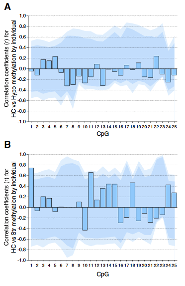
“No umbilical cord or amniotic measurements were available in this study and we therefore cannot control for testosterone levels in utero, a period during which significant testosterone-related changes in brain structure are thought to occur.”

There’s evidence that too much testosterone for a female fetus and too little testosterone for a male fetus both have lifelong adverse effects. The researchers dismissed this etiologic line of inquiry with a “supporting the notion” referral to noncausal studies.


The researchers were keen to establish:

“A very specific, aggression-related structural brain phenotype.”

This putative phenotype hinged on:

  • Older subjects’ behavioral self-reports, and
  • Parental assessments of younger subjects’ behavior

exhibited during the previous six months, and within six months of their fMRI scan.

These self-reports and interested-party observations were the entire bases for the “aggressive behavior” and “anxious–depressed” associations! The researchers disingenuously provided multiple references and models for the reliability of these assessments.


Experimental behavioral measurements – such as those done to measure performance in decision studies – may have been more accurate and informative than what the older subjects chose to self-report about their own behavior over the previous six months.

People of all ages have an imperative to NOT be completely honest about their own behavior. One motivation for this condition is that some of our historical realities are too painful to enter our conscious awareness and inform us about our own behavior. As a result, our feelings, thoughts, and behavior are sometimes driven by our histories without us being aware of it.

For example, would a teenager/young adult subject self-report an impulsive act, even if they didn’t fully understand why they acted that way? Maybe they would if the act could be viewed as prosocial, but what if it was antisocial?

What are the chances that the lives of these teenager/young adult subjects were NOT filled with impulsive actions during the six months before their fMRI scans? Could complete and accurate self-reports of such behaviors be expected?

Experimental behavioral measurements may have also been more accurate and informative than second-hand, interested-party observations of the younger subjects. Could a parent who provided half of the genes and who was responsible for many of their child’s epigenetic changes make anything other than subjective observations of their handiwork’s behavior?


Epigenetic studies have shown that adaptations to environments are among the long-lasting causes for effects that include behavior, hormones, and brain measurements. Why, in 2015, did researchers spend public funds developing what they knew or should have known would be noncausal associations, while not investigating possible causes for these effects?

Why weren’t the researchers interested enough to gather and assess etiologic genetic and epigenetic evidence? Was it that difficult to get blood samples at the same time the subjects gave saliva samples, and perform selected genetic and DNA methylation analyses?

What did the study contribute towards advancing science? Who did the study really help?

My judgment: less than nothing; and nobody. The researchers only wasted public funds advancing a meme, giving it an imprimatur of science.

http://www.psyneuen-journal.com/article/S0306-4530%2815%2900924-5/fulltext “A testosterone-related structural brain phenotype predicts aggressive behavior from childhood to adulthood”

Epigenetic effects of cow’s milk

This 2015 German paper with 342 references described:

“Increasing evidence that milk is not “just food” but represents a sophisticated signaling system of mammals.

This paper highlights the potential role of milk as an epigenetic modifier of the human genome paying special attention to cow milk-mediated overactivation of FTO [a gene associated with fat mass and obesity] and its impact on the transcriptome of the human milk consumer.”

The author declared “no competing interests” and “There are no sources of funding.” He presumably wasn’t pressured into writing this paper.

The paper wasn’t agenda-free, however. The main thesis was:

“Persistent milk-mediated epigenetic FTO signaling may explain the epidemic of age-related diseases of civilization.”

There were separate sections on how milk may promote:

  • Breast cancer
  • Prostate cancer
  • Obesity
  • Metabolic syndrome
  • Coronary heart disease
  • Early menarche
  • Type 2 diabetes
  • Neurodegenerative diseases

I don’t eat or drink dairy products because I’m lactose-intolerant. I coincidentally don’t have any of the diseases mentioned in the paper.

My life experiences haven’t led me to share the author’s sense of alarm, or to attribute other people’s problems to their consumption of milk products. However, more than a few problems I’ve had are things I’ve done to myself through actions or inaction that may have turned out differently if I had better information.

So I curated this article in case we’re insufficiently informed about the harmful epigenetic effects of milk. What do you think?

http://www.ncbi.nlm.nih.gov/pmc/articles/PMC4687119/ “Milk: an epigenetic amplifier of FTO-mediated transcription? Implications for Western diseases”

Using twins to estimate the extent of epigenetic effects

This 2015 international study of intellectual disability used human twins to estimate the impact of genetic, shared-environment, and non-shared-environment on the study’s subjects:

  1. “Estimate of 0.46 (95% CI: 0.32–0.60) can be ascribed to genetic factors.
  2. Estimate of 0.30 (95% CI: 0.19–0.41) may be due to environmental factors involved in growing up in the same environment.
  3. The remaining 24% (95% CI: 0.18–0.29) of the difference is due to error of measurement and nonshared environmental influences.”

The primary causes of individual differences in DNA methylation are environmental factors used analysis of the study’s twin subjects’ CpG methylation compared to “CpGs displaying differential methylation in a healthy population (pDMCs)” to estimate:

  1. “37 % of the pDMCs genetic effects
  2. 3 % of the pDMCs had shared environment
  3. The remaining proportion of the non-genetic variance was due to non-shared environment and/or stochastic factors.”

Those researchers performed several additional tests to find and confirm:

“Non-shared environmental DMCs account for 64% of all detected DMCs.”


Comparing the two studies, the current study’s 32%-60% estimate of genetic effects encompassed the second study’s 37% estimate. However, the current study’s researchers treated their 18%-29% non-shared environment estimate as a remainder not warranting further investigation, whereas the second study’s researchers validated their 64% non-shared environment estimate.

Bringing in a third study, a relevant citation from Epigenetic consequences of early-life trauma: What are we waiting for? confirmed the second study’s estimates with a 2000 twin study that found:

“Environmental effects specific to the individual (63%), whilst genetic effects accounted for 37%. Subsequent studies have produced similar results.”


The Increased epigenetic brain capacity is an evolved human characteristic study found:

“The human brain is extensively shaped by its environment no matter its genetics.”

The epigenetic effects of each of our unique experiences of our non-shared environment predominately determine our individual physiology.

http://www.pnas.org/content/early/2015/12/23/1508093112.full.pdf “Discontinuity in the genetic and environmental causes of the intellectual disability spectrum”