Common features of autoimmune diseases

This 2018 French review subject was mechanisms of autoimmunity:

“Autoimmune diseases (AIDs) encompass more than 80 distinct chronic disorders characterized by inflammatory reactions that can either be systemic or organ specific. In all cases, the disease development is the consequence of the effects of environmental factors in predisposed individuals.

Most of the genes identified by genome-wide association studies (GWAS) on AIDs are related to immunity. However, functional immune parameters that are commonly dysregulated in AIDs do not necessarily stem from these genetic variants. Rather than performing even larger GWAS, understanding complex traits, such as human diseases, may require meticulous analysis or cell-specific gene networks and take into account not only core genes but also seemingly irrelevant genes that may overall have an impact on the disease.

Treg cell defects have been considered a primary cause of AIDs. However, one could ask whether the Treg cell dysfunction exists before the onset of the disease or is provoked by the inflammatory event induced by the triggering components. The defect of Treg cells generally coexists with the inflammatory processes, suggesting several hypotheses:

  1. The inflammation might develop because of a poor regulation of the immune system,
  2. The Treg cells could become inefficient because of the inflammatory environment, or
  3. A common factor concomitantly leads to both effects.

It is likely that autoimmunity results from a chronic imbalance involving both environmental and intrinsic factors. It is now clear that polygenic explanations did not fulfill expectations and that more efforts are needed to understand how the interplay of environmental clues may have a phenotypic impact.”

https://nyaspubs.onlinelibrary.wiley.com/doi/full/10.1111/nyas.13560 “Pathophysiological mechanisms of autoimmunity” (not freely available) Thanks to Dr. Julien Verdier for providing a copy.

A dietary supplement that reversed age-related hearing problems in the brainstem

This 2018 Nevada rodent study was on acetyl-L-carnitine’s action in the brainstem:

“We examined age-related changes in the efficiency of synaptic transmission at the calyx of Held, from juvenile adults (1-month old) and late middle-age (18- to 21-month old) mice. The calyx of Held synapse has been exploited as a model for understanding excitation-secretion coupling in central glutamatergic neurons, and is specialized for high-frequency transmission as part of a timing circuit for sound localization.

Our observations suggest that during aging, there is neuronal cell loss in the MNTB [Medial nucleus of the trapezoid body, a collection of brainstem nuclei in an area that’s the first recipient of sound and equilibrium information], similar to previous reports. In remaining synapses of the MNTB, we observed severe impairments in transmission timing and SV [synaptic vesicle] recycling, resulting in timing errors and increased synaptic depression in the calyx of Held synapse. These defects reduce the efficacy of this synapse to encode temporally sensitive information and are likely to result in diminished sound localization.

We orally administered ALCAR for 1 month and found that it reversed transmission defects at the calyx of Held synapse in the older mice.

These results support the concept that facilitators of mitochondrial metabolism and antioxidants may be an extremely effective therapy to increase synaptic function and restore short-term plasticity in aged brains, and provide for the first time a clear mechanism of action for ALCAR on activity-dependent synaptic transmission.


Human brainstem research is neglected, as noted by Advance science by including emotion in research. Evidence from such research doesn’t play well with beliefs in the popular models and memes of human cerebral dominance.

Do you know any “late middle-age” people who have obvious auditory and synaptic deficits? What if some of the neurobiological causes of what’s wrong in their brains could be “reversed by ALCAR?”

Before using this study as a guide, however, I asked the study’s researchers about the “daily dose of ~2.9 g/kg/d.” An equivalent for a 70 kg human is (2.9 g x 70) x .081 = 16 grams daily, compared with the 500 mg to 1 g dietary supplement dose of acetyl-L-carnitine.

The study’s corresponding coauthor replied:

“This is indeed much larger than that normally consumed by humans via dietary supplementation. We are currently working to determine the effective ‘minimal’ dose of ALCAR and alpha lipoic acid, to better assist guidelines for human application of this supplement.”

https://www.researchgate.net/publication/323941877_Age-related_defects_in_short-term_plasticity_are_reversed_by_acetyl-L-carnitine_at_the_mouse_calyx_of_Held “Age-related defects in short-term plasticity are reversed by acetyl-L-carnitine at the mouse calyx of Held”

Immune memory in the brain

This 2018 German rodent study was a proof-of-principle for immune epigenetic memory in the brain:

“Innate immune memory is a vital mechanism of myeloid [bone marrow] cell plasticity that occurs in response to environmental stimuli and alters subsequent immune responses.

Two types of immunological imprinting can be distinguished – training and tolerance. These are epigenetically mediated and enhance or suppress subsequent inflammation, respectively.

Certain immune stimuli train blood monocytes to generate enhanced immune responses to subsequent immune insults. By contrast, other stimuli induce immune tolerance – suppression of inflammatory responses to subsequent stimuli.

Microglia (brain-resident macrophages) are very long-lived cells. This makes them particularly interesting for studying immune memory, as virtually permanent modification of their molecular profile appears possible. Immune memory in the brain is predominantly mediated by microglia.

In a mouse model of Alzheimer’s pathology, immune training exacerbates cerebral β-amyloidosis and immune tolerance alleviates it; similarly, peripheral immune stimulation modifies pathological features after stroke. Our results identify immune memory in the brain as an important modifier of neuropathology.

Immune memory in the brain could conceivably affect the severity of any neurological disease that presents with an inflammatory component, but this will need to be studied for each individual condition.”


The researchers performed multiple experiments to test different hypotheses about how immune-response experiences are remembered. Modifications to histone methylation and acetylation were targeted.

The stimulus dosage needed to produce immune tolerance – “suppression of inflammatory responses to subsequent stimuli” – was usually four times the dosage used for immune training – “enhanced immune responses to subsequent immune insults.”

https://www.nature.com/articles/s41586-018-0023-4 “Innate immune memory in the brain shapes neurological disease hallmarks” (not freely available)

The epigenetic clock theory of aging

My 400th curation is a 2018 US/UK paper by coauthors of Using an epigenetic clock to distinguish cellular aging from senescence. They reviewed the current state of epigenetic clock research, and proposed a new theory of aging:

“The proposed epigenetic clock theory of ageing views biological ageing as an unintended consequence of both developmental programmes and maintenance programmes, the molecular footprints of which give rise to DNAm [DNA methylation] age estimators.

It is best to interpret epigenetic age estimates as a higher-order property of a large number of CpGs much in the same way that the temperature of a gas is a higher-order property that reflects the average kinetic energy of the underlying molecules. This interpretation does not imply that DNAm age simply measures entropy across the entire genome.

To date, the most effective in vitro intervention against epigenetic ageing is achieved through expression of Yamanaka factors, which convert somatic cells into pluripotent stem cells, thereby completely resetting the epigenetic clock. In vivo, haematopoietic stem cell therapy resets the epigenetic age of blood of the recipient to that of the donor.

Future epidemiological studies should consider other sources of DNA (for example, buccal cells), because more powerful estimates of organismal age can be obtained by evaluating multiple tissues. Other types of epigenetic modifications such as adenine methylation or histone modifications may lend themselves for developing epigenetic age estimators.”


https://www.nature.com/articles/s41576-018-0004-3 “DNA methylation-based biomarkers and the epigenetic clock theory of ageing” (not freely available)


I curated four other papers cited in this review:

Do you want your quality of life to be under or over this curve?

What are you doing to reverse epigenetic processes and realize what you want?

  • Do you have ideas and/or behaviors that interfere with taking constructive actions to change your phenotype?
  • If you aren’t doing anything, are you honest with yourself about feelings of helplessness?
  • Do your beliefs in fate, or in technology, or in divine interventions justify inactions?

The lifelong impact of maternal postpartum behavior

This 2018 French/Italian/Swiss rodent study was an extension of the work done by the group of researchers who performed Prenatal stress produces offspring who as adults have cognitive, emotional, and memory deficiencies and Treating prenatal stress-related disorders with an oxytocin receptor agonist:

“Reduction of maternal behavior [nursing behavior, grooming, licking, carrying pups] was predictive of behavioral disturbances in PRS [prenatally restraint stressed] rats as well as of the impairment of the oxytocin and its receptor gene expression.

Postpartum carbetocin [an oxytocin receptor agonist unavailable in the US] corrected the reduction of maternal behavior induced by gestational stress as well as the impaired oxytocinergic system in the PRS progeny, which was associated with reduced risk-taking behavior.

Moreover, postpartum carbetocin had an anti-stress effect on HPA [hypothalamic-pituitary-adrenal] axis activity in the adult PRS progeny and increased hippocampal mGlu5 [type 5 metabotropic glutamate] receptor expression in aging.

Early postpartum carbetocin administration to the dam enhances maternal behavior and prevents all the pathological outcomes of PRS throughout the entire lifespan of the progeny..proves that the defect in maternal care induced by gestational stress programs the development of the offspring.


This chart from Figure 4 summarized the behavioral performance of aged adult male progeny in relation to the experimental variables of:

  1. Stress administered to the mothers three times daily every day during the second half of pregnancy up until delivery; and
  2. The effects on the mothers’ behavior of daily carbetocin administration during postpartum days 1 through 7.

The symbols denote which of these relationships had statistically significant effects:

  • “* p [Pearson’s correlation coefficient] < 0.05 PRS-Saline vs. CONT-Saline;
  • # p < 0.05 PRS-Carbetocin vs. the PRS-Saline group.”

There are many interesting aspects to this study. Ask the corresponding coauthor Dr. Sara Morley-Fletcher at sara.morley-fletcher@univ-lille1.fr for a copy.

One place the paper referenced the researchers’ previous studies was in this context:

“Postpartum carbetocin administration reversed the same molecular and behavioral parameters in the hippocampus, as does adult chronic carbetocin treatment, i.e. it led to a correction of the HPA axis negative feedback mechanisms, stress and anti-stress gene expression, and synaptic glutamate release. The fact that postpartum carbetocin administration [to the stressed mothers in this study] had the same effect [on the PRS infants in this study] as adult carbetocin treatment [to the PRS offspring in the previous study] indicates a short-term effect of carbetocin when administered in adulthood and a reprogramming (long-term) effect lasting until an advanced age when administered in early development.”

This group’s research seems to be constrained to treatments of F0 and F1 generations. What intergenerational and transgenerational effects would they possibly find by extending research efforts to F2 and F3 generations?


As the study may apply to humans:

The study demonstrated that stresses during the second half of pregnancy had lifelong impacts on both the mothers’ and offsprings’ biology and behavior. Studies and reviews that attribute similar human biological and behavioral conditions to unknown causes, or shuffle them into the black box of individual differences, should be recognized as either disingenuous or insufficient etiological investigations.

The study showed that prevention of gestational stress was a viable strategy. The control group progeny’s biology and behavior wasn’t affected by carbetocin administration to their mothers because neither they nor their mothers had experience-dependent epigenetic deficiencies.

The study demonstrated a biological and behavioral cure for the PRS offspring by changing their stressed mothers’ behaviors during a critical period of their development. The above excerpt characterized improving the mothers’ behaviors as a long-term cure for the PRS descendants, as opposed to the short-term cure of administering carbetocin to the PRS children when they were adults.

What long-term therapies may be effective for humans who had their developmental trajectories altered by their mothers’ stresses during their gestation, or who didn’t get the parental care they needed when they needed it?

https://www.sciencedirect.com/science/article/pii/S0161813X18301062 “Reduced maternal behavior caused by gestational stress is predictive of life span changes in risk-taking behavior and gene expression due to altering of the stress/anti-stress balance” (not freely available)

Prenatal stress produces offspring who as adults have cognitive, emotional, and memory deficiencies

This 2018 French/Italian/Swiss rodent study used a prenatally restraint stressed (PRS) model to create problems that could be resolved by various chemicals:

“S 47445 is a positive modulator of glutamate AMPA-type receptors, possessing neurotrophic and enhancing synaptic plasticity effects as well as pro-cognitive and anti-stress properties.

Most of studies examining the antidepressant effects of new molecules are carried out using behavioral tests performed in unstressed animals.

Corticosterone-treated mice and rats exposed to chronic stress are models that do not recapitulate the early programming of stress-related disorders, which likely originates in the perinatal period. The PRS rat model is characterized by a prolonged corticosterone response to stress and by abnormal behavior.

All the behavioral alterations induced by PRS were corrected by chronic S 47445 administration at both doses.”


The paper included a section comparing S 47445 to ketamine:

“Ketamine, however, causes severe cognitive impairment and psychotomimetic [mimics the symptoms of psychosis, reference not freely available] effects that are direct consequences of the prolonged inhibition of NMDA receptors in cortical and hippocampal interneurons, and seriously limit the chronic administration of the drug in the clinical setting. [reference not freely available]

S 47445 by inducing a direct activation of AMPARs displayed an antidepressant activity without the adverse effect of ketamine. Indeed, contrary to ketamine, S 47445 presented no psychotomimetic effects and induced no occurrence of spontaneous epileptic seizures. [reference freely available] Moreover, S 47445 also presented pro-cognitive properties.”

Compare the above with this April 2018 Chicago Tribune story that had opinions with no linked references:

“ketamine, an anesthetic used to sedate both people and animals before surgery. It’s also a notorious street drug, abused by clubgoers seeking a trancelike, hallucinatory high. But in recent years, numerous studies have found that ketamine can be an effective and speedy treatment for people with depression.”

Which coverage better informed us?


Treating prenatal stress-related disorders with an oxytocin receptor agonist was performed by several of this paper’s coauthors. One references to it was:

“We have already reported that depolarization-evoked glutamate release in the ventral hippocampus is negatively correlated with risk-taking behavior of PRS rats, and that such correlation can be corrected by chronic treatment with monoaminergic/ melatoninergic antidepressants or oxytocin receptor agonist. Thus, an impairment of glutamatergic transmission in the ventral hippocampus lies at the core of the pathological phenotype of PRS rats.”

Looking at the above graphic of the experimental design, I’m not sure why the term perinatal (occurring during or pertaining to the phase surrounding the time of birth) was used in the paper’s title and content to describe the stress period. The pregnant females were stressed three times every day during the second half of pregnancy up until delivery, so the prenatal (previous to birth) term was more applicable.


So, how does this study help humans?

One takeaway is to avoid stressing pregnant mothers-to-be if her children will be expected to become adults without cognitive, emotional, and behavioral problems.

The study demonstrated one way prenatal events cause lifelong effects. The PRS model provides another example of why it’s useless to ask adult humans to self-report causes of epigenetic problems in their lives when these originated before birth, during infancy, or in early childhood, well before humans develop sufficient cognitive capability to recognize such situations. It’s incomprehensible that this unreliable paradigm is still given significant weight in stress studies, especially when experimental designs:

“Do not recapitulate the early programming of stress-related disorders, which likely originates in the perinatal period.”

Also, a relevant difference between humans and PRS rats is that we can ourselves individually change our responses to experiential causes of ongoing adverse effects. Standard methodologies can only apply external treatments such as those mentioned above.

https://www.sciencedirect.com/science/article/pii/S0028390818301291 “The reduction in glutamate release is predictive of cognitive and emotional alterations that are corrected by the positive modulator of AMPA receptors S 47445 in perinatal stressed rats” (not freely available) Thanks to coauthors Stefania Maccari and Dr. Jerome Mairesse for providing a copy.

The role of DNMT3a in fear memories

This 2018 Chinese rodent study found:

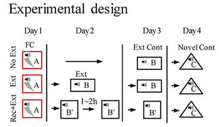
“Elevated Dnmt3a [a DNA methyltransferase] level in the dorsal dentate gyrus (dDG) of hippocampus was associated with the absence of fear renewal in an altered context after extinction training. Overexpression and knockdown of Dnmt3a in the dDG regulated the occurrence of fear renewal in a bi-directional manner.

We found that renewal of remote fear memory can be prevented, and the absence of renewal was concurrent with an elevated Dnmt3a level.

Our results indicate that Dnmt3a in the dDG is a key regulator of fear renewal after extinction, and Dnmt3a may play a critical role in controlling fear memory return and thus has therapeutic values.”


The study was a collection of five experiments investigating causes and effects of biology and behavior. The researchers used different techniques to achieve their goals. I’ve quoted extensively below to show some background and results.

“Alterations in histone acetylation and DNA methylation are involved in the formation and extinction of long-term memory. DNMTs catalyze the cytosine methylation and are required to establish and maintain genomic methylation.

Dnmt3a and Dnmt3b are de novo DNA methyltransferases. Dnmt1 is the maintenance DNA methyltransferase.

  1. Dnmt3a expression was elevated in the dDG after extinction training followed by a brief memory retrieval (Rec+Ext), which was associated with the absence of fear renewal when tested in an altered context.
  2. Increasing Dnmt3a expression in the dDG using AAV [recombinant adeno-associated virus] expression led to the prevention of fear renewal following a standard extinction training protocol. 
  3. Knockdown of Dnmt3a in the dDG using CRISPR/Cas9 resulted in fear renewal following Rec+Ext protocol.
  4. Renewal of remote fear memory can be prevented using the Rec+Ext protocol.
  5. The absence of renewal was concurrent with an elevated Dnmt3a level.

Current exposure therapy, although effective in many patients, suffers from the inability to generalize its efficacy over time, or is limited by the potential return of adverse memory in the new/novel contexts. These limitations are caused by the context-dependent nature of extinction which is widely viewed as the biological basis of exposure therapy.

Achieving a context-independent extinction may significantly reduce fear renewal to improve the efficacy of exposure therapy. Our current study suggests that the effectiveness of these approaches, and ultimately the occurrence of fear renewal, is determined by the level of Dnmt3a after extinction training, especially in the dDG.

There are two potential mechanisms underlying extinction, one is erasure or updating of the formed memory, and the other is the formation of a new extinction memory which suppresses or competes with the existing memory in a context-dependent manner. While most studies favor the suppression mechanism in the adult, limited studies do suggest that erasure occurs in the immature animals.

We propose that if Dnmt3a level is elevated with extinction training (such as with Rec+Ext protocol), modification to the existing memory occurs and as a consequence extinction does not act as a separate mechanism or form a new memory; but if Dnmt3a level is unaltered with extinction training, a separate extinction memory is formed which acts to suppress or compete with the existing memory.”


The relevant difference between humans and lab rats is that we can ourselves individually change our responses to experiential causes of ongoing adverse effects. Standard methodologies can only apply external treatments such as exposure therapy and manipulating Dnmt3a levels.

https://www.nature.com/articles/s41598-018-23533-w “Dnmt3a in the dorsal dentate gyrus is a key regulator of fear renewal”

Placebo is better than these drugs

Consider this post a reblog of Neuroskeptic’s informative About that New Antidepressant Study.

“Here’s why the new study doesn’t tell us much new. The authors..conclude that “all antidepressants were more effective than placebo,” but the benefits compared to placebo were “mostly modest.” Using the Standardized Mean Difference (SMD) measure of effect size, Cipriani et al. found an effect of 0.30, on a scale where 0.2 is considered ‘small’ and 0.5 ‘medium.’

The thing is, “effective but only modestly” has been the established view on antidepressants for at least 10 years. Just to mention one prior study, the Turner et al. (2008) meta-analysis found the overall effect size of antidepressants to be a modest SMD=0.31 – almost exactly the same as the new estimate.”


From the comments section:

“I put his data in a Forest plot and ALL of the positive effect[s] by CBT [cognitive behavior therapy] could be explained by publication bias.

Paroxetine was developed in 1975 and FDA approved for MDD in 1992. It was 2017 before we discovered the true data behind suicides in these trials. That is 25 years. The order of SSRI approval is fluoxetine-> sertraline-> paroxetine-> citalopram-> escitalopram. We know from court cases and other efforts that the suicide data for the first three are false.

PhRMA never got serious about studying clinically meaningful subtypes of “depression” so most data in the meta-analysis just bear on a weak construct called “major depression.”

The reality is these drugs do not help depression much (if any) at all – their effect is to numb the emotions in most people.

The only thing worse than Paxil is Paxil withdrawal.”

Another review of the study, Rewarding the Companies That Cheated the Most in Antidepressant Trials, from which this post is titled, had these comments:

“Patients who take part in these drug trials have been on an antidepressant before the trial. They are then put on placebo for 10 days, a so-called washout. Then half the group, now in cold turkey wit[h]drawal, is now put back on a similar drug to what they had 10 days earlier, and the other group gets to continue their Cold turkey withdrawal.

The fact that these studies are just testing relief from abstinence symptoms by taking a similar drug, could explain why there is no effect in children and young adults.

Most people don’t realize that we are talking about statistical significance, and not clinical significance. The so-called significant difference between drug and placebo is approximately two points on the Hamilton depression scale. The difference has to be at least three for either patient or therapist to notice a difference.

According to this study (https://www.ncbi.nlm.nih.gov/pubmed/23357658), changes of three points or less on the HAM-D correspond to ratings of “no change” on clinician‐rated global symptom severity.

What this study has confirmed is that antidepressants can create a totally insignificant difference compared to a placebo pill. The placebo pill is often combined with attention and close follow up with a professional, and this has a very positive effect.”

A study of gene-environment interactions

This 2018 Hungary/UK study used Bayesian analysis to better understand gene-environment interactions that produce depression:

“Most genetic studies do not consider the effect of stressors which may be one reason for the lack of replicable results in candidate gene studies, GWAS [genome-wide association studies] and between human studies and animal models. Animal models of depression usually imply environmental factors, such as chronic unpredictable stress or learned helplessness.

Relevance of functional polymorphisms in seven candidate genes previously implicated in animal and human studies on a depression-related phenotype given various recent stress exposure levels was assessed with Bayesian relevance analysis in 1682 subjects.

Our data support the strong causative role of the environment modified by genetic factors, similar to animal models.”

From the Methods and Materials section:

“In order to identify recent negative life events (RLE) we used the List of Threatening Experiences questionnaire which queried problems related to illnesses/injuries, financial difficulties, problems related to intimate relationships, and social network occurring in the last year. Based on corresponding items the number of RLEs was counted for each subject, and categorized (low = 0–1, moderate = 2, high = 3/more).”

One item from the findings, and two from the cited references were:

“5-HTTLPR [serotonin transporter], the most extensively investigated polymorphism with respect to interaction with life events, showed only very low relevance.

Compared to heritability which accounts for 37–42% in the variance in general population samples, influence of environmental effects is estimated at 63% in depression.

Etiologically relevant distal and proximal stressors are relatively common, and while frequency of severe life events is estimated to be one in every 3–4 years, depression is triggered in only about one fifth of those with acute stress exposure.”


The methods of this study bypassed problems with GWAS and provided evidence for the lasting effects of “Etiologically relevant distal..stressors.” This was another way of saying that traumatic experiences beginning from the earliest parts of our lives can affect our lifelong biology and behavior.

As mentioned in Changing an individual’s future behavior even before they’re born, GWAS:

“Focuses on the average effect of alternative alleles averaged in a population.”

What this methodology often missed was:

“When phenotypic variation results from alleles that modify phenotypic variance rather than the mean, this link between genotype and phenotype will not be detected.”

The problems found in GWAS may also be found in epigenome-wide association studies. Researchers conducting DNA methylation analyses in particular may benefit from changing their approach if what they’re doing follows the GWAS paradigm.

Using twins to estimate the extent of epigenetic effects summarized three studies’ methods that showed:

“The epigenetic effects of each of our unique experiences of our non-shared environment predominately determine our individual physiology.”

This study’s approach should be considered, given the almost 2:1 relative impacts of environmental over genetic factors in influencing our biology and behavior. It’s especially indicated when human studies don’t replicate animal studies’ findings from strictly controlled experimental environments.


It wasn’t the study’s purpose to evaluate effective treatments for depression. Yet the abstract ended with:

“Galanin-2 receptor, BDNF and X-type purin-7 receptor could be drug targets for new antidepressants.”

The researchers were very careful to document the benefits of using a different approach to a problem. I hope that in the future, they will maintain their carefulness and independence in their approach to solutions, and not be influenced by:

“Consultancy, speaking engagements and research for Bristol-Myers Squibb, AstraZeneca, Eli Lilly, Schering Plough, Janssen-Cilag and Servier..share options in P1vital..consultancy fees from Alkermes, Lundbeck-Otsuka Ltd., Janssen-Cilag Ltd and fees for speaking from Lundbeck.”

https://www.nature.com/articles/s41598-018-22221-z “Significance of risk polymorphisms for depression depends on stress exposure”

An example of researchers changing their field’s paradigms

This 2018 German review subject was retroviruses:

“Initial indications that retroviruses are connected to neoplastic transformation were seen more than a century ago. 43% of the human genome is made up of such elements and 8% of the genome is comprised of retroviruses that infected human ancestors, entering cells of the germ line or proliferating thereafter by retrotransposition.

Endogenized retroviruses (ERVs) are abundantly expressed in many transformed cells. In healthy cells, ERV expression is commonly prevented by DNA methylation and other epigenetic control mechanisms.

A recent string of papers has described favorable outcomes of increasing human ERV (HERV) RNA and DNA abundance by treatment of cancer cells with methyltransferase inhibitors. Analogous to an infecting agent, the ERV-derived nucleic acids are sensed in the cytoplasm and activate innate immune responses that drive the tumor cell into apoptosis.”


Some researchers weren’t satisfied with the status quo of this century-old field:

“Chiappinelli et al. (2015) and Roulois et al. (2015) demonstrated a link between DNMTi-induced activation of HERV expression and innate sensing of transcribed viral RNAs and activation of innate immunity signaling pathways leading to an inhibition of tumor cell growth. These results represent a paradigm shift in our comprehension of the antitumor activity of demethylating agents.”

There are opportunities for any researcher whose field can be related to epigenetics to update the way studies are done. Why should researchers settle for mediocrity when they can make a difference?

https://www.ncbi.nlm.nih.gov/pmc/articles/PMC5816757/pdf/fmicb-09-00178.pdf “HERVs New Role in Cancer: From Accused Perpetrators to Cheerful Protectors”

The influence of donor age on induced pluripotent stem cell functionality

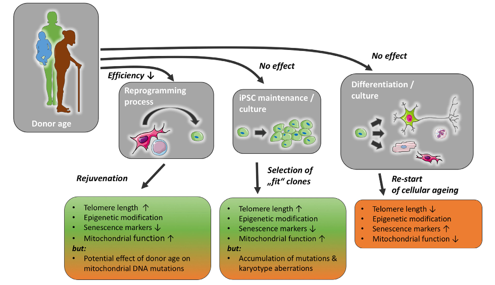
This 2018 German review subject was the influence of donor age on induced pluripotent stem cell functionality:

“Induced pluripotent stem cells (iPSCs) avoid many of the restrictions that hamper the application of human embryonic stem cells. Also, the donor’s clinical phenotype is often known when working with iPSCs.

Typical signs of cellular ageing are reverted in the process of iPSC reprogramming, and iPSCs from older donors do not show diminished differentiation potential nor do iPSC-derived cells from older donors suffer early senescence or show functional impairments when compared with those from younger donors.”

The reviewers discussed limitations in the current research:

  • “Mutations in nuclear and mitochondrial DNA acquired over the donor’s lifespan and during the reprogramming process might persist.
  • It is not yet known how strongly the variable genetic background of individual donors affects the reprogramming process and the quality of resulting iPSCs.
  • A low number of donors and cell lines is a general problem in almost all research articles on the topic of iPSCs. This combined with the lack of a standardised protocol for optimal iPSC derivation, culture and quality control makes any comparison between different publications very difficult if not impossible. Especially, since it has been shown that many factors influence the quality of iPSCs and iPSC-derived cells, such as time and cell type used for reprogramming, time in culture, or reprogramming modality.
  • A problem lies in the retention of tissue-specific epigenetic alterations which in part could be caused by incomplete reprogramming and might be improved by vigorous quality testing and careful selection of iPSC colonies during reprogramming and passaging.
  • The question regarding tumourigenicity will most likely only be answered satisfactorily once 1) the differentiation methods are further improved, 2) iPSC-derived cell-based therapies have made their way further into clinical practice, and 3) patients receiving treatments have been observed for multiple years.”

https://www.ncbi.nlm.nih.gov/pmc/articles/PMC5790033/pdf/fcvm-05-00004.pdf “Age Is Relative-Impact of Donor Age on Induced Pluripotent Stem Cell-Derived Cell Functionality”

Sex-specific impacts of childhood trauma

This 2018 Canadian paper reviewed evidence for potential sex-specific differences in the lasting impacts of childhood trauma:

“This paper will provide a contextualized summary of neuroendocrine, neuroimaging, and behavioral epigenetic studies on biological sex differences contributing to internalizing psychopathology, specifically posttraumatic stress disorder and depression, among adults with a history of childhood abuse.

Given the breadth of this review, we limit our definition [of] trauma to intentional and interpersonal experiences (i.e., childhood abuse and neglect) in childhood. Psychopathological outcomes within this review will be limited to commonly explored internalizing disorders, specifically PTSD and depression.

Despite the inconsistent and limited findings in this review, a critical future consideration will be whether the biological effects of early life stress can be reversed in the face of evidence-based behavioral interventions, and furthermore, whether these changes may relate to potentially concurrent reductions in susceptibility to negative mental health outcomes.”


It was refreshing to read a paper where the reviewers often interrupted the reader’s train of thought to interject contradictory evidence, and display the scientific method. For example, immediately after citing a trio of well-respected studies that found:

“Psychobiological research on relationships linking impaired HPA axis functioning and adult internalizing disorders are suggestive of lower basal and afternoon levels of plasma cortisol in PTSD phenotype.”

the reviewers stated:

“However, a recent meta-analysis suggests no association between basal cortisol with PTSD.”

and effectively ended the cortisol discussion with:

“Findings are dependent upon variance in extenuating factors, including but not limited to, different measurements of:

  • early adversity,
  • age of onset,
  • basal cortisol levels, as well as
  • trauma forms and subtypes, and
  • presence and severity of psychopathology symptomology.”

The reviewers also provided good summaries of aspects of the reviewed subject. For example, the “Serotonergic system genetic research, childhood trauma and risk of psychopathology” subsection ended with:

“Going forward, studies must explore the longitudinal effects of early trauma on methylation as well as comparisons of multiple loci methylation patterns and interactions to determine the greatest factors contributing to health outcomes. Only then, can we start to consider the role of sex in moderating risk.”


I didn’t agree with the cause-ignoring approach of the behavior therapy mentioned in the review. Does it make sense to approach one category of symptoms:

“the biological effects of early life stress”

by treating another category of symptoms?

“can be reversed in the face of evidence-based behavioral interventions.”

But addressing symptoms instead of the sometimes-common causes that generate both biological and behavioral effects continues to be the direction.

After receiving short-term symptom relief, wouldn’t people prefer treatments of originating causes so that their various symptoms don’t keep bubbling up? Why wouldn’t research paradigms be aligned accordingly?

I was encouraged by the intergenerational and transgenerational focus of one of the reviewer’s research:

“Dr. Gonzalez’s current research focus is to understand the mechanisms by which early experiences are transmitted across generations and how preventive interventions may affect this transmission.”

This line of hypotheses requires detailed histories, and should uncover causes for many effects that researchers may otherwise shrug off as unexplainable individual differences. Its aims include the preconception through prenatal periods when both the largest and the largest number of epigenetic changes occur, and is when our susceptibility and sensitivity to our environment is greatest. There are fewer opportunities for effective “preventive interventions” in later life compared with these early periods.

Unlike lab rats, women and men can reach some degree of honesty about our early lives’ experiential causes of ongoing adverse effects. Experiential therapies that allow humans to potentially change their responses to these causes deserve more investigation than do therapies that apply external “interventions.”

https://www.sciencedirect.com/science/article/pii/S0272735817302647 “Biological alterations affecting risk of adult psychopathology following childhood trauma: A review of sex differences” (not freely available) Thanks to lead author Dr. Ashwini Tiwari for providing a copy.

Lysine acetylation is gnarly and dynamic

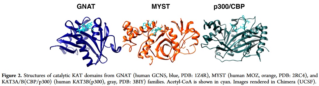
This 2018 UC San Francisco cell review provided details of lysine acetylation:

“Lysine acetylation has moved from being a specialized mark on histones to a critical modification controlling cell fate, proliferation, and metabolism.

During the lifetime of a protein there are many points at which an acetyl group may be added to influence function. The dynamic interplay between the writers, erasers, and readers of acetylation regulates critical epigenomic and metabolic processes, in addition to other major cellular functions.

Acetylation sites are well conserved, in contrast to methylation, where species-specific differences exist.”

The review included a section on mitochondrial protein acetylation:

“Mitochondria have emerged as organelles in which acetylation is more prominent than phosphorylation and plays a key role in integrating metabolic cues with the bioenergetic equilibrium of the cell.

Increased mitochondrial protein acetylation is associated with physiological conditions that result in higher levels of acetyl-CoA (e.g., fasting, calorie restriction, high-fat diet, and ethanol intoxication).”

https://pubs.acs.org/doi/full/10.1021/acs.chemrev.7b00181 “Lysine Acetylation Goes Global: From Epigenetics to Metabolism and Therapeutics” (not freely available) Thanks to lead author Ibraheem Ali for providing a full copy.

A review of human pluripotent stem cell research

Starting the fourth year of this blog with a 2018 Belgian review of human pluripotent stem cells (hPSCs):

“hPSCs are now starting to live up to the great expectations they created after their first derivation nearly twenty years ago. The first results of clinical trials to treat macular degeneration are being published, and an increasing number of clinical or preclinical trials are being started for conditions such as spinal cord injury, diabetes, and heart disease.

This imminent transition of pluripotent stem cells to the clinic has resulted in researchers and clinicians becoming acutely aware of the problems related to the genetic and epigenetic diversity of these cells, included acquired mutations.”

The review included a section on mitochondrial processes that impact the differentiation capacity of pluripotent stem cells, summarized by:

“From this overview, we also observe a more ample contribution of mtDNA in cell fate determination than is represented in many studies tackling the topic.

The transition from aerobic glycolysis to aerobic phosphorylation plays a vital role in cells’ ability to correctly proceed through differentiation, though the mtDNA is rarely evaluated.”

https://academic.oup.com/humupd/advance-article-abstract/doi/10.1093/humupd/dmx042/4825062?redirectedFrom=fulltext “Genetic and epigenetic factors which modulate differentiation propensity in human pluripotent stem cells” (not freely available) Thanks to lead author Alexander Keller for providing a copy.

Using an epigenetic clock to assess liver disease

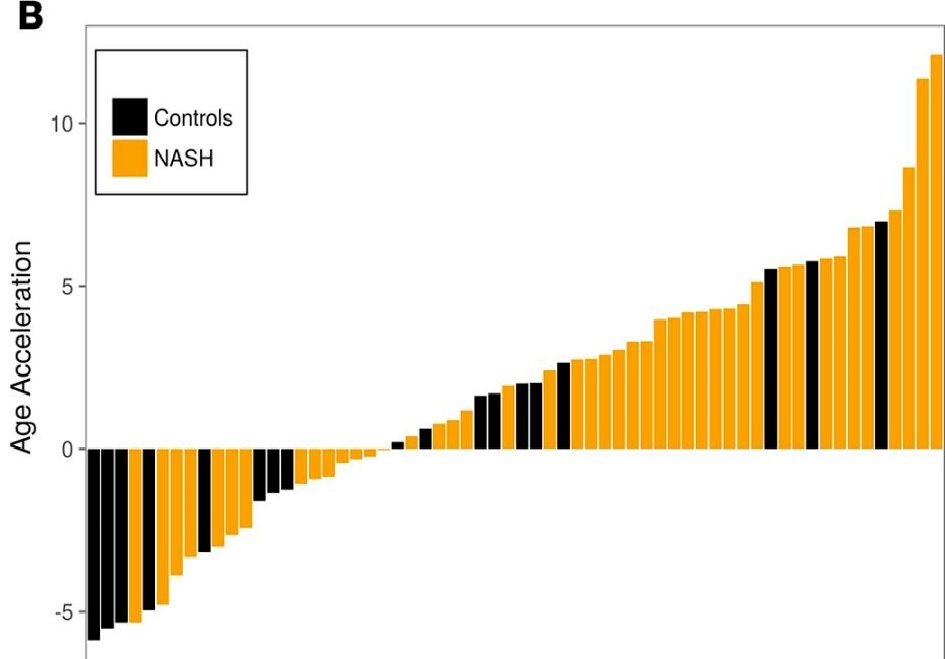
This 2018 UC San Diego human study investigated the capability of the epigenetic clock methodology to detect biological aging with nonalcoholic steatohepatitis (NASH) patients:

“The ability to measure a surrogate marker of liver aging from a peripheral blood sample has broad implications for assessing clinically “silent” chronic diseases, such as NASH, and, potentially, their response to interventions.

In the current study, we validate the utility of the Horvath clock in measuring age acceleration in a defined cohort of NASH patients with moderate to severe liver fibrosis.”

The study demonstrated several aspects of age acceleration and disease conditions, including:

– Use of clinical trial data.

“This study, however, included patients who were part of a clinical trial in which protocol-obtained biopsies were read by a central pathologist (ZG) and morphometric quantification of collagen standardized.”

– Continuous measures were more relevant than “Stage X” measures.

“The findings in the current work are in contrast to an earlier study that found no association of DNAm with the NAFLD activity score or stage of liver fibrosis in patients with NASH. Importantly, that study assessed liver fibrosis based on conventional histological staging only, using the ordinal METAVIR classification. Similarly, we also found no difference in age acceleration between patients with stage 2 and 3 fibrosis according to the NASH Clinical Research Network (CRN) classification.

On the contrary, by evaluating two continuous measures of fibrosis (hepatic collagen content by morphometry and the serum ELF test), which have a greater dynamic range than traditional histological staging, we found that patients with higher age acceleration have increased hepatic fibrosis.”

– Causalities may not necessarily be ascribed.

“Although these reports establish a potential relationship between fibrosis and specific epigenetic modifications, targeted individual CpG sites may not accurately reflect the complex interaction between causal and compensatory measures in chronic diseases such as NASH.”

https://insight.jci.org/articles/view/96685 “DNA methylation signatures reflect aging in patients with nonalcoholic steatohepatitis”