Fetal exposure to sex hormones and female anxiety

This 2015 Swedish rodent study found:

“Women with polycystic ovary syndrome (PCOS) display high circulating androgen levels that may affect the fetus and increase the risk of mood disorders in offspring.

Although clinical data are inconsistent, there are indications that androgens play a crucial role in behavior and mood regulation in females.

Studies on the link between testosterone and anxiety behavior in males have generated inconsistent results.

Higher circulating testosterone has previously been reported in female rat PNA [prenatal androgen] offspring. This discrepancy may be a result of the higher doses of maternal testosterone (5 mg) used in the previous study compared with the present study (0.5 mg).

Although the anxiety-like behavior observed in the female PNA offspring in the present study cannot be directly explained by high circulating androgens, the reduced AR [androgen receptor] expression in the amygdala suggests a compensatory response to the high prenatal testosterone exposure, a result implicating the amygdala as the CNS site underlying the changes in anxiety in the PNA offspring. This idea is further strengthened by our experiment showing that subchronic testosterone exposure into amygdala is sufficient to produce anxiety-like behavior in adult females.

Maternal testosterone exposure causes anxiety-like behavior in female, and to a lesser extent male offspring, an effect that seems to occur during fetal life and to be mediated via AR in the amygdala, together with changes in ER [estrogen receptor] and in the serotonergic and GABAergic pathways in the amygdala and hippocampus of female PNA rats.”

The news coverage – too much testosterone caused anxiety-like symptoms in females whether they are adults or fetuses – was NOT what the study found. The headlines disregarded its caveat:

“The anxiety-like behavior observed in the female PNA offspring in the present study cannot be directly explained by high circulating androgens.”

I look forward to research on floor levels of testosterone, below which there are also adverse effects on females. There is such evidence, but would it play well with popular memes?

See Sex hormone exposure to the developing female fetus causes infertility in adulthood for another study that used the PCOS phenotype.

http://www.pnas.org/content/112/46/14348.full “Maternal testosterone exposure increases anxiety-like behavior and impacts the limbic system in the offspring”

A study of DNA methylation and age

This 2014 Finnish human study compared and contrasted the DNA methylation levels of young adults with people age 90:

“We identified 8540 high-confidence CpG [cytosine and guanine separated by only one phosphate link] sites that show a large difference in methylation levels between nonagenarians and young controls and that present high statistical significance in a regression model adjusted for the leukocyte proportion.

The majority of frequently reported CpG sites are hypermethylated with increasing age.

Ageing-associated hypermethylation is concentrated in genes associated with developmental processes as well as DNA-binding and transcription of genes, whereas hypomethylation is not enriched among a specific set of genes.

The largest percentage of the variation in our methylation data was associated with the proportions of different leukocyte subtypes.

We found that only a minority of ageing-associated CpG sites showed an association between methylation and expression levels. Furthermore, only a minority of these genes have been identified as differentially expressed between nonagenarians and young individuals.”

The finding concerning:

“Ageing-associated hypermethylation is concentrated in genes associated with developmental processes as well as DNA-binding and transcription of genes”

was in concert with a referenced 2013 review Aging is not programmed that stated:

“Aging is not and cannot be programmed. Instead, aging is a continuation of developmental growth, driven by genetic pathways.

Aging is a shadow. Its shape is determined by the developmental growth.

Genetic programs determine developmental growth and the onset of reproduction. When these programs are completed, they are not switched off.

Aging has no purpose (neither for individuals nor for group), no intention. Nature does not select for quasi-programs. It selects for robust developmental growth.

Whereas the growth of the body is programmed, the emergence of the shadow is not. Natural selection cannot eliminate the shadow without hurting the “body”.”


The researchers made several points relating the current study and other epigenetic studies.

Regarding DNA methylation and gene expression:

“Due to the methods applied in the present study, not all the effects of DNA methylation on gene expression could be detected; this limitation is also true for previously reported results.

The textbook case of DNA methylation regulating gene expression (the methylation of a promoter and silencing of a gene) remains undetected in many cases because in an array analysis, an unexpressed gene shows no signal that can be distinguished from background and is therefore typically omitted from the analysis.”

Regarding biological age and chronological age:

“It remains to be investigated whether these [CpG] sites are only associated with chronological age or if there are also associations with phenotypic changes related to (successful) ageing.

If these frequently reported sites are only markers of chronological age, markers of biological age are yet to be identified.”

Regarding the rapid progression of technology used in epigenetic studies, they noted several times how what they used was significantly improved over pre-2014 technologies with statements such as:

“Global hypomethylation has been associated with an increasing risk of frailty, but very few other associations between phenotype and DNA methylation have been reported. However, this may be due to technical concerns, as the study by Bell et al. was performed with the 27K array, which almost exclusively contains promoter-associated probes that are not methylated at baseline and can therefore primarily acquire hypermethylation. Phenotype association studies performed with the 450K array or using sequencing techniques are necessary to clarify if hypomethylation is associated with typical ageing-associated phenotypes.”

Compare that with the limitations of the same 450K array acknowledged in A human study of changes in gene expression 2015 study:

“This array queries only 1.6% of all CpGs in the genome and the CpG selection is biased towards CpG islands. Other techniques – whole-genome bisulfite sequencing and methylC-capture (MCC) sequencing, for example – have definite technical advantages (higher resolution and no CpG island selection bias).”

http://www.biomedcentral.com/1471-2164/16/179 “Ageing-associated changes in the human DNA methylome: genomic locations and effects on gene expression”

Is the purpose of research to define opportunities for interventions?

In this 2014 review, a social scientist first presented an interpretive history of what he found to be important in the emergence of epigenetics. He proceeded into his ideas of “a possible agenda of the social studies of the life-sciences” in the “postgenomic age” with headings such as “Postgenomic biopolitics: “upgrade yourself” or born damaged for ever?”

This perspective included:

“The upgradable epigenome may become the basis for a new motivation to intervene, control and improve it through pharmacological agents or social interventions.

An important trend is the use of epigenetic and developmental findings in the so-called early-intervention programmes.

It is possible that epigenetic findings will become increasingly relevant in social policy strategies.”


In this blog I often highlight research that may help us understand details of how each of us is a unique individual. It’s my view that insofar as research helps each of us understand our unique, real self, we may be able to empathetically understand others’ unique qualities.

Click individual differences for a sample of how researchers explain away uniqueness in order to converge on a study’s desired objectives. There’s seldom an attempt to further understand what caused each subject to develop their unique qualities.

Why would this reviewer advocate that

  • Researchers,
  • People working in the social sciences,
  • People employed or involved in social services, and
  • Their sponsors and employers

intentionally disregard another individual’s unique qualities?

I’ll answer this question from a perspective that explains how this common, reflexive action derives from a person being unable to face the facts of their own life. Pertinent fundamentals of Dr Arthur Janov’s Primal Therapy are:

  1. Pain motivates a person’s unconscious act-outs of their underlying problems.
  2. The behavior that caused a problem is sometimes also the act-out behavior.
  3. Act-outs enable a person to re-experience the feelings of their historical struggles, in a vain attempt to resolve them.
  4. Due to pain barriers, people seldom become consciously aware of and – more importantly – address the causes for their own problematic behavior.
  5. “The patient has the power to heal himself.”

A consequent hypothesis is that a person will often glorify their unconscious act-outs and surround themself with justifications for these actions. For example, a person who can’t sit still may refer to their incessant activity with socially acceptable phrases such as “I’m always busy” or “I love to travel.” They’ll structure their life to enable their unconscious behavior, never questioning how they were attracted to an always-on-the-go occupation such as flight attendant, only vaguely feeling that they were made for it.

The behavior relevant to the current review may be exhibited by a person with a history of having no control over their own life. Following the above first two fundamentals, the pain of historically not having control over their life may motivate them to control other people’s lives.

Unfortunately for everyone who’s affected, such unconscious act-outs don’t resolve anything:

  1. The initiator may achieve some symbolic satisfaction by controlling others’ lives.
  2. This temporary satisfaction doesn’t make the initiator’s underlying problems less painful.
  3. The motivation impelling these unconscious act-outs isn’t thereby reduced.
  4. So the initiator soon repeats their controlling behavior, stuck in a loop of unresolved feelings.
  5. Since the self-chosen interests of someone who’s being controlled are lesser concerns to the initiator than exercising control, the controlled person may or may not be helped by the controller’s act-outs.

Research provides abundant evidence that we are unique individuals.

This is a strong indicator of who is best qualified to direct each of our unique lives.

A person who is driven to control others’ lives won’t accept epigenetic research as instructive for understanding, honoring, and respecting others as unique individuals. They’ll use research as a way to enable their own unconscious act-outs, and view it as offering opportunities for interventions into the lives of others.

This is the way that “pharmacological agents or social interventions” are often the intended “use of epigenetic and developmental findings.” Interventions receive justifications with “a possible agenda of the social studies of the life-sciences.”

Becoming aware of one’s own act-outs – and then individually addressing one’s own underlying problems – often take backseats to employment and other concerns to keep enabling one’s own behavior. That makes it likely that interventions justified by “epigenetic findings..in social policy” will continue, whether or not the subjects agree that they’re being helped.

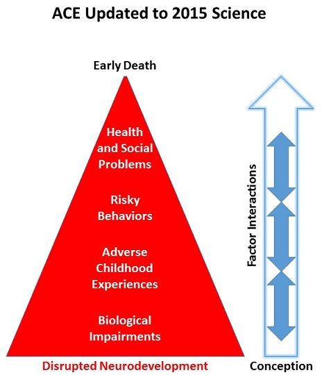
For examples, take a look at a few of the YouTube presentations by people employed in the social sciences and social services on a topic of epigenetics. Compare them with the current state of epigenetic research in Grokking an Adverse Childhood Experiences (ACE) score.

What did you notice? How many presentations emphasized disrupted prenatal development – a period when problems can be prevented? Did you instead see that many more of the presentations emphasized controlling behavior?

http://journal.frontiersin.org/article/10.3389/fnhum.2014.00309/full “The social brain meets the reactive genome: neuroscience, epigenetics and the new social biology

Transgenerational epigenetic programming with stress and microRNA

This 2015 Pennsylvania rodent study found:

“Sperm miRs [microRNAs, a small non-coding RNA that has a role in gene expression] function to reduce maternal mRNA [messenger RNA, a large RNA that carries codes for protein production] stores in early zygotes, ultimately reprogramming gene expression in the offspring hypothalamus and recapitulating the offspring stress dysregulation phenotype.”

These researchers caused stress-induced changes at an early stage of embryonic development with microRNA injections. Resultant adverse effects weren’t observed until subjects were adults!

Most news coverage focused on it being a male’s stress, not a female’s, that affected a developing embryo. Either or both sexes can epigenetically disadvantage a fetus – okay.

Demonstrating how a damaging influence can begin immediately after conception, but symptoms didn’t present until adulthood made this study newsworthy.


Although the term “transgenerational” was used in the study’s title, abstract, and elsewhere, studied epigenetic effects were intergenerational rather than transgenerational. Per A review of epigenetic transgenerational inheritance of reproductive disease, for the term to apply, researchers need to provide evidence in at least the next 2 male or non-gestating female generations and/or 3 gestating female generations of:

“Altered epigenetic information between generations in the absence of continued environmental exposure.”


From a press release, a study coauthor who also coauthored How to make a child less capable even before they are born: stress the pregnant mother-to-be stated:

“Bale suspects that when a male experiences stress it may trigger the release of miRs contained in exosomes from epithelial cells that line the epididymis, the storage and maturation site for sperm between the testes and the vas deferens. These miRs may be incorporated into maturing sperm and influence development at fertilization.”

Not all stress-related gene expression in pituitary and adrenal glands differed.

http://www.pnas.org/content/112/44/13699.full “Transgenerational epigenetic programming via sperm microRNA recapitulates effects of paternal stress”

A review of the epigenetic basis for mental illness

This 2015 New York combined animal and human review of epigenetic studies noted:

“While genetic factors are important in the etiology of most mental disorders, the relatively high rates of discordance among identical twins, particularly for depression and other stress-related syndromes, clearly indicate the importance of additional mechanisms.

Environmental factors such as stress are known to play a role in the onset of these illnesses.

Exposure to such environmental insults induces stable changes in gene expression, neural circuit function, and ultimately behavior, and these maladaptations appear distinct between developmental versus adult exposures.

Increasing evidence indicates that these sustained abnormalities are maintained by epigenetic modifications in specific brain regions.”

Placing the “maladaptations” and “sustained abnormalities” phrases into their contexts:

  • A fetus biologically adapted to their environment – however toxic it was – in order to best survive.
  • These adaptations for survival were subsequently viewed as Disrupted Neurodevelopment and “maladaptations” from the perspectives of normal development and environments.
  • The “sustained abnormalities” caused within the earlier environments “are maintained by epigenetic modifications.” An improved environment wasn’t impetus enough to change developmental “maladaptations.”

Per the below link, it’s been a month since this review was published. Why has there been ZERO news coverage of it?

One reason may be that the Friedman Brain Institute, Icahn School of Medicine at Mount Sinai, didn’t issue a press release or otherwise publicize it. Another reason may be the groups that are opposed to its findings:

  • Parents who provided harmful environments for their children, beginning at conception;
  • People who feel threatened when scientific causal evidence resonates with what happened in their own lives, and in response, limit their empathetic understanding of others’ problems;
  • Social workers, psychologists, and others in industries whose paychecks depend on efforts that aren’t directed towards ameliorating the causes for these later-life effects;
  • Psychiatrists and medical personnel whose livelihoods depend on pharmaceutical and other treatments that only alleviate symptoms;
  • Researchers whose funding depends on producing non-etiologic findings.

Despite resistance to this review’s findings, a large number of people would benefit from publicizing evidence for:

“These sustained abnormalities are maintained by epigenetic modifications in specific brain regions.”

http://nro.sagepub.com/content/early/2015/09/24/1073858415608147 “Epigenetic Basis of Mental Illness”

Grokking an Adverse Childhood Experiences (ACE) score

What does it take to empathetically understand, to make a part of oneself, to grok an ACE score?

The ACE effort was initiated in 1985 in an era before epigenetics was well-studied. Its artifacts included the ACE pyramid:

The_ACE_PyramidThe historical ACE lifespan continuum on the left began at conception. The pyramid on the right promoted a limited view of ACE that assigned childhood as the pyramid’s base.

Current official depictions of the ACE pyramid assign an expanded view of ACE as the pyramid’s base. The viewer’s attention is directed to “Scientific Gaps” between pyramid layers, but the largest gap remains: the continuum starts at conception but the pyramid still starts at childhood. The narrative claims:

“To provide scientific information that would be useful for developing new and more effective prevention programs.”

The official ACE pyramid doesn’t accurately reflect current science documented in, for example, Epigenetic effects of early life stress exposure. By downplaying Disrupted Neurodevelopment that may begin at conception, governing agencies implicitly endorse approaches that fail to address prenatal causes for later-life adverse effects.


If the ACE diagram was drawn thirty years later in 2015 to incorporate evidence for epigenetics, Disrupted Neurodevelopment wouldn’t be a consequent layer to an ACE base. The potential start of Disrupted Neurodevelopment would coincide with conception:Updated for 2015 to show Disrupted Neurodevelopment

What’s an example of current ACE-related scientific evidence that wasn’t present three decades ago and also isn’t represented in the official ACE pyramid? Prenatal Disrupted Neurodevelopment may be considered today as a possible consequence of a “Yes” answer to half of the original ACE questions:

  • Were your parents were too drunk or high to take care of you or take you to the doctor?
  • Were your parents ever separated or divorced?
  • Was your mother often or very often pushed, grabbed, slapped, or had something thrown at her?
  • Did you live with anyone who was a problem drinker or alcoholic or who used street drugs?
  • Was a household member depressed or mentally ill?

These threats and other stresses cause a fetus to biologically adapt. When such adaptations occur during prenatal development, they may:

  • Have much larger impacts and
  • Cause Biological Impairments that
  • Don’t unassistedly disappear over time.

Emphasizing Disrupted Neurodevelopment that may begin at conception would encourage:

  • Research that’s directed toward producing causal evidence for adaptations that largely occur during the early periods of an individual’s lifespan; and
  • Research on how these adaptations consistently influence our later-life ideas, biology, and behavior.

The above recommendations for research are neither the current focus of ACE research nor the direction of related efforts to assist affected individuals. Relevant studies that I’ve curated on this blog often only produced symptomatic evidence:

  • If a study couched its findings in non-etiologic phrases such as “is associated with” or “is linked to” or “may relate to,” it didn’t address ACE originating causes.
  • “New and more effective prevention programs” seldom address Disrupted Neurodevelopment and Biological Impairments with efforts to reduce the source of the damage.
  • If a program’s presentation showed multivariate analyses with ACE score probabilities and percentages, it didn’t address originating causes.

Here’s a YouTube search of ACE + adverse. Evaluate the current focus of ACE efforts by people employed in the social sciences and services. What did you notice?

How many presentations emphasized prenatal Disrupted Neurodevelopment, a period during which problems may be prevented by addressing causes? Did you instead see that these were outnumbered by many more presentations that emphasized Health and Social Problems symptom interventions?


So, what does it take to empathetically understand, to make a part of oneself, to grok a person’s ACE score?

Regarding empathy – it’s best to avoid the advice of studies such as:

People who are helped may not recognize it at first, but over time, they’ll sense whether the helper’s empathy is genuine.

Regarding understanding – I feel that people first need to ameliorate the origins of their own problems. Then they may be able to help others therapeutically address causes for ACE symptoms.

Need proof? Think of someone you’ve met whose thoughts and feelings and behavior were caught up in and motivated by their own problems:

  • Did you feel they could empathetically understand others?
  • Wasn’t the welfare of the people who may have been helped truly incidental and secondary to someone who was acting out their own problems?

Genetic causes for epigenetic symptoms

This 2015 human summary study was of 44 genetic disorders that disrupt the maintenance of epigenetic modifications:

“..making them likely to have significant downstream epigenetic consequences. Interestingly, these patients often demonstrate neurological dysfunction, suggesting that precise epigenetic regulation may be critical for neuronal homeostasis. However, at the same time, it is important to keep in mind that many of these proteins have additional non-epigenetic roles.

Mutations in many of these components have now been linked to a number of well-known causes of intellectual disability. Intellectual disability is generally defined as deficits of intellectual function and adaptive behavior that occur during the developmental period.

Given the opposing activity of many of the components of the epigenetic machinery, the pathogenic sequence in these disorders involves an imbalance of chromatin states. Keeping a subset of genes under “pressure” from two opposing systems may allow the cellular system to rapidly respond to environmental stimuli.

These disorders, on average, have unusual phenotypic breadth. Similarly, there is a shift in distribution toward a higher number of organ systems affected.

In addition to developmental phenotypes (multiple congenital anomalies), in some cases there appear to be ongoing defects that remain consequential in post-natal life. An example of the latter is the hippocampal memory defects seen in many of the mouse models.

This raises the question whether cells undergoing neurogenesis and synaptogenesis are particularly sensitive to subtle defects of the epigenetic machinery and downstream epigenetic abnormalities. A major remaining question is whether neurogenesis defects and/or abnormalities of synaptic plasticity are a unifying pathophysiological process.”

The researchers represented the 44 genetic disorders on a wheel graph:

F1.large

I look forward to further research that includes non-genetic disruptors of epigenetic modifications.

http://genome.cshlp.org/content/25/10/1473.full “The Mendelian disorders of the epigenetic machinery”

Identifying epigenetic DNA changes with blood tests

This 2015 Chinese human study found:

“With reference to methylation profiles of different tissues, we developed a general approach for studying the major tissue contributors to the circulating DNA pool. This development has opened up numerous research avenues and diagnostic applications.

Our study takes advantage of the recent availability of reference methylomes of a number of tissues. It is likely that such reference databases would be continually updated to include more sample types and from more individuals.”

Up to 41% of plasma DNA in pregnant women was from the placenta. However, I didn’t understand why the non-pregnant women in the control group had measurable placental DNA of up to 2.9%. Maybe it was leftover from a prior pregnancy?

http://www.pnas.org/content/112/40/E5503.full “Plasma DNA tissue mapping by genome-wide methylation sequencing for noninvasive prenatal, cancer, and transplantation assessments”

Countering the epigenetic effects of obese mothers on their fetuses

This 2015 Colorado rodent study found:

“Maternal ADN [adiponectin, a hormone produced by fat cells, that regulates fat and glucose metabolism] supplementation reversed the adverse effects of maternal obesity on placental function and fetal growth.

Babies of mothers with obesity and/or gestational diabetes mellitus (GDM) are often large at birth and have increased adiposity, which predisposes them to the development of metabolic disease later in life.

Maternal ADN infusion in obese dams from E14.5 to E18.5 [the last 4 days of pregnancy, a period that accounts for 70% of the total fetal growth] normalized maternal insulin sensitivity, placental insulin/mTORC1 and PPARα signaling, nutrient transport, and fetal growth without affecting maternal fat mass.”

As the study may apply to humans:

“This hormone or a similar agent could feasibly do the same thing for humans that it did for mice,” Jansson said.

Jansson said more work needs to be done to track the long-term effects of the hormone treatment on the mice.”

The study focused on epigenetic effects of the mothers’ environment on fetuses, and didn’t assess possible genetic contributions.

As alternatives to adiponectin supplementation:

http://www.pnas.org/content/112/41/12858.full “Adiponectin supplementation in pregnant mice prevents the adverse effects of maternal obesity on placental function and fetal growth”

Leaky gates, anxiety, and grocery store trips without buying list items

An interview with Jeff Link, the editor of Dr. Arthur Janov’s 2011 book “Life Before Birth: The Hidden Script that Rules Our Lives” with Ken Rose:

“Even further confirmation for some of the views of Janov, that maybe weren’t widely accepted for a time, it’s new research now being done into memory and what a lot of scientist are seeing, a lot of different studies is that memory reactivates the same neuroimpulses that were initially firing off when the event happened.

So a traumatic event when you remember it, the act of remembering it is actually creating a neuromirror of what went on initially.

In a lot of ways that is what Primal Therapy is attempting to do; is to go back to that place and reconnect, or as it’s sometimes referred to, reconsolidate the brain state so that real healing can take place.”

Transcript (part 4 of 6): http://cigognenews.blogspot.com/2015/09/ken-rose-on-life-before-birth-part-46.html

MP3: http://www.pantedmonkey.org/podcastgen/download.php?filename=2011-12-15_1300_what_now_jeff_link.mp3

Epigenetic changes in the developing brain change behavior

This 2015 review cited 143 studies to tie together findings in epigenetic chemistry and behavioral neuroscience.

In addition to studies I’ve previously curated, other research included:

  • a 2012 study which completely abolished mouse maternal behavior by silencing a gene encoding an estrogen receptor;
  • a 2012 study which found that stress-induced changes in the rat hippocampus were heritable;
  • a 2014 study that distinguished between transgenerational and intergenerational epigenetic effects such as:

    in utero exposure to nutritional status, stress, or toxic environmental factors that act on the developing embryo and its germ line”

  • a 2013 study that showed how genomic imprinting coordinated:

    “Genetic coadaptation where beneficially interacting alleles evolve to become coinherited.”

The current status of research incorporating both epigenetic chemistry and behavioral neuroscience was summed up as:

“A large number of behavioral epigenetic studies attempt to correlate epigenetic marker changes at global levels and in mixed populations of cells with phenotypic changes.

Specific changes at specific gene levels and at single cell levels correlating with behavioral changes remain largely unknown.”

http://www.pnas.org/content/112/22/6789.full “Epigenetic changes in the developing brain: Effects on behavior”

RNA as a proxy signal for context-specific biological activity

This 2015 Harvard/MIT rodent study was of long (more than 200 nucleotides) noncoding (non-protein coding) RNAs (ribonucleic acids). These are of interest because:

“Within the mammalian body, the largest repertoire and diversity of lncRNA genes outside the germ line occurs in the brain, where lncRNAs exhibit regional and cell-specific localization.

The expression patterns of lncRNAs may serve as a proxy signal for important, context-specific biological activity.”

The researchers explained what they could and couldn’t determine with current techniques and technologies:

“The whole-gene ablation method used here is often a first approach to determine the functionality of a locus.

Although each of these loci contains a lncRNA, it is important to consider that any observation resulting from this strategy could reflect the loss of any regulatory element in the deleted region.

The rate of lncRNA gene discovery has significantly outpaced our ability to evaluate both the physiological significance and function of these genes. It is difficult to predict whether the loss of any particular lncRNA locus will present a phenotype, but crucial information on the spatiotemporal dynamics of expression from each locus can provide significant direction and focus to downstream mechanistic studies by highlighting those loci most likely to have a physiological impact.

It is important to stress that no single method exists that can account for all possible mechanisms of action of a noncoding locus. Within these limits, the phenotypes observed after ablation of specific lncRNA loci confirm that expression of this class of noncoding RNAs can serve as a proxy signal to identify functional genomic loci with physiological relevance to disease and development, independent of whether this activity is directly ascribed to a functional lncRNA molecule.”

http://www.pnas.org/content/112/22/6855.full “Spatiotemporal expression and transcriptional perturbations by long noncoding RNAs in the mouse brain”

Stress in early life can alter physiology and behavior across the entire lifespan

I’ll quote a few sections of this 2014 summary of 111 studies concerning stress, including the authors’ research:

“The brain is the central organ of stress and adaptation to stressors because:

  • It not only perceives what is threatening or potentially threatening and initiates behavioral and physiological responses to those challenges,
  • But also is a target of the stressful experiences and the hormones and other mediators of the stress response.

The stress history of parents is a significant factor in the resilience of their offspring.

Environmental stress transduces its effects into lasting changes on physiology and behavior, which can vary even among genetically identical individuals.

Stress in early life can alter physiology and behavior across the entire lifespan.

Structural stress memory is even more apparent with regard to gene expression in stress-sensitive brain regions like the hippocampus.

Individual history is important and that there is a memory of stress history retained by neurons at the cellular level in regions like the hippocampus.

Stress has a number of known effects on epigenetic marks in the brain, producing alterations in DNA methylation and histone modifications in most of the stress-sensitive brain regions examined, including the hippocampus, amygdala, and prefrontal cortex.”


It seemed to be taboo to note that most of – and the largest of – detrimental effects of stress occurred during womb-life in the mother’s environment. The authors instead opted for a politically correct “the stress history of parents” phrase.

Referenced studies had findings relevant to the earliest periods of life, including Figure 1:

interactions

“Those organs that show the highest levels of retrotransposon [a repeat element (mobile DNA sequences often involved in mutations) type formed by copy-and-paste mechanisms] activity, such as the brain and placenta, also seem to be both steroidogenic and steroid-sensitive.”

However, Figure 1 was given a beneficial context, and other studies’ findings weren’t mentioned in their contexts of detrimental effects on fetuses of mothers who were stressed while pregnant.

http://www.pnas.org/content/112/22/6828.full “Stress and the dynamic genome: Steroids, epigenetics, and the transposome”

Changing an individual’s future behavior even before they’re born

This 2015 Harvard fruit fly research was a companion of the Is what’s true for a population what’s true for an individual? study.

The researchers began with the question:

“If we could rear genetically identical individuals from a variety of genetic backgrounds and rear them in the same environment, how much phenotypic variation between individuals of the same genotype would we see?”

They answered with:

“We show that different genotypes vary dramatically in their propensity for variability, that phenotypic variability itself, as a trait, can be heritable, and that loci affecting variability can be mapped.”


The specific problem that probably prompted this study was that the methodology of genome-wide association studies (GWAS) usually:

“Focuses on the average effect of alternative alleles averaged in a population.”

What this methodology often missed was:

“When phenotypic variation results from alleles that modify phenotypic variance rather than the mean, this link between genotype and phenotype will not be detected.”


The researchers altered the environment during a critical period of fruit flies’ development in order to induce epigenetic changes in the fruit fly pupae brains:

“Disruption of Ten-a [the synaptic target recognition gene Tenascin accessory] expression in midpupa affects behavioral variance [the standard statistical dispersion parameter].

In all cases, disrupting Ten-a increased the variability [the median of the absolute deviation from each observation’s median] in turning bias with no effect on the mean.”

I fully expect researchers to demonstrate that this finding has general applicability for humans, especially during womb-life. Research such as:

are steps in this direction just for one factor in the human fetal environment – stress. The effects of stressing a human fetus should be at least as significant as the effects produced on the study’s subjects with increased temperature during pupation.

http://www.pnas.org/content/112/21/6706.full “Behavioral idiosyncrasy reveals genetic control of phenotypic variability”

Separating genetic from environmental factors when assessing educational achievement

This 2014 UK study of identical and fraternal twins found that an average of 62% of the differences among their scores on a significant test given at age 16 were due to genetic factors:

“Genetic influence is greater for achievement than for intelligence, and other behavioral traits are related to educational achievement largely for genetic reasons.”

However, the “genetic reasons” term didn’t mean that the researchers actually took genetic samples. From one news article:

“Identical twins share 100 percent of their genes while non-identical twins share just 50 percent of their genes. Because these sets of twins share the same environment, the scientists were able to compare identical and non-identical twins to estimate the relative contributions of genetic and environmental factors.”

This estimation method produced an artificial divide between genetic and environmental factors. Identical twins start out sharing 100% of their genes, but then their genes become expressed differently – often because of environmental factors – to produce unique individuals even before birth.

The sets of identical twins were definitely not the 100% same genetic makeup between themselves at age 16 as they were at conception, and that assumption was the foundation of the researchers’ model:

F2

“Bivariate estimates for additive genetic (A), shared environmental (C), and nonshared environmental (E) contributions to the correlations between GCSE and nine predictors. The total length of the bar indicates the phenotypic correlations.”

The researchers didn’t provide evidence that “genetic reasons” were causal factors to the stated extent. Although the model’s numbers may have indicated that the method’s results were valid, that didn’t necessarily mean that the reality of genetic and epigenetic influences on the subjects were represented to the stated precision by the results.

The weather analogy of Scientific evidence applies to this study’s methods:

“We can think about what we mean by evidence. For example, that when you see dark storm clouds overhead, that’s strong evidence that it’s about to rain. If you smell a certain scent, that’s maybe weak evidence that it’s about to rain. And if we see the dark storm clouds and then we smell the scent, the evidence doesn’t get weaker: if anything, it gets stronger.

But P-values in a circumstance like that, where you have a very small P-value in one dataset and a not-so-small P-value in a second dataset, you put the data together and the P-value will tend to sort of average.

So the P-value is not behaving like evidence.”

Better methods of estimating “the relative contributions of genetic and environmental factors” are available with actual genetic sampling. One way is to measure the degree of DNA methylation of genes as did:


The study and its news coverage were full of politically-correct buzzwords – for example, the researchers’ statement:

“The results also support the trend in education toward personalized learning.”

This “personalized learning” is a teacher not telling a student:

“You’re doing poorly at math. You need to pay attention in class and do the homework.”

but instead saying:

“You have a different learning style. We’ll tailor the math lessons to your style.”

The funniest thing I saw in the study’s news coverage was this one where someone argued that the researchers were wrong and that they needed educational psychologists on their staff to interpret the data. Guess the profession of the arguer!

http://www.pnas.org/content/111/42/15273.full “The high heritability of educational achievement reflects many genetically influenced traits, not just intelligence”