Improving epigenetic clocks’ signal-to-noise ratio

This 2021 computational study investigated several methods of improving epigenetic clock reliability:

“Epigenetic clocks are widely used aging biomarkers calculated from DNA methylation data. Unfortunately, measurements for individual CpGs can be surprisingly unreliable due to technical noise, and this may limit the utility of epigenetic clocks.

Noise produces deviations up to 3 to 9 years between technical replicates for six major epigenetic clocks. Elimination of low-reliability CpGs does not ameliorate this issue.

We present a novel computational multi-step solution to address this noise, involving performing principal component analysis (PCA) on the CpG-level data followed by biological age prediction using principal components as input. This method extracts shared systematic variation in DNAm while minimizing random noise from individual CpGs.

Our novel principal-component versions of six clocks show agreement between most technical replicates within 0 to 1.5 years, equivalent or improved prediction of outcomes, and more stable trajectories in longitudinal studies and cell culture. This method entails only one additional step compared to traditional clocks, does not require prior knowledge of CpG reliabilities, and can improve the reliability of any existing or future epigenetic biomarker.

PC-based clocks showed greatly improved agreement between technical replicates, with 90+% agreeing within 1-1.5 years. The median deviation ranged from 0.3 to 0.8 years, whereas CpG clocks ranged from 0.9-2.4 years.

PCPhenoAge vs. PhenoAge

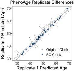
The most dramatic improvement was in PhenoAge. CpG-trained PhenoAge has a median deviation of 2.4 years, 3rd quartile of 5 years, and maximum of 8.6 years. In contrast, PCPhenoAge has a median deviation of 0.6 years, 3rd quartile of 0.9 years, and maximum of 1.6 years. PCPhenoAge was trained directly on phenotypic age based on clinical biomarkers rather than DNAm.

Correlations between different PC clocks was stronger than between CpG clocks. This may be partly due to the shared set of CpGs used to train PCs, or due to the reduction of noise that would have biased correlations towards the null. Correlations between PC clocks and CpG clocks tended to be stronger compared to correlations between CpG clocks and CpG clocks, consistent with a reduction of noise.

PC clocks preserve relevant aging signals unique to each of their CpG counterparts. They reduce technical variance but maintain relevant biological variance.

PCA is a commonly used tool and does not require specialized knowledge. High reliability of principal component-based epigenetic clocks will make them particularly useful for applications in personalized medicine and clinical trials evaluating novel aging interventions.”

https://doi.org/10.1093/geroni/igab046.015 “A Computational Solution to Bolster Epigenetic Clock Reliability for Clinical Trials and Longitudinal Tracking”


I appreciate that a coauthor – who is the originator of PhenoAge – is open to evidence and improvements. There’s a fun do-it-yourself demo of PCA at https://setosa.io/ev/principal-component-analysis/.

I found this study from it citing a 2021 review:

https://www.sciencedirect.com/science/article/abs/pii/S1084952121000094 “Aging biomarkers and the brain” (not freely available)

I found that review from it citing a 2020 study:

https://www.cell.com/iscience/fulltext/S2589-0042(20)30384-9 “Human Gut Microbiome Aging Clock Based on Taxonomic Profiling and Deep Learning”

Maybe this last study could be improved from its “mean absolute error of 5.91 years” with PCA? See Part 2 for another view.


PXL_20210704_092829847

Take acetyl-L-carnitine for early-life trauma

This 2021 rodent study traumatized female mice during their last 20% of pregnancy, with effects that included:

  • Prenatally stressed pups raised by stressed mothers had normal cognitive function, but depressive-like behavior and social impairment;
  • Prenatally stressed pups raised by control mothers did not reverse behavioral deficits; and
  • Control pups raised by stressed mothers displayed prenatally stressed pups’ behavioral phenotypes.

Acetyl-L-carnitine (ALCAR) protected against and reversed depressive-like behavior induced by prenatal trauma:

alcar regime

ALCAR was supplemented in drinking water of s → S mice either from weaning to adulthood (3–8 weeks), or for one week in adulthood (7–8 weeks). ALCAR supplementation from weaning rendered s → S mice resistant to developing depressive-like behavior.

ALCAR supplementation for 1 week during adulthood rescued depressive-like behavior. One week after ALCAR cessation, however, the anti-depressant effect of ALCAR was diminished.

Intergenerational trauma induces social deficits and depressive-like behavior through divergent and convergent mechanisms of both in utero and early-life parenting environments:

  • We establish 2-HG [2-hydroxyglutaric acid, a hypoxia and mitochondrial dysfunction marker, and an epigenetic modifier] as an early predictive biomarker for trauma-induced behavioral deficits; and
  • Demonstrate that early pharmacological correction of mitochondria metabolism dysfunction by ALCAR can permanently reverse behavioral deficits.”

https://www.nature.com/articles/s42003-021-02255-2 “Intergenerational trauma transmission is associated with brain metabotranscriptome remodeling and mitochondrial dysfunction”


This study had an effusive endorsement of acetyl-L-carnitine in its Discussion section, ending with:

“This has the potential to change lives of millions of people who suffer from major depression or have risk of developing this disabling disorder, particularly those in which depression arose from prenatal traumatic stress.”

I take a gram daily. Don’t know about prenatal trauma, but I’m certain what happened during my early childhood.

I asked both these researchers and those of Reference 70 for their estimates of a human equivalent to “0.3% ALCAR in drinking water.” Will update with their replies.


No word from those researchers, so here’s what I calculate:

  • (.003 x .081) x 70 kg = 1.7% human equivalent dose.
  • 1 liter water = 1 kg, so .017 x 1000 g = 17 g per liter of water.

We all drink at least a liter of water every day. A 17 gram/liter dose is way too high for humans, considering:

I downgraded this study to Required further work. It’s likely these researchers overdosed mice to ensure their treatment produced an effect. That’s counterproductive to the purpose of animal studies: to help humans.


PXL_20210704_095621886

Part 2 of Week 63 of Changing to a youthful phenotype with sprouts

To follow up Part 1, received Thursday’s lab results yesterday. Downloaded the workbook at https://michaellustgarten.com/2019/09/09/quantifying-biological-age/ and filled it in. Went to http://aging.ai/, selected 3.0, and entered values.

My starting point’s calculated values were:

biological age 1

A biological age snapshot from a year ago‘s video included optimal ranges:

optimizing biological age

Values in these optimal ranges were:

  • Albumin: 46;
  • Creatinine: 1.07;
  • high-sensitivity C-reactive protein: 0.24;
  • Red cell distribution width: 11.8; and
  • White blood cell count: 4.6.

I have some work to do on the other four. Good health while aging seldom happens on its own.

Reading more about Phenotypic age and its biological relationships. It definitely doesn’t mean I can do things I did 9.5 years ago like play golf and Frisbee football on the weekends.

I’d probably use DNAm PhenoAge to compare with other epigenetic clocks. Not sure how to use Aging.ai 3.0 calculations.

Sometime over the past year, Labcorp changed their adult alkaline phosphatase reference range from 39-117 to 48-121. Don’t know whether alkaline phosphatase’s optimal range will change with Labcorp’s new range, since < 48 was based on all-cause-mortality data.

PXL_20210622_093759263

Smoke and die early, while your twin lives on

A 2021 human twin study investigated epigenetic clocks:

“This study showed that accelerated epigenetic aging is associated with increased mortality, and smoking plays a role in explaining this association. Present findings suggest that DNAm GrimAge is a strong predictor of mortality independent of genetic influences among female twin pairs.

An invitation to participate in the study was sent to 414 female twin pairs, aged 63–76 years. Of 199 twin pairs, at least one twin died in 112 pairs during follow-up:

twins

This epigenetic age estimate that measures biological age and runs alongside, but not always in parallel with chronological age, may inform life expectancy predictions. Further research is needed to determine whether results apply to men, and the extent to which DNA methylation age can be used as a clinical biomarker of lifespan.”

https://clinicalepigeneticsjournal.biomedcentral.com/articles/10.1186/s13148-021-01112-7 “Does the epigenetic clock GrimAge predict mortality independent of genetic influences: an 18 year follow-up study in older female twin pairs”


If you don’t have a twin, substitute yourself as an analogous entity who has opposite behaviors. Don’t assume that smoking cannabis will produce different results from tobacco.

Meanwhile, Dr. Steven Horvath, who has a twin, continued his 2021 torrent of coauthored studies last week with Epigenetic clock and methylation studies in elephants. Amazing epigenetic clock information being published this year. Dr. Horvath is completely open to evidence, IAW, a real scientist.

All about the betaine

A trio of papers on betaine, the first being a 2021 series of thorough rodent experiments relating betaine and gut microbiota, and cause and effect:

“Compared with lean individuals, adipose tissues in obese individuals secrete high levels of pro-inflammatory cytokines such as TNF-α, IL-1β, and IL-6, inducing:

  • Systemic inflammation;
  • Insulin resistance;
  • Large amounts of carcinogenic factors; and
  • Increasing risk of certain types of cancer such as melanoma, colon cancer, and liver cancer.

Prebiotics obtained from fruits and vegetables can regulate host lipid metabolism and glucose homeostasis by reversing gut dysbiosis in obese individuals.

kgmi_a_1862612_f0005_oc

Results of this study show that dietary betaine alleviated gut microbiota imbalance in obese mice, and reduced development of obesity and obesity-related complications. Regulation of the miR-378a-YY1 regulatory axis by gut microbial acetate and butyrate was a critical mechanism for modulating:

  • White adipose tissue browning;
  • Classical brown adipose tissue activation; and
  • Lipid and glucose homeostasis

in obese mice after betaine supplementation.

These findings offer novel insights into underlying mechanisms by which gut microbiota affect host metabolism and host immune system, and demonstrate that the betaine-gut microbiota-derived signal axis is a potential therapeutic target in obesity and metabolic syndrome.”

https://www.tandfonline.com/doi/full/10.1080/19490976.2020.1862612 “Dietary betaine prevents obesity through gut microbiota-drived microRNA-378a family”


A second 2021 paper was a meta-analysis of effects on human cardiovascular biomarkers:

“Betaine supplementation had a significant effect on concentrations of:

  • Betaine;
  • Total cholesterol;
  • Low-density lipoprotein (LDL);
  • Homocysteine [negative effect]; and
  • Methionine.

Betaine supplementation did not affect serum concentrations of:

  • Triglycerides;
  • High-density lipoprotein (HDL);
  • Fasting blood glucose;
  • C-reactive protein;
  • Liver enzymes alanine aminotransferase (ALT), aspartate aminotransferase (AST), gamma-glutamyl transferase (GGT); and
  • Blood pressure.

Our meta-analysis supports the advantage of a lower dose of betaine supplementation (<4 g/d) on homocysteine concentrations without the lipid-augmenting effect observed with a higher dosage.”

https://www.tandfonline.com/doi/abs/10.1080/10408398.2021.1902938 “Effects of betaine supplementation on cardiovascular markers: A systematic review and Meta-analysis” (not freely available)


A third paper was a 2014 cereal analysis of betaine and its precursor choline that found a 224% increase in betaine from 62 to 139 μg/g and a 31% increase in choline from microwaving oats:

“Betaine and its precursor choline are important components of one-carbon metabolism, remethylating homocysteine into methionine and providing methyl groups for DNA methylation. Cereals are the main source of betaine in diet.

During cooking processes which did not involve removal of water (in this case oat porridge microwaved using instant oats) appeared to lead to creation of betaine. Explanations for this phenomenon could be that betaine is synthesised during the process, or that heating with water liberates betaine from cereal matrix, enhancing efficiency of extraction.”

https://www.sciencedirect.com/science/article/abs/pii/S0308814613012247 “Cereal foods are the major source of betaine in the Western diet – Analysis of betaine and free choline in cereal foods and updated assessments of betaine intake” (not freely available)


Another 2021 betaine (aka trimethyl glycine) study was curated in Ride the waves of gene expression with betaine for its role in preventing nerve disease. I take 1.5 grams of a betaine supplement every morning and evening when eating hulled Avena sativa 3-day-old oat sprouts.

I found the first two papers from their citing a 2016 human and rodent study Dietary Betaine Supplementation Increases Fgf21 Levels to Improve Glucose Homeostasis and Reduce Hepatic Lipid Accumulation in Mice, which was linked in a comment on this 2021 video:

Ride the waves of gene expression with betaine

This 2021 cell study investigated a dietary supplement’s role in preventing nerve disease:

“A loss of epigenetic control has been implicated in development of neurodegenerative diseases. Previous studies have implicated aberrant DNA and histone methylation in multiple sclerosis (MS) disease pathogenesis.

We have previously reported that methyl donor betaine is depleted in MS and is linked to changes in histone H3 trimethylation (H3K4me3) in neurons. We have also shown that betaine increases histone methyltransferase activity by activating chromatin bound betaine homocysteine S-methyltransferase (BHMT).

A hallmark of MS is the death of oligodendrocytes, the cells responsible for wrapping axons in myelin in the central nervous system and maintaining a healthy sheath. In demyelinating diseases like MS, oligodendrocyte progenitor cells (OPCs) fail to differentiate and make more myelin, resulting in sclerotic lesions.

Promoting differentiation of OPCs and generation of myelin is of great interest as a novel MS therapy. Waves of gene regulation (repression and activation) need to occur to promote myelination.

This BHMT-betaine methylation pathway ensures availability of S-adenosylmethionine (SAM) for a variety of DNA and histone methylation processes. OPC survival and differentiation are dependent upon DNA and histone methylation, and both processes require SAM.

journal.pone.0250486.g001

BHMT uses betaine to remethylate homocysteine to methionine. Betaine can be taken in through the diet or synthesized through the oxidation of choline in mitochondria.

We demonstrated that oligodendrocyte gene expression can be modulated by betaine supplementation through the BHMT-betaine methylation pathway. Our study suggests that dietary betaine supplementation may prove to be a therapeutic agent for MS and other demyelinating disorders.”

https://journals.plos.org/plosone/article?id=10.1371/journal.pone.0250486 “The BHMT-betaine methylation pathway epigenetically modulates oligodendrocyte maturation”


I started taking betaine 16 years ago. Didn’t know of these effects until reading this study.

Treating psychopathological symptoms will somehow resolve causes? had more on betaine (aka trimethyl glycine). Current dose is 1.5 grams twice daily.

Giving children allergies with pets

This 2021 human study investigated development and persistence of allergies:

“Allergic rhinitis (AR) is a common IgE-mediated disorder involving troublesome symptoms of nasal congestion, nasal itch, sneezing, and associated eye symptoms. Like many chronic health conditions, AR stems from complex gene–environment interactions.

130 subjects with AR were recruited. Control population included 154 healthy children who underwent a regular physical examination in the same ear, nose and throat clinic as AR patients. Individuals with history of asthma or atopic dermatitis were excluded.

AR analysis

Plenty of contradictory associations exist as whether furred pet exposure (cats and dogs) may be a risk or a protective factor for AR development. Discrepancies are likely due to the ubiquitous nature of pet allergens, while pet owners are more concerned about sanitation and many other hygiene-related reasons.

Interaction of early-life pet exposure with methylation level of ADAM33 increased the risk for AR onset 1.423 times more in children. This study provides evidence that:

  • Early-life pet exposure and low methylation level of ADAM33 increase AR risk in children; and
  • The interaction between pet exposure and methylation level of ADAM33 may play an important role in development of AR.”

https://aacijournal.biomedcentral.com/articles/10.1186/s13223-021-00526-5 “Interaction between early-life pet exposure and methylation pattern of ADAM33 on allergic rhinitis among children aged 3–6 years in China”


There’s nothing children can do about who their parents were. Exposing them to pet allergens, though, may be another example of early-life experiences causing lifelong effects.

Happy Mothers Day

This 2021 rodent study investigated effects on offspring of maternal high-fat diet (HFD) during gestation and lactation, and offspring HFD during young adulthood:

“We found that gestation was the most sensitive period to induce obesity in late life, and there was no difference between sexes in chance of obesity. Furthermore, we found that lactation and administration of a HFD post‐weaning increased incidence of lipid metabolism disorders and obesity in offspring.

gestational hfd effects on offspring

There are different windows of opportunity for programming epigenetically labile genes. Some studies support the alteration of epigenetic status during development as an important cause induced adult obesity.

Gestation is considered as the most sensitive period because high DNA synthesis and DNA methylation patterns are established for normal tissue development during the embryonic period. These two programming events are the times when the epigenetic state changes most widely in the life cycle.”

https://onlinelibrary.wiley.com/doi/10.1111/jcmm.16551 “Gestational high-fat diet impaired demethylation of Pparα and induced obesity of offspring”


Hey mothers! Do what you please. But don’t turn around and deny consequences of your behavior and choices on your descendants’ physiology and behavior, and possibly those of further descendants.

Gestation, birth, infancy, and early childhood are critical periods for humans. There’s no going back to correct errors and problems.

An overlooked gut microbiota product

This 2021 review subject was histone crotonylation:

“Histone crotonylation is a newly identified epigenetic modification that has a pronounced ability to regulate gene expression. It belongs to an expanding group of short chain lysine acylations that also includes the extensively studied mark histone acetylation.

Histone Kcr was first identified in 2011 where it was found to be mainly associated with active chromatin. Kcr occurs on the ε-amino group of the lysine side chain, where it neutralizes the positive charge of this residue. The loss in positive charge on histone Lys residues weakens DNA interaction, thus making chromatin less compact and accessible to DNA-binding factors.

Crotonate, like other short chain fatty acids (SCFAs), is mainly produced by gut microbiota during fermentation of partially and nondigestible carbohydrates. Circulating SCFAs (acetate, crotonate, butyrate, and propionate) can be taken up by tissues and converted into their cognate short-chain acyl-CoAs, the direct donors of histone Lys acylations.

fcell-09-624914-g001

Crotonyl-CoA is generated as a by-product of fatty acid and amino acid metabolism. Synthesis of crotonyl-CoA can occur in mitochondria or the cytoplasm. Evidence suggests that histone acylations are directly sensitive to changes in concentrations of their corresponding acyl-CoA metabolites, and therefore can act as indicators of cellular metabolic state.

Only a small number of Kcr sites in human histones have been identified so far. This is in part due to a lack of commercially available Kcr site-specific antibodies, which has meant much of the research in this field has focused on studying total histone crotonylation. This is likely to limit our understanding of the importance of histone Kcr, as functional impact of modification at specific sites cannot be readily assessed.”

https://www.frontiersin.org/articles/10.3389/fcell.2021.624914/full “The Regulation and Function of Histone Crotonylation”


At first I thought I had missed recent studies of gut microbiota producing crotonate. Searching again for “crotonate” “microbiota” 2020 2021, I didn’t find any that weren’t cited by this paper.

A lack of research could be due to factors mentioned above. It may also be that researchers just don’t look for evidence of the circulating SCFA crotonate.

Repositioning DNA methylation

This 2021 human study found:

“We report on a randomized controlled clinical trial conducted among 43 healthy adult males between the ages of 50-72. The 8-week treatment program included diet, sleep, exercise and relaxation guidance, and supplemental probiotics and phytonutrients.

This is the first randomized controlled study to suggest that specific diet and lifestyle interventions may reverse Horvath DNAmAge (2013) epigenetic aging in healthy adult males. Larger-scale and longer duration clinical trials are needed to confirm these findings, as well as investigation in other human populations.

aging-v13iundefined-202913-figure-f3

In both treatment and control groups, there was no net increase or decrease in methylation of 353 sites that compose the Horvath clock. This finding suggests that intervention did not lead to an overall increase in methylation of Horvath clock sites, but rather it prompted a repositioning of clock CpG methylation patterns consistent with a younger biological age.

One significant limitation of this pilot trial is limited statistical power due to relatively small sample size. It is not yet fully established whether interventions that slow any methylation clocks necessarily curtail risks of age-related disease.”

https://www.aging-us.com/article/202913/text “Potential reversal of epigenetic age using a diet and lifestyle intervention: a pilot randomized clinical trial”


Baffled as to why these researchers relied on 2013 research rather than at least Dr. Horvath’s improved 2018 skin and blood clock, a review of which noted:

“Although the skin-blood clock was derived from significantly less samples (~900) than Horvath’s clock (~8000 samples), it was found to more accurately predict chronological age, not only across fibroblasts and skin, but also across blood, buccal and saliva tissue. A potential factor driving this improved accuracy in blood could be related to the approximate 18-fold increase in genomic coverage afforded by using Illumina 450k/850k beadarrays.”

Which would you prefer? A 2013 flip phone, or a 2018 smartphone?

A bat epigenetic clock

This 2021 study subject was bats:

“Exceptionally long-lived species, including many bats, rarely show overt signs of aging, making it difficult to determine why species differ in lifespan. Here, we use DNA methylation (DNAm) profiles from 712 known-age bats, representing 26 species, to identify epigenetic changes associated with age and longevity.

Hypermethylated age- and longevity-associated sites are disproportionately located in promoter regions of key transcription factors (TF) and enriched for histone and chromatin features associated with transcriptional regulation. Predicted TF binding site motifs and enrichment analyses indicate that:

  • Age-related methylation change is influenced by developmental processes, while
  • Longevity-related DNAm change is associated with innate immunity or tumorigenesis genes, suggesting that
  • Bat longevity results from augmented immune response and cancer suppression.

Molossus molossus [a short-lived species] age genes are not enriched for immunity genes or genes that frequently mutated in cancer. However, M. molossus longevity genes exhibit significant overlap with genes involved in immunity and genes frequently mutated in human tumors.

Similar overlap patterns among immunity, longevity, and tumor-mutated genes also exist for long-lived bats.

Two species’ genetic adaptations for tumor suppression have been described to help explain their extreme longevity. Bats also have genetic mechanisms that enable strong antiviral immune responses without inducing damaging inflammatory reactions that may enable them to tolerate high levels of viral exposure.

Our results are consistent with an epigenetic clock theory of aging that connects beneficial developmental and cell maintenance processes to detrimental processes causing tissue dysfunction.”

https://www.nature.com/articles/s41467-021-21900-2 “DNA methylation predicts age and provides insight into exceptional longevity of bats”


The founder of the epigenetic clock has been busy, coauthoring more published studies than there have been weeks in this year! I’ve read five other 2021 studies he’s coauthored on dogs, horses, mammals (2), and humans in DNA methylation biomarker for cumulative lead exposure is associated with Parkinson’s disease. This one stood out for its “longevity results from augmented immune response and cancer suppression” findings.

If we’re interested in longevity, this clarity can direct efforts to both improve our immune systems and avoid problems like cancer. Symptoms may be subclinical, but that doesn’t provide adequate rationale to not address causes.

Peer review comments and responses were informative:

Reviewer #1 – “Developing an aging clock that works for a diverse set of bat species is a spectacular achievement.”

Reviewer #2 – “This is a tour de force study.”

Replies to Reviewer #3:

“Difference in recorded lifespans between three long-lived species and two short-lived species that we used to identify longevity DMPs [differentially methylated positions] is 20 years or more, even though they have similar body sizes (20-40 g). The three long-lived species [maximum ages 29.9, 30.5, and 37.1 years] also represent three different phylogenetic lineages.

CpG sites that undergo hypomethylation with age do so largely at random. In contrast, sites that undergo hypermethylation with age are highly nonrandom, and as has been noted before, are near genes associated with development. So yes, we believe there are predictable methylation changes with age.”

Our first 1000 days

This 2021 review subject was a measurable aspect of our early lives:

“The first 1000 days from conception are a sensitive period for human development programming. During this period, environmental exposures may result in long-lasting epigenetic imprints that contribute to future developmental trajectories.

The present review reports on effects of adverse and protective environmental conditions occurring on glucocorticoid receptor gene (NR3C1) regulation in humans. Thirty-four studies were included.

The hypothalamic-pituitary-adrenal (HPA) axis is key in regulating mobilization of energy. It is involved in stress reactivity and regulation, and it supports development of behavioral, cognitive, and socio-emotional domains.

The NR3C1 gene encodes for specific glucocorticoid receptors (GRs) in the mammalian brain, and it is epigenetically regulated by environmental exposures.

When mixed stressful conditions were not differentiated for their effects on NR3C1 methylation, no significant results were obtained, which speaks in favor of specificity of epigenetic vestiges of different adverse conditions. Specific maternal behaviors and caregiving actions – such as breastfeeding, sensitive and contingent interactive behavior, and gentle touch – consistently correlated with decreased NR3C1 methylation.

If the neuroendocrine system of a developing fetus and infant is particularly sensitive to environmental stimulations, this model may provide the epigenetic basis to inform promotion of family-centered prevention, treatment, and supportive interventions for at-risk conditions. A more ambiguous picture emerged for later effects of NR3C1 methylation on developmental outcomes during infancy and childhood, suggesting that future research should favor epigenome-wide approaches to long-term epigenetic programming in humans.”

https://www.sciencedirect.com/science/article/abs/pii/S0149763421001081 “Glucocorticoid receptor gene (NR3C1) methylation during the first thousand days: Environmental exposures and developmental outcomes” (not freely available). Thanks to Dr. Livio Provenci for providing a copy.


I respectfully disagree with recommendations for an EWAS approach during infancy and childhood. What happened to each of us wasn’t necessarily applicable to a group. Group statistics may make interesting research topics, but they won’t change anything for each individual.

Regarding treatment, our individual experiences and needs during our first 1000 days should be repeatedly sensed and felt in order to be therapeutic. Those memories are embedded in our needs because cognitive aspects of our brains weren’t developed then.

To become curative, we first sense and feel early needs and experiences. Later, we understand their contributions and continuations in our emotions, behavior, and thinking.

And then we can start to change who we were made into.

Treat your gut microbiota as one of your organs

Two 2021 reviews covered gut microbiota. The first was gut microbial origins of metabolites produced from our diets, and mutual effects:

“Gut microbiota has emerged as a virtual endocrine organ, producing multiple compounds that maintain homeostasis and influence function of the human body. Host diets regulate composition of gut microbiota and microbiota-derived metabolites, which causes a crosstalk between host and microbiome.

There are bacteria with different functions in the intestinal tract, and they perform their own duties. Some of them provide specialized support for other functional bacteria or intestinal cells.

Short-chain fatty acids (SCFAs) are metabolites of dietary fibers metabolized by intestinal microorganisms. Acetate, propionate, and butyrate are the most abundant (≥95%) SCFAs. They are present in an approximate molar ratio of 3 : 1 : 1 in the colon.

95% of produced SCFAs are rapidly absorbed by colonocytes. SCFAs are not distributed evenly; they are decreased from proximal to distal colon.

Changing the distribution of intestinal flora and thus distribution of metabolites may have a great effect in treatment of diseases because there is a concentration threshold for acetate’s different impacts on the host. Butyrate has a particularly important role as the preferred energy source for the colonic epithelium, and a proposed role in providing protection against colon cancer and colitis.

There is a connection between acetate and butyrate distinctly, which suggests significance of this metabolite transformation for microbiota survival. The significance may even play an important role in disease development.

  • SCFAs can modulate progression of inflammatory diseases by inhibiting HDAC activity.
  • They decrease cytokines such as IL-6 and TNF-α.
  • Their inhibition of HDAC may work through modulating NF-κB activity via controlling DNA transcription.”

https://www.hindawi.com/journals/cjidmm/2021/6658674/ “Gut Microbiota-Derived Metabolites in the Development of Diseases”


A second paper provided more details about SCFAs:

“SCFAs not only have an essential role in intestinal health, but also enter systemic circulation as signaling molecules affecting host metabolism. We summarize effects of SCFAs on glucose and energy homeostasis, and mechanisms through which SCFAs regulate function of metabolically active organs.

Butyrate is the primary energy source for colonocytes, and propionate is a gluconeogenic substrate. After being absorbed by colonocytes, SCFAs are used as substrates in mitochondrial β-oxidation and the citric acid cycle to generate energy. SCFAs that are not metabolized in colonocytes are transported to the liver.

  • Uptake of propionate and butyrate in the liver is significant, whereas acetate uptake in the liver is negligible.
  • Only 40%, 10%, and 5% of microbial acetate, propionate, and butyrate, respectively, reach systemic circulation.
  • In the brain, acetate is used as an important energy source for astrocytes.

Butyrate-mediated inhibition of HDAC increases Nrf2 expression, which has been shown to lead to an increase of its downstream targets to protect against oxidative stress and inflammation. Deacetylase inhibition induced by butyrate also enhances mitochondrial activity.

SCFAs affect the gut-brain axis by regulating secretion of metabolic hormones, induction of intestinal gluconeogenesis (IGN), stimulation of vagal afferent neurons, and regulation of the central nervous system. The hunger-curbing effect of the portal glucose signal induced by IGN involves activation of afferents from the spinal cord and specific neurons in the parabrachial nucleus, rather than afferents from vagal nerves.

Clinical studies have indicated a causal role for SCFAs in metabolic health. A novel targeting method for colonic delivery of SCFAs should be developed to achieve more consistent and reliable dosing.

The gut-host signal axis may be more resistant to such intervention by microbial SCFAs, so this method should be tested for ≥3 months. In addition, due to inter-individual variability in microbiota and metabolism, factors that may directly affect host substrate and energy metabolism, such as diet and physical activity, should be standardized or at least assessed.”

https://www.hindawi.com/journals/cjidmm/2021/6632266/ “Modulation of Short-Chain Fatty Acids as Potential Therapy Method for Type 2 Diabetes Mellitus”


Eat broccoli sprouts for your kidneys

Starting Year 7 of curating research with a 2021 review of kidney disease and sulforaphane:

“Many chronic kidney disease (CKD) patients progress to end-stage kidney disease – the ultimate in failed prevention. While increased oxidative stress is a major molecular underpinning of CKD progression, no treatment modality specifically targeting oxidative stress has been established clinically.

Pathophysiologic effects occur when there is an imbalance between oxidation and reduction – an altered redox state in which excess free radicals react with other molecules, including lipids, proteins, and nuclear DNA. Mitochondrial DNA is also susceptible to oxidative damage.

All mechanisms discussed above have been shown to be present in CKD. When levels of antioxidant agents such as SOD, CAT, GPx/glutathione, and NRF2 are reduced, harmful effects of oxidation and generation of ROS cannot be appropriately mitigated.

Data suggest continued SFN [sulforaphane] administration is needed to maintain activation of the NRF2 pathway to confer protection against oxidative damage of diabetes. Renal protective effect of SFN has been demonstrated in many other models of kidney injury.

SFN may have therapeutic potential in kidney disease by stimulating the NRF2 pathway.”

https://www.mdpi.com/2072-6643/13/1/266/htm “Eat Your Broccoli: Oxidative Stress, NRF2, and Sulforaphane in Chronic Kidney Disease”


Didn’t see where these researchers intended to perform a suggested “clinical study to assess the effect of SFN in CKD.” Keep reading before experimentally treating patients, please. Targets they missed included:

  • Parameters of myrosinase hydrolizing glucoraphanin;
  • “Consumption of broccoli strains with more glucoraphanin leads to higher plasma levels of SFN” and
  • “It follows that SFN could also pose similar adverse effects, particularly if taken in an isolated preparation.”

Also missing from this kidney review were connections to broccoli sprouts’ effectiveness in preventing bladder disease. Isothiocyanate metabolites accumulate in the bladder.

I came across this paper from it citing Sulforaphane: Its “Coming of Age” as a Clinically Relevant Nutraceutical in the Prevention and Treatment of Chronic Disease. I curated it due to informatively citing Microwave broccoli to increase sulforaphane levels.

Gut microbiota and aging

This 2020 review explored the title subject:

“The human body contains 1013 human cells and 1014 commensal microbiota. Gut microbiota play vital roles in human development, physiology, immunity, and nutrition.

Human lifespan was thought to be determined by the combined influence of genetic, epigenetic, and environmental factors including lifestyle-associated factors such as exercise or diet. The role of symbiotic microorganisms has been ignored.

Age-associated alterations in composition, diversity, and functional features of gut microbiota are closely correlated with an age-related decline in immune system functioning (immunosenescence) and low-grade chronic inflammation (inflammaging). Immunosenescence and inflammaging do not have a unidirectional relationship. They exist in a mutually maintained state where immunosenescence is induced by inflammaging and vice versa.

Immunosenescence changes result in both quantitative and qualitative modifications of specific cellular subpopulations such as T cells, macrophages and natural killer cells as opposed to a global deterioration of the immune system. Neutrophils and macrophages from aged hosts are less active with diminished phagocytosing capability.

Gut microbiota transform environmental signals and dietary molecules into signaling metabolites to communicate with different organs and tissues in the host, mediating inflammation. Gut microbiota modulations via dietary or probiotics are useful anti-inflammaging and immunosenescence interventions.

The presence of microbiomic clocks in the human body makes noninvasive, accurate lifespan prediction possible. Prior to occurrence of aging-related diseases [shown above], bidirectional interactions between the gut and extraenteric tissue will change.

Correction of accelerated aging-associated gut dysbiosis is beneficial, suggesting a link between aging and gut microbiota that provides a rationale for microbiota-targeted interventions against age-related diseases. However, it is still unclear whether gut microbiota alterations are the cause or consequence of aging, and when and how to modulate gut microbiota to have anti-aging effects remain to be determined.”

https://www.tandfonline.com/doi/abs/10.1080/10408398.2020.1867054 “Gut microbiota and aging” (not freely available; thanks to Dr. Zongxin Ling for providing a copy)


1. The “Stable phase” predecessor to this review’s subject deserved its own paper:

“After initial exposure and critical transitional windows within 3 years after birth, it is generally agreed that human gut microbiota develops into the typical adult structure and composition that is relatively stable in adults.

gut microbiota by age phenotype

However, the Human Microbiome Project revealed that various factors such as food modernization, vaccines, antibiotics, and taking extreme hygiene measures will reduce human exposure to microbial symbionts and led to shrinkage of the core microbiome, while the reduction in microbiome biodiversity can compromise the human immune system and predispose individuals to several modern diseases.”

2. I looked for the ten germ-free references in the “How germ-free animals help elucidate the mechanisms” section of The gut microbiome: its role in brain health in this review, but didn’t find them cited. Likewise, the five germ-free references in this review weren’t cited in that paper. Good to see a variety of relevant research.

There were a few overlapping research groups with this review’s “Gut-brain axis aging” section, although it covered only AD and PD research.

3. Inflammaging is well-documented, but is chronic inflammation a condition of chronological age?

A twenty-something today who ate highly-processed food all their life could have gut microbiota roughly equivalent to their great-great grandparents’ at advanced ages. Except their ancestors’ conditions may have been byproducts of “an unintended consequence of both developmental programmes and maintenance programmes.

Would gut microbiota be a measure of such a twenty-something’s biological age? Do we wait until they’re 60, and explain their conditions by demographics? What could they do to reset themself back to a chronological-age-appropriate phenotype?