A PhD in oats

The lead researcher of Eat oat sprouts for AVAs‘s second study made their PhD thesis freely available. It’s still informative 13 years later:

“The main objective of this research project was to obtain new knowledge on how to treat raw oat material of oat-based products in order to sustain or even increase levels of endogenous phenolic compounds, with emphasis on avenanthramides, in the final food product. Germination of oats proved to be a potential processing method for use on oats since it is relatively easy to perform, although time-consuming.”

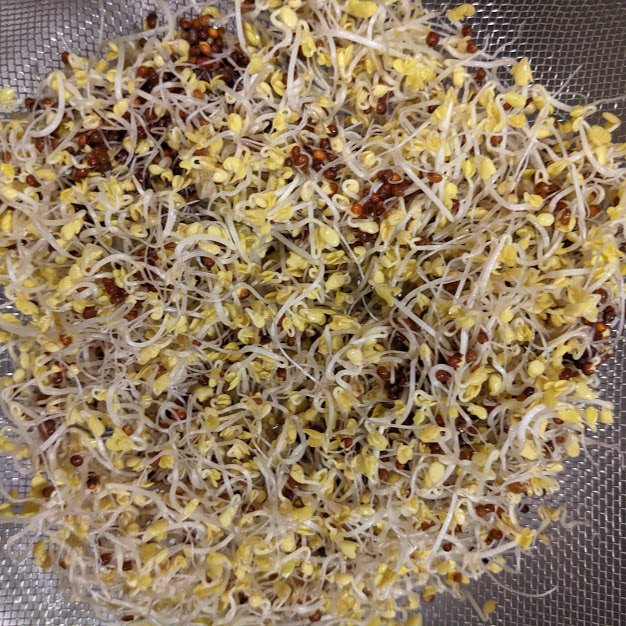
Which of these may be better for you? 44.1 grams of 4-day-old hulless oat sprouts at the top:

Or 53.2 g of 3-day-old hulled oat sprouts at the bottom?

They both started from 20.0 g seeds, and germinated the same way up through three days. 20 grams was over 1,300 hulled oat seeds, and close to 700 hulless oat seeds.

3-day-old hulled oat sprouts taste better, have a higher germination rate, and supply more quantity of their nutrients by weight. Characteristics of hulls from the thesis:

“The hull constitutes on average approximately 25% of the total grain weight. Protein, oil, starch and water-soluble carbohydrate levels are overall relatively low.

A large number of bioactive phenolic compounds can be found, among them p-coumaric acid, ferulic acid, vanillic acid, tricin and avenanthramides. Hull constituents remain unaffected during germination.

Activity of β-glucanase increases during germination of oats, resulting in almost total degradation of β-glucan. Since β-glucan is known to have health beneficial effects in humans, degradation during germination is not desirable if oats are intended for use in food products rather than for brewing.”

I’m not going to deal with hulls. Humans can’t digest oat hulls anyway.

A case for 4-day-old hulless oat sprouts:

“Germination of oats can be a good method to sustain or increase avenanthramides and other potentially health beneficial phenolic compounds. Levels of avenanthramides can increase during germination, sometimes to a high degree.

There was no indication that the increase in avenanthramide content had reached a plateau for any cultivars at 120 h of germination, indicating that further increase could take place.

Total protein content in oats increases slightly during germination. Even though the increase is small, it is important, since essential amino acids lysine and tryptophan increase and therefore improve nutritional value.

Lipid content in oats decreases slightly during germination while content of free fatty acids increases, although there are differences between cultivars as well as between hulled and hulless cultivars.”

Eat oat sprouts for AVAs

Here are three oat studies, two of them specifically on oat sprouts. The first from 2019 was cited in Don’t brew oat sprouts – eat them! for oat sprouts having “up to 25-fold increase” in avenanthramides (AVAs):

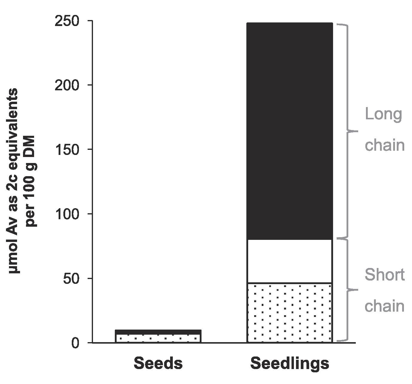
“Oat seeds were germinated, extracted, and analysed, finding 28 unique AVAs. AVAs 2p, 2c, and 2f, which are commonly described as the major AVAs, represented less than 20% of total content in seedlings.

The germinator program was: soak for 20 h at 20 °C (aeration 1 min every 10 min), followed by germination for 72 h at 25 °C (RH ≥ 99%). After the first 72 h of germination, oat seedlings were incubated for another 96 h at 30 °C (RH 70–80%).

AVA content was boosted by germination, resulting in around 25 times larger quantities found in oat seedlings.
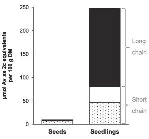
Previous studies also showed an increase in AVA content upon germination, but to a lesser extent than in our current experiment. It used different growth conditions, namely a shorter soaking time (10–14 vs. 20 h) and their seed germination phase was performed at lower temperature and different duration (120 h at 16 °C or 72 h at 20 °C vs 96 h at 30 °C).

Additionally, they quantified AVAs 2p, 2c, 2f, and 3f but also observed a large number of unknown peaks in the UV 340 nm chromatogram. Based on the wide variety of AVAs annotated in our work, many of these peaks probably corresponded to other AVAs, but they were not identified and quantified as such.”

https://www.sciencedirect.com/science/article/pii/S0308814618319411 “Mass spectrometric characterisation of avenanthramides and enhancing their production by germination of oat (Avena sativa)”

No measurements were taken at three days when germination parameters changed. These researchers didn’t bother to take any samples between Hours 0 and 168. This lack of germination-stage evidence limited findings’ utility to other researchers.

Was this study designed to create a headline rather than useful germination-stage information? Why obliquely and directly fault another study in the Abstract, Results and Discussion, and Conclusion sections for being performed more than a decade earlier, without subsequent advancements in science and technology?

Contrast this study’s design with 2020 Oat sprouts analysis which took measurements under 18 different conditions (hulled / dehulled seeds of two oat varieties, for 1-to-9 days, at 12-to-20°C). Those researchers produced evidence to support many further studies, such as:

“Presence / absence of hull might determine different effects of germination conditions on α-amylase, protease, and lipase activities.”


The referenced disparaged 2007 study:

“..investigated the effect of a steeping and germination process, using a pilot plant malting system, on content of AVAs and other phenolic compounds. This was performed to gain a more collective and comparative picture of what happens to phenolic compounds in the oat kernel during germination.

Steeping and germination was performed at two different temperatures, 16 and 20°C. Three closely related North American covered oat cultivars were steeped to 45% moisture, which took 10, 12, and 14 h for Vista, Gem, and Dane, respectively.

After steeping, oat grains were drained and samples were germinated at 16°C for 120 h or at 20°C for 72 h (due to a machine breakdown, the 20°C experiment had to be stopped at 72 h of germination). Sampling during germination was carried out at set hours (for 16°C at 12, 24, 48, 72, 96 and 120 h and for 20°C at 12, 24, 48 and 72 h).
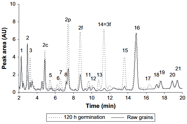
Chromatogram of AVAs and other phenols from cultivar Dane (16°C treatment). Peaks for AVAs 2c, 2p, 2f and 3f are identified. Unknown peaks are numbered in order of appearance (from 1-21). For isolation and identification of AVA 3f the commercial product SPC-flakes, purchased in a health food shop, was used.

An increase in AVA content of germinated seeds, as compared to raw grains, was observed for Dane (125%, p < 0.001) and for Vista (29%, p = 0.007). HHT [avenanthramide-synthesizing enzyme] activity increased 62% (p = 0.014) in Dane, whereas no change was detected in Vista and Gem. This increase started early in germination to reach its maximum at 96 h of germination.

Effects of temperature on AVAs 2c, 2p and 2f, and activities of HHT and PO, was less important than time of steeping, or time of germination, or cultivar. However, almost all unknown compounds were affected by temperature, indicating the importance of this factor.”

https://www.sciencedirect.com/science/article/abs/pii/S0733521007001762 “Avenanthramide content and related enzyme activities in oats as affected by steeping and germination” (not freely available)

This study’s HHT (avenanthramide-synthesizing enzyme) maximum-at-96-hours finding may be what I tasted in Sprouting hulless oats, where that variety’s sprouts improved their sweetness and enzymes between Days 3 and 4. Did an extra day improve AVA content?

So their pilot malting system broke down. They had to get an analysis standard from a health food store. And there were many unknowns due to 2007 science and technology.

Despite difficulties, germination-stage samples produced evidence and analyses other people could use more than a decade later.


A 2020 study cited the first study for basic, not headline, information:

“There are various potential mechanisms for AVAs anti-inflammatory effects, including inhibition of lipoxygenases (LOX), which catalyse oxygenation of polyunsaturated fatty acids into potent signal molecules involved in inflammatory processes.

It was found that AVAs comprising caffeic or sinapic acid exhibited significant lipoxygenase inhibition (60–90%), whereas low or no inhibition was observed with AVAs containing p-coumaric or ferulic acid.

Corresponding free cinnamic acids, AVA analogue Tranilast® and LOX inhibitor trans-resveratrol were included for comparison. Trans-resveratrol showed inhibition, whereas no difference in inhibition was seen on comparing AVAs with their free corresponding cinnamic acids, which implies that the anthranilic acid part of the avenanthramide molecule does not affect inhibition.

Whether dietary AVAs at intake levels normally achieved through consumption of oat products exert LOX inhibitory activity in vivo, and thereby inhibit production of pro-inflammatory compounds, remains to be elucidated. In this context it may also be of importance to emphasize that lipid content in groats of various oat cultivars is comparably high (49–135 g kg−1) and that LA [linoleic acid (C18:2, n-6)] and ALA [α-linolenic acid (C18:3, n-3)] comprise about 40% and 1%, respectively, of fatty acids.

Consumption of oats has been linked to a decreased risk of several chronic diseases, and AVAs contribute to protective effects. This study suggests that avenanthramides comprising caffeic acid or sinapic acid partly exert their antioxidant and anti-inflammatory effects via lipoxygenase inhibition.”

https://www.sciencedirect.com/science/article/pii/S2405844020311488 “Avenanthramides as lipoxygenase inhibitors”


Tampa’s Riverwalk

Don’t brew oat sprouts – eat them!

This 2020 study chemically analyzed four grains and their brew-processing products:

“Side-stream products of malting, particularly rootlet, are currently treated as animal feed. Instead of ending up in final products (e.g., malt and beer), a substantial portion of phytochemicals end up in side streams.

Rootlets are being increasingly investigated to overcome their bitter taste and to unleash their potential. Adding the fact that side-stream products produced in high quantity are also rich in protein, their nutritional value may be too high to justify usage as feed rather than food.

Grains were steeped for 26 to 30 h with a wet–dry–wet steeping program. Oats were wet steeped for 4 h at 13 °C before and after 18 h of dry steeping at 15 °C.

All grains were germinated for 6 days at 15 °C, after which they were dried with a gentle kilning program to a final temperature of 83 °C and moisture of 4%. Rootlets were separated from malt after drying.

Statistically significant changes occurred in abundance of all 285 annotated phytochemicals during malting, when comparing whole grain with malted grain or rootlet. In oats, cumulative levels of avenanthramides increased by 2.6-fold in the malted grain compared to intact whole grain. Up to 25-fold increase has been reported previously after a slightly longer germination.

Phenolamides cumulative levels in oats increased in both malted grain (11-fold) and rootlet (50-fold). Cumulative flavonoid levels were nearly 3-fold higher in malted grain and rootlet compared to whole grain.

Avenanthramides and phenolamides had much lower extractability into the water extract and wort.

To our knowledge, this is the first time avenanthramides are reported from any other species than oats, suggesting that the synthesis pathway for avenanthramides evolved before oats diverged from the other cereals. Furthermore, benzoxazinoids are herein reported for the first time in oats.

Several previously uncharacterized saponins were found in oats in addition to the previously known avenacins and avenacosides. However, because of limited reference data currently available, their identity could not be determined beyond compound class and molecular formula in this study.

Plants can synthetize up to hundreds of thousands of secondary metabolites, and current spectral databases only contain a fraction of them to allow identification. Compounds found in this study do not represent the complete range of phytochemicals existing in cereals.”

https://www.nature.com/articles/s41538-020-00081-0 “Side-stream products of malting: a neglected source of phytochemicals”


Twice a day for six weeks I’ve eaten oat sprouts 3-to-6-days old from two species and three varieties. I’ve never noticed any “bitter taste” of rootlets mentioned.

Maybe “a final temperature of 83 °C and moisture of 4%” had something to do with it? Oat sprouts I ate never got above 25°C, and I doubt their moisture content was < 80%.

Maybe “Oats were wet steeped for 4 h at 13 °C before and after 18 h of dry steeping at 15 °C” gave oat sprouts a bitter taste? I process oat sprout batches the same way I do broccoli sprout batches. A new batch soaks to start germination every 12 hours, then is rinsed three times every 24 hours on a 6 hours – 6 hours – 12 hours cycle. Temperature in my kitchen is 21°C (70°F) because it’s winter outside.

The above graphic is a heat map of 29 studied C-type avenanthramides. Don’t know why 26 known A-type avenanthramides described in Eat oats today! weren’t analyzed. The second study of Sprouting oats stated:

“There is a higher concentration of A-type AVAs [avenanthramides] than C-type AVAs in sprouted oats.”

Reference 33’s “up to 25-fold increase” is curated in Eat oat sprouts for AVAs.

Go with the Alzheimer’s Disease evidence

This 2021 study investigated gut microbiota differences between 100 AD patients and 71 age- and gender-matched controls:

“Structural changes in fecal microbiota were evident in Chinese AD patients, with decreased alpha-diversity indices and altered beta-diversity ones, evidence of structurally dysbiotic AD microbiota.

Interestingly, traditionally beneficial bacteria, such as Bifidobacterium and Akkermansia, increase in these AD patients while Faecalibacterium and Roseburia decrease significantly. Different species of Bifidobacterium may have different effects that can explain why Bifidobacterium spp. are commonly associated with healthy and diverse microbiota but sometimes also isolated in other conditions. We needed to re-examine the therapeutic potential of Bifidobacterium in terms of maintaining cognitive function and treating dementia.

Surprisingly, our data indicate that Akkermansia was among the most abundant genera in AD-associated fecal microbiota. Similar to Bifidobacterium, Akkermansia was negatively correlated with clinical indicators of AD, such as MMSE, WAIS, and Barthel, and anti-inflammatory cytokines such as IFN-γ.

Based on our present observations, Akkermansia cannot always be considered a potentially beneficial bacterium. It might be harmful for the gut–brain axis in the context of AD development in the elderly.

Aging is associated with an over-stimulation of both innate and adaptive immune systems, resulting in a low-grade, chronic state of inflammation defined as inflammaging. This can increase gut permeability and bacterial translocation.

Characteristics of AD microbial profiles changed from butyrate producers, such as Faecalibacterium, into lactate producers, such as Bifidobacterium. These alterations contributed to shifts in metabolic pathways from butyrate to lactate, which might have participated in pathogenesis of AD. Specific roles of AD-associated signatures and their functions should be explored in further studies.”

https://www.frontiersin.org/articles/10.3389/fcell.2020.634069/full “Structural and Functional Dysbiosis of Fecal Microbiota in Chinese Patients With Alzheimer’s Disease”


The control group’s 73-year-olds were better off than AD patients. How were they compared with their previous life stages?

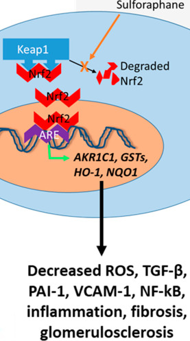
Since we’re all aging, how do we each prepare ourselves? I’ll return to evidence including 2020 A rejuvenation therapy and sulforaphane, recently amplified in Part 2 of Switch on your Nrf2 signaling pathway:

“A link between inflammation and aging is the finding that inflammatory and stress responses activate NF-κB in the hypothalamus and induce a signaling pathway that reduces production of gonadotropin-releasing hormone (GnRH) by neurons.

The case is particularly interesting when we realize that the aging phenotype can only be maintained by continuous activation of NF-κB. So here we have a multi-level interaction:

  1. Activation of NF-κB leads to
  2. Cellular aging, leading to
  3. Diminished production of GnRH, which then
  4. Acts (through cells with a receptor for it, or indirectly as a result of changes to GnRH-receptor-possessing cells) to decrease lifespan.

Cell energetics is not the solution, and will never lead to a solution because it makes the assumption that cells age. Cells take on the age-phenotype the body gives them.

Aging is not a defect – it’s a programmed progressive process, a continuation of development with the body doing more to kill itself with advancing years. Progressive life-states where each succeeding life-stage has a higher mortality (there are rare exceptions).

Cellular aging is externally controlled (cell non-autonomous). None of those remedies that slow ‘cell aging’ (basically all anti-aging medicines) can significantly extend anything but old age.

For change at the epigenomic/cellular level to travel up the biological hierarchy from cells to organ systems seems to take time. But the process can be repeated indefinitely (so far as we know).”

We may express concern about others. But each of us should also take responsibility for our own one precious life.

Treat your gut microbiota as one of your organs

Two 2021 reviews covered gut microbiota. The first was gut microbial origins of metabolites produced from our diets, and mutual effects:

“Gut microbiota has emerged as a virtual endocrine organ, producing multiple compounds that maintain homeostasis and influence function of the human body. Host diets regulate composition of gut microbiota and microbiota-derived metabolites, which causes a crosstalk between host and microbiome.

There are bacteria with different functions in the intestinal tract, and they perform their own duties. Some of them provide specialized support for other functional bacteria or intestinal cells.

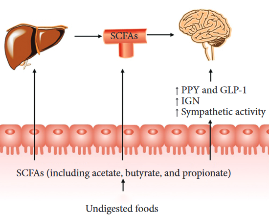
Short-chain fatty acids (SCFAs) are metabolites of dietary fibers metabolized by intestinal microorganisms. Acetate, propionate, and butyrate are the most abundant (≥95%) SCFAs. They are present in an approximate molar ratio of 3 : 1 : 1 in the colon.

95% of produced SCFAs are rapidly absorbed by colonocytes. SCFAs are not distributed evenly; they are decreased from proximal to distal colon.

Changing the distribution of intestinal flora and thus distribution of metabolites may have a great effect in treatment of diseases because there is a concentration threshold for acetate’s different impacts on the host. Butyrate has a particularly important role as the preferred energy source for the colonic epithelium, and a proposed role in providing protection against colon cancer and colitis.

There is a connection between acetate and butyrate distinctly, which suggests significance of this metabolite transformation for microbiota survival. The significance may even play an important role in disease development.

  • SCFAs can modulate progression of inflammatory diseases by inhibiting HDAC activity.
  • They decrease cytokines such as IL-6 and TNF-α.
  • Their inhibition of HDAC may work through modulating NF-κB activity via controlling DNA transcription.”

https://www.hindawi.com/journals/cjidmm/2021/6658674/ “Gut Microbiota-Derived Metabolites in the Development of Diseases”


A second paper provided more details about SCFAs:

“SCFAs not only have an essential role in intestinal health, but also enter systemic circulation as signaling molecules affecting host metabolism. We summarize effects of SCFAs on glucose and energy homeostasis, and mechanisms through which SCFAs regulate function of metabolically active organs.

Butyrate is the primary energy source for colonocytes, and propionate is a gluconeogenic substrate. After being absorbed by colonocytes, SCFAs are used as substrates in mitochondrial β-oxidation and the citric acid cycle to generate energy. SCFAs that are not metabolized in colonocytes are transported to the liver.

  • Uptake of propionate and butyrate in the liver is significant, whereas acetate uptake in the liver is negligible.
  • Only 40%, 10%, and 5% of microbial acetate, propionate, and butyrate, respectively, reach systemic circulation.
  • In the brain, acetate is used as an important energy source for astrocytes.

Butyrate-mediated inhibition of HDAC increases Nrf2 expression, which has been shown to lead to an increase of its downstream targets to protect against oxidative stress and inflammation. Deacetylase inhibition induced by butyrate also enhances mitochondrial activity.

SCFAs affect the gut-brain axis by regulating secretion of metabolic hormones, induction of intestinal gluconeogenesis (IGN), stimulation of vagal afferent neurons, and regulation of the central nervous system. The hunger-curbing effect of the portal glucose signal induced by IGN involves activation of afferents from the spinal cord and specific neurons in the parabrachial nucleus, rather than afferents from vagal nerves.

Clinical studies have indicated a causal role for SCFAs in metabolic health. A novel targeting method for colonic delivery of SCFAs should be developed to achieve more consistent and reliable dosing.

The gut-host signal axis may be more resistant to such intervention by microbial SCFAs, so this method should be tested for ≥3 months. In addition, due to inter-individual variability in microbiota and metabolism, factors that may directly affect host substrate and energy metabolism, such as diet and physical activity, should be standardized or at least assessed.”

https://www.hindawi.com/journals/cjidmm/2021/6632266/ “Modulation of Short-Chain Fatty Acids as Potential Therapy Method for Type 2 Diabetes Mellitus”


Increasing soluble fiber intake with inulin

From a 2015 USDA technical report:

“Inulin is a naturally-occurring carbohydrate found in roots of chicory and many other food plants. Oligofructose is derived from inulin.

Inulin is a polymer chain of multiple fructose molecules with a glucose molecule at one end. Length of the fructose chain of inulin can range from 2–60 fructose molecules.

Inulin is mostly indigestible by human enzymes due to its shape, but is digestible by microbes in the large intestine. It can serve as a prebiotic, a nutrient source for microflora in the human digestive system.”


From a 2021 review Friend or foe? The roles of inulin-type fructans (not freely available):

“Inulin-type fructans are a mixture of inulin, oligofructose and fructooligosaccharide (FOS). They aren’t absorbed in the stomach and small intestine. They can be completely fermented by bacteria in the large intestine.

They treat digestive diseases, metabolic syndrome, immune system and inflammatory diseases, endothelial dysfunction, and prevent infection and cancer.

A 2010 gastrointestinal tolerance of chicory inulin products study indicated that 10 g/day of native inulin or 5 g/day of oligofructose were well-tolerated in healthy, young adults. Over this dose would induce mild gastrointestinal symptoms.”


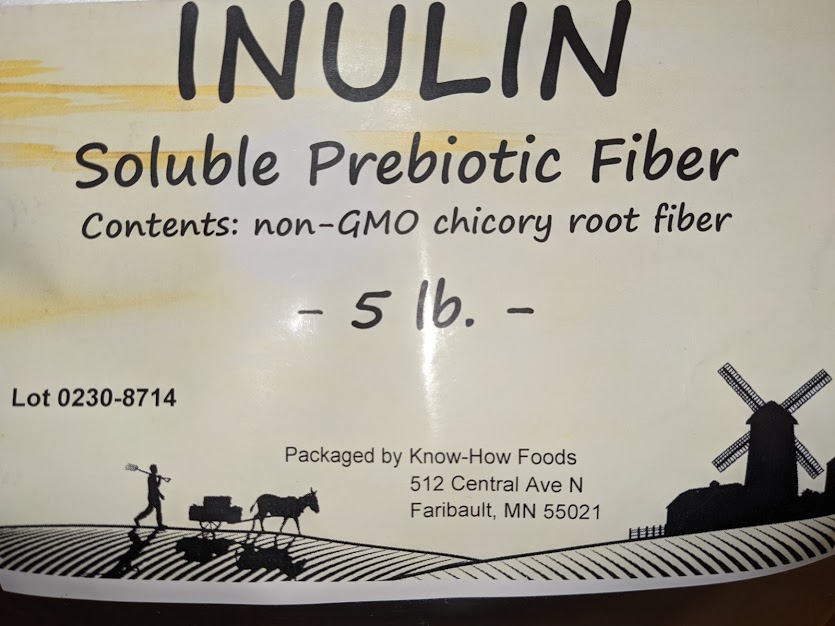
I bought this last month:

From the manufacturer:

“A powdered food ingredient based on chicory inulin with a high level of oligofructose 1 (DP2-DP10). This product is characterized by a high solubility.

Inulin from chicory is a polydisperse mixture of linear fructose polymers with mostly a terminal glucose unit, coupled by means of beta (2-1) bonds. The number of units (degree of polymerization) can vary between 2 and 60.

It is a fine, white powder with 30% the sweetness of sucrose. It has >=85% inulin/oligofructose and <15% fructose, glucose, sucrose. It has 2.2 kcal/gram and a glycemic response of 20.”

From the vendor:

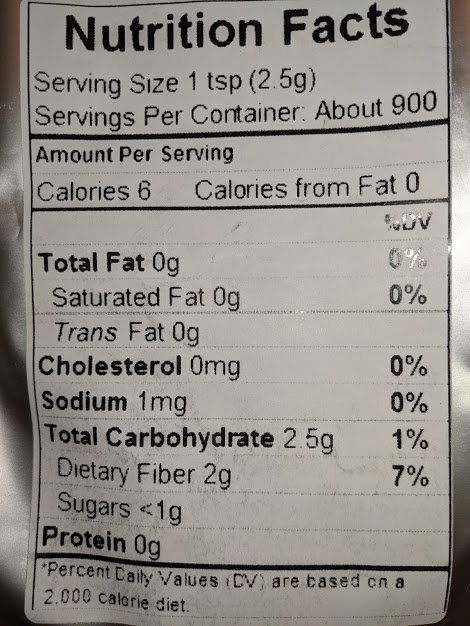
“You pay for the product… not the product packaging! Each teaspoon (tsp) delivers 2g fiber.

Inulin is hygroscopic so will take on moisture, especially in humid environments. Store in a dry place and remove as much air from the pouch as able before resealing after each use. Alternately, you could store in several smaller air-tight containers. This will limit exposure to possible humidity. Room temperature or cooler is ideal.”


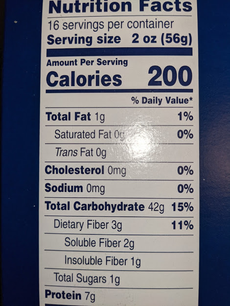
It tastes like cotton candy. 🙂 Its first use was to replace 2 grams of soluble fiber I got from eating 56 grams of noodles:

Probably won’t reorder FOS when I run out. I’ve taken 1.5 grams FOS every day for 16 years.

Eat broccoli sprouts for your kidneys

Starting Year 7 of curating research with a 2021 review of kidney disease and sulforaphane:

“Many chronic kidney disease (CKD) patients progress to end-stage kidney disease – the ultimate in failed prevention. While increased oxidative stress is a major molecular underpinning of CKD progression, no treatment modality specifically targeting oxidative stress has been established clinically.

Pathophysiologic effects occur when there is an imbalance between oxidation and reduction – an altered redox state in which excess free radicals react with other molecules, including lipids, proteins, and nuclear DNA. Mitochondrial DNA is also susceptible to oxidative damage.

All mechanisms discussed above have been shown to be present in CKD. When levels of antioxidant agents such as SOD, CAT, GPx/glutathione, and NRF2 are reduced, harmful effects of oxidation and generation of ROS cannot be appropriately mitigated.

Data suggest continued SFN [sulforaphane] administration is needed to maintain activation of the NRF2 pathway to confer protection against oxidative damage of diabetes. Renal protective effect of SFN has been demonstrated in many other models of kidney injury.

SFN may have therapeutic potential in kidney disease by stimulating the NRF2 pathway.”

https://www.mdpi.com/2072-6643/13/1/266/htm “Eat Your Broccoli: Oxidative Stress, NRF2, and Sulforaphane in Chronic Kidney Disease”


Didn’t see where these researchers intended to perform a suggested “clinical study to assess the effect of SFN in CKD.” Keep reading before experimentally treating patients, please. Targets they missed included:

  • Parameters of myrosinase hydrolizing glucoraphanin;
  • “Consumption of broccoli strains with more glucoraphanin leads to higher plasma levels of SFN” and
  • “It follows that SFN could also pose similar adverse effects, particularly if taken in an isolated preparation.”

Also missing from this kidney review were connections to broccoli sprouts’ effectiveness in preventing bladder disease. Isothiocyanate metabolites accumulate in the bladder.

I came across this paper from it citing Sulforaphane: Its “Coming of Age” as a Clinically Relevant Nutraceutical in the Prevention and Treatment of Chronic Disease. I curated it due to informatively citing Microwave broccoli to increase sulforaphane levels.

Harnessing endogenous defenses with broccoli sprouts

This 2019 article was by the author of Sulforaphane: Its “Coming of Age” as a Clinically Relevant Nutraceutical in the Prevention and Treatment of Chronic Disease. It isn’t widely available, so I’ll quote liberally:

“Demand for solutions to digestive health issues is accelerating, especially since both scientific literature and popular press dedicate significant resources to promoting awareness of what has come to be known as ‘gut health’. In considering available therapies and the possibility that a somewhat different approach may more comprehensively optimise function of the gut ecosystem, a number of questions which do not yet have satisfactory answers are ponderable dilemmas:

  1. If diet alone can dramatically shift composition of the microbiome within 24 hours, what do we expect of a probiotic supplement?
  2. Even though probiotics as food or supplements demonstrate favourable clinical outcomes, they typically don’t colonise the gut. How do we expect them to restore diversity and lost species to the gut microbiome after antibiotics? If no trace of an administered probiotic organism can be found a few weeks later, is there any sustained benefit?
  3. Presence of obesity and other diseases is indirectly proportional to diversity of microbial organisms inhabiting the human gut. What can we expect of a few selected probiotic strains in helping to solve this problem?
  4. No antimicrobial approach selectively destroys a pathogen without impacting commensals to some degree. If we select a tool to eradicate gut pathogens, pathobionts or rogue commensals, how do we avoid damaging protective commensals with which we live symbiotically?
  5. The value of using a probiotic supplement after antibiotic therapy to recolonise the gut is uncertain. A 2018 multi-centre study showed that probiotic supplementation after antibiotics delayed gut microbiome reconstitution by around five months.
  6. If the gut can harbour around 1,000 different species, why do we expect a probiotic supplement harbouring just a few species to favourably modify a human microbiome?
  7. If Lactobacilli make up <0.1% of total microbes, why do we so readily choose them as probiotic supplements?
  8. If L-glutamine is a preferred energy source for the small intestine and not the colon, why is it used almost universally in gut repair programmes regardless of the affected region?

Removal of gluten and administration of probiotics have lesser impact than endogenous factors like elevated HbA1c:

Shift emphasis closer to optimising colonocyte metabolism as the primary driver of dysbiosis in the colon. Since these mechanisms within the human gut ecosystem already exist, intervene at this level, as distinct from using antimicrobials and exogenous probiotic strains to influence host cell function.

Phytonutrients that potently activate these core processes have been identified and are sufficiently bioavailable to achieve this end. Restoring homeostasis to intestinal epithelial cells can be readily justified as a key initial step.

Sulforaphane is a potent inducer of hundreds of genes associated with cellular defences mechanisms. In this context, these genes include those that code for antioxidant and phase II detoxification enzymes, glutathione and metallothionein.

Sulforaphane exhibits other more specific gut and immune-related effects. As the most potent single food-derived activator of Nrf2, sulforaphane is capable of upregulating protective genes in colonocytes and other cells.

A growing body of work has identified the colonocyte as the driver of dysbiosis. Targeting colonocyte function provides an alternative to targeting microbes for remediation of dysbiosis.”

https://www.researchgate.net/publication/336578800_Restoring_Gut_Ecology_Harnessing_the_Inbuilt_Defence_Mechanisms_of_the_Gut_Epithelium “Restoring Gut Ecology: Harnessing the Inbuilt Defence Mechanisms of the Gut Epithelium” (registration required)


If you can’t access this paper, read The future of your brain is in your gut right now. If you can’t access that paper, listen to Switch on your Nrf2 signaling pathway.

Sprouting hulless oats

I finished a 3-lb. bag of hulled Avena sativa oats used in Sprouting hulled oats after starting 20 gram batches twice a day. Amazon said that Montana farmer’s products were “Currently unavailable. We don’t know when or if this item will be back in stock.” I went to their website and emailed an inquiry.

Turns out it’s Amazon’s problem in restocking pallets that are already received! I placed an order directly with the farmer.

In the meantime, I’m trying another oat species, Avena nuda, from an Illinois farmer. I’ll reuse Degree of oat sprouting as the model, since it was also an Avena nuda oat variety.

  • Oat seed size was 7-9 mm x 2-3 mm. The model used “huskless oat ‘Gehl’” which may be a different variety.
  • 100 seeds weighed 2.9 grams. There were close to 700 seeds per 20 g batches.
  • Oat sprout batches were processed the same way I do broccoli sprout batches. A new batch started soaking to start germination every 12 hours, then was rinsed three times every 24 hours on a 6 hours – 6 hours – 12 hours cycle.
  • Temperature in my kitchen was 21°C (70°F) because it’s snowing outside. The model findings included “Temperatures between 20° and 25°C yielded the most dramatic changes in properties of sprouted oats.”

I evaluated germination results per the model’s Degree of Sprouting finding:

“Length of the coleoptile [shoot] was selected as a criterion of categorization of degree of sprouting. Grains of degree 0 do not show any radicle [root] or coleoptile growth. Degree:

  1. Has visible embryos (small white point), while radicles and coleoptile are not visible;
  2. Shows a developed embryo emerging from the seed coat;
  3. Coleoptile lengths of at least half the oat grain length;
  4. Coleoptile lengths between half and a full grain length; and
  5. Coleoptile longer than a full grain length.”

Here’s what this hulless oat variety’s seeds and 3-day-old sprouts looked like:

The tedious part was evaluating degrees of sprouting. I took as large a bottom-to-top sample as I could tolerate sorting (160 seeds / sprouts, about 23%), with these results:

A 91% germination rate. 🙂 Average weight of 3-day-old batches was 42.5 grams, for a 213% weight gain. That wasn’t as much as 3-day-old hulled oats’ 97% germination rate and 260% weight gain.

For degree-of-sprouting comparisons, here are my eyeball estimates of the model study’s 3-day-old hulless oats:

These 3-day-old hulless oat sprouts taste starchier with less enzyme aftertaste than 3-day-old hulled oat sprouts. Will extending their growth to four days increase degree-of-sprouting categories 4 and 5, and change their taste?

An extra day from 5 to 6 didn’t make a difference in Sprouting whole oats germination rate. I don’t expect non-germinated percentages to change from 3 to 4 days, but we’ll see.

I expect similar overall increases in antioxidants, GABA, phenolic compounds, protein, amino acids, β-glucan, and polyunsaturated fatty acids as hulled oat sprouts.

Update: Four-day-old hulless oat sprouts have a little more sweetness and enzyme aftertaste. Their degree-of-sprouting and germination rate didn’t change much, though.

Mid-life gut microbiota crisis

This 2019 rodent study investigated diet, stress, and behavioral relationships:

“Gut microbiome has emerged as being essential for brain health in ageing. We show that prebiotic supplementation with FOS-Inulin [a complex short- and long-chain prebiotic, oligofructose-enriched inulin] is capable of:

  • Dampening age-associated systemic inflammation; and
  • A profound yet differential alteration of gut microbiota composition in both young adult and middle-aged mice.

Middle-aged mice exhibited an increased influx of inflammatory monocytes into the brain. However, neuroinflammation at this stage was not significant enough to manifest in major cognitive impairments.

A much longer exposure to prebiotics might be needed to achieve significant effects, suggesting that supplementation may have to start earlier to be effectively preventative before alterations in the brain occur. This is particularly evident for behaviour.

Targeting gut microbiota, as we have done with a prebiotic, can affect the brain and subsequent behaviour through a variety of potential pathways including SCFAs [short-chain fatty acids], amino acids and immune pathways. All of these are interconnected. Future studies are needed to better deconvolve [figure out] such pathways in eliciting beneficial effects of inulin.

Modulatory effects of prebiotic supplementation on monocyte infiltration into the brain and accompanied regulation of age-related microglia activation highlight a potential pathway by which prebiotics can modulate peripheral immune response and alter neuroinflammation in ageing. Our data suggest a novel strategy for the amelioration of age-related neuroinflammatory pathologies and brain function.”

https://www.nature.com/articles/s41380-019-0425-1 “Mid-life microbiota crises: middle age is associated with pervasive neuroimmune alterations that are reversed by targeting the gut microbiome” (not freely available)


This study’s experiments subjected young and middle-aged mice to eight stress tests. I appreciated efforts to trace causes to behavioral effects, since behavior provided stronger evidence.

I’m in neither life stage investigated by this study. Still, per Reducing insoluble fiber, I’ll start taking inulin next week. See Increasing soluble fiber intake with inulin.

I came across this study through its citation in How will you feel?

Inauguration day

Don’t take Beano if you’re stressed

This 2021 rodent study investigated diet and stress relationships:

“We show that dietary raffinose metabolism to fructose couples stress-induced gut microbial remodeling to intestinal stem cells (ISC) renewal and epithelial homeostasis. Chow diet (CD) and purified diet (PD) confer distinct vulnerability to gut epithelial injury, microbial alternation and ISC dysfunction in chronically restrained mice.

raffinose

  • We hypothesized that CD components might provide a favorable condition to sustain the expansion of Lactobacillus spp. during stress. We performed a thorough chemical analysis of the diets with special attention to oligosaccharide and polyphenol compounds.
  • To understand whether raffinose could underlie diet-shaped epithelial response to stress, we fed mice with raffinose-supplemented PD (RD) and examined effects of chronic restraint stress (RS) on gut epithelial integrity. Mice receiving RD had noticeably increased density of stem cells in the intestine and colon after stress.
  • We next investigated whether dietary supplementation with raffinose could recapitulate the effect of CD to increase resilience to epithelial injury. Dietary raffinose abundance appears to be the major factor driving gut microbial and epithelial response to stress.
  • A striking change in fructo-oligosaccharide (FOS) and raffinose utilization was intensified after stress. Given the specific increase of fructose after raffinose supplementation to mice, we further explored effects of fructose on intestinal epithelial renewal in stressed mice.

Dietary components and chronic stress interactively modulate gut microbial metabolism and its crosstalk with ISCs. In particular, we identify that dietary raffinose and L. reuteri constitute a metabolic feedforward circuit promoting ISC proliferation via fructose-augmented and engaged glycolysis.

Our data shed light on the dynamic nature of psychological stress-gut microbe crosstalk in adaption to host diets, which highlights diet-microbe interplay in dictating gut response to psychological stress.”

https://www.ncbi.nlm.nih.gov/pmc/articles/PMC7801547/ “A diet-microbial metabolism feedforward loop modulates intestinal stem cell renewal in the stressed gut”


These researchers conducted more than a dozen-and-a-half experiments, with each successively investigating previous ones’ outcomes. One that caught my interest identified raffinose as a major difference between chow and purified diets, which was further investigated.

Our gut microbiota will handle raffinose better than us eating Beano to make raffinose immediately digestible. Lookup raffinose, and you’ll see many more articles condemning it for social purposes than praising it for health purposes.

Experiments weren’t done with soluble fiber as It’s the fiber, not the fat did. There may have been unstudied effects of soluble fiber:

  • The two studies’ chow diets were similar; and
  • Soluble fiber contents of both purified diet (this study) and refined diets (“It’s the fiber”) were zero, as they contained only insoluble cellulose.

I came across this study by it citing 2018’s Colonocyte metabolism shapes the gut microbiota, which was a notable citation in The future of your brain is in your gut right now.

And 2021 will look like..?

Week 42 of Changing to a youthful phenotype with broccoli sprouts

1. I had two wake-up calls on scale this week. The first started with A follow-on study to 3-day-old broccoli sprouts have the optimal yields that found:

“Activity of free MYR [myrosinase enzyme] was the highest at pH 5.0, and it decreased rapidly when pH was less or higher.”

myrosinase pH

Bought a pH meter and ReaLemon to adjust water pH when immersing broccoli sprouts for microwaving.

It turns out that only 3 drops of pH 3.5 ReaLemon is needed to change 100 ml of pH 7.0 filtered water to pH 5. A 100-fold pH change with a ReaLemon amount too small for my scale to measure.

The second came from Broccoli sprouts activate the AMPK pathway translating mouse experimental time frames to humans. One effect wasn’t realized after an equivalent 10 human years, and required another 12 human-equivalent years to manifest.

Patience with broccoli sprout efforts may stretch way past what I’ve imagined so far. 42 wasn’t the answer:

Thanks for all the fish!

n’t: A reader pointed out that the sulforaphane effect requiring another 12 human-equivalent years to manifest occurred in human-equivalent 42-year-olds. So 42 was the answer – in years, not weeks.

2. I finished the 3-lb. bag of Avena sativa oats used in Sprouting hulled oats after starting 20 gram batches twice a day. Amazon said that Montana farmer’s products were “Currently unavailable. We don’t know when or if this item will be back in stock.” I went to their website and emailed an inquiry.

Turns out it’s Amazon’s problem in restocking pallets that are already received! I placed an order directly with the farmer.

In the meantime, I’m trying another oat species, Avena nuda, from an Illinois farmer. I’ll reuse Degree of oat sprouting as the model, since it was also an Avena nuda oat variety. Results posted in Sprouting hulless oats.

I’ve had a 97% germination rate with these Avena sativa hulled oats. Too bad for vendors who:

  • Appear to sell substantially the same Avena sativa hulled oats I’ve sprouted, but put a ‘Not for sprouting’ disclaimer in product descriptions without explaining exactly why their product can’t be sprouted; and
  • Are clueless about what they sell, writing silliness like “Our Organic Gluten Free Oat Groats can not be sprouted due to the hull being removed.”

3. The first link of Item 1 above was also the blog post I made better this week after reader feedback. I included an important point previously excluded:

“Sulforaphane was extremely unstable during storage and it was best to enzymatically convert to sulforaphane before oral intake.

I also modified analysis of this graph:

myrosinase activity temperatures

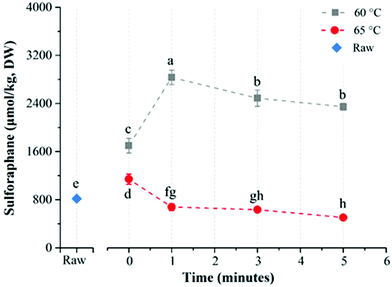
I thought about adjusting microwave practices for 3-day-old broccoli sprouts from ≤ 60°C to 55°C (131°F) in consideration of both the ESP and the 55-to-65°C decline in myrosinase activity.

But myrosinase activity at unmeasured 60°C isn’t at the graph’s straight line drawn between measured 55°C and 65°C. A substantial decline begins after 60°C, not after 55°C as drawn.

Consider this graphic from Enhancing sulforaphane content:

c9fo02089f-f4
Myrosinase robustly hydrolyzes glucoraphanin into sulforaphane at 60°C. There’s clearly a myrosinase deactivation cliff between 60°C and 65°C. Go up to the cliff edge if you must, but don’t go further.

cliff

Broccoli sprouts activate the AMPK pathway

I’ll curate this 2020 rodent study through its summary graphic and caption:

“Type 2 diabetes exhibits elevated levels of circulating fatty acids and CD36. This results in excessive fatty acids binding with CD36 to suppress AMPK [adenosine 5′ monophosphate-activated protein kinase, a key player in regulating energy metabolism].

Inactivation of AMPK breaks homeostasis in lipid metabolism and the antioxidative system, and subsequently induces cardiac oxidative stress, inflammation, and fibrosis. These damages contribute to diabetic cardiomyopathy.

SFN [sulforaphane] treatment significantly induces AMPK activation, which:

  • Enhances mitochondrial fatty acids oxidation via PPARα/CPT-1B and PGC1-α pathways; and
  • Inhibits SCD-1 to down-regulate lipid synthesis.

This greatly alleviates cardiac lipid accumulation.

NRF2-mediated antioxidative effects can be activated via AMPK/AKT/GSK3β pathway, developing another pathway to confront cardiac oxidative damage.

AMPK is indispensable in SFN-mediated cardiac prevention against T2D.”

https://www.metabolismjournal.com/article/S0026-0495(19)30217-3/fulltext “Protective effects of sulforaphane on type 2 diabetes-induced cardiomyopathy via AMPK-mediated activation of lipid metabolic pathways and NRF2 function” (not freely available)


1. A human-mouse relative age perspective:

  • Experiments started with subjects at 2-months-old, equivalent to 20 human years. Treatment subjects ate a high-fat diet.
  • Sulforaphane was injected subcutaneously at 0.5 mg/kg every working day. It didn’t have significant effects on cardiac lipid accumulation at 5 months (a 30-year-old human), but did at 8 months (a 42-year-old human).

2. This study demonstrated that for sulforaphane to produce evidenced Nrf2 pathway effects, it first activated the AMPK/AKT/GSK3β pathway. For 5 days a week, over periods of human-equivalent decades.

3. CPT-1B pictured above is carnitine palmitoyltransferase-1B, an enzyme in the outer membrane of mitochondria. It controls transfer of long-chain fatty acyl CoA into mitochondria to convert fat into energy.

AMPK pathway activation also subsequently activates “PPARα/CPT-1B and PGC1-α pathways.” See A case for carnitine supplementation for a review.


Can a prebiotic help you feel better?

This 2019 rodent study investigated an inulin-type fructo-oligosaccharide (FOS):

“The microbiota-gut-brain axis was used to investigate anti-depressive properties of FOS at the interface of gut microbiota. FOS was introduced via gavage to rats exposed to chronic unpredictable mild stress:

  • FOS alleviated depression-like behaviors and repaired intestinal epithelia damages.
  • FOS treatment lowered corticosterone level.
  • FOS-induced modulation of gut microbiota was more anti-depressive compared to fluoxetine, the standard antidepressant drug.

  • N-Ctrl and M-Ctrl were normal and model control groups which received only water.
  • N-FOS and M-FOS were normal and model rats administrated FOS (50 mg/kg) [human equivalent (50 mg x .162) x 70 kg = 567 mg].
  • M-Flx and M-DP5 rats were model rats given fluoxetine hydrochloride (10 mg/kg) and DP5 compound of FOS (15 mg/kg).

Villi structure was broken for rats in a depression-like state. Mucosal erosion was increased, and the crypt in the small intestinal epithelium was disrupted. Treatment with FOS, DP5 and fluoxetine relieved this damage.

However, a severe side effect was found in the colon of rats that demonstrated apposition to fluoxetine:

  • There was obvious goblet cell loss and inflammatory cells infiltration in the colonic epithelium of fluoxetine treated rats, which showed more severity than in model control rats. Although fluoxetine has high bioavailability, its irritation to gastrointestinal tract may cause inflammation reaction thus lead to colonic destruction.
  • These pathological changes in the intestine were investigated to compare the influence of stress and possible drug irritation to the gastrointestinal tract. Stress had negatively affected microstructure of the small intestine.

Anti-depressant efficacy of FOS was inseparable from and strongly associated with modulation of the host’s gut microbiota.”

https://www.sciencedirect.com/science/article/abs/pii/S0944711319304738 “Fructo-oligosaccharides from Morinda officinalis remodeled gut microbiota and alleviated depression features in a stress rat model” (not freely available)


Forcing people to learn helplessness explored human equivalents of this study’s chronic, unpredictable stress experiments. Related phenotypes and symptoms in humans and animals include:

  • “Social defeat
  • Social avoidance behavior
  • Irritable bowel syndrome
  • Depression
  • Anxiety
  • Anhedonia
  • Increased hypothalamic-pituitary-adrenal (HPA)-axis sensitivity
  • Visceral hypersensitivity.”

These researchers spent a lot of time and effort comparing microbiota categories. The point for people, though, is how we feel.

PXL_20210122_122029867

It’s the fiber, not the fat

I came across this 2020 fiber-vs-fat rodent study from its citation in Gut microbiota and aging:

“Dietary intervention studies largely revolve around altering fat content. Little consideration has been given to amount of fiber and whether or not it is soluble.

We examined age- and sex-specific effects of a refined high-fat/low soluble fiber diet (rHFD) on body weight and gut microbiota composition relative to mice fed a refined low-fat diet (rLFD) that is nutritionally and compositionally matched to rHFD.

Chow diet supplied energy as 13.4% fat, 28% protein, 57.9% carbohydrates, and 15% dietary fiber (range of total dietary fiber between 15 and 25% with 15–20% insoluble and 2–5% soluble fiber).

Two refined diets were used: rLFD supplying energy as 12% fat, 21% protein, and 67% carbohydrates; and rHFD supplying energy as 45% fat, 20% protein, and 35% carbohydrates. [Both rLFD and rHFD contained] 5% fiber in the form of insoluble cellulose.

Young adult animals consumed chow diet for 17 weeks, and 1-year aged animals consumed chow diet for 60 weeks. We included a 1-week transition period wherein all mice were fed rLFD. For the following 4 weeks, half of the animals remained on rLFD while the other half consumed rHFD.

After 4 weeks, young adult female mice showed resistance to weight gain to rHFD, consistent with previous reports. Aged females fed rHFD showed rapid body weight gain relative to rLFD-fed aged females.

Young adult and 1-year aged males showed a significant gain in body weight that was independent of refined diet formulation, suggesting that other components of the refined diet contribute to body weight gain that is independent of dietary fat.

Transition from chow diet to rLFD resulted in changes to microbiota community structure and composition in all groups, regardless of sex and age. This dietary transition was characterized by a loss within phylum Bacteroidetes and a concomitant bloom of Clostridia and Proteobacteria in a sex- and age-specific manner.

No changes to gut microbiota community structure and composition were observed between mice consuming either rLFD or rHFD, suggesting that transition to rLFD that lacks soluble fiber is the primary driver of gut microbiota alterations, with limited additional impact of dietary fat on gut microbiota.”

https://microbiomejournal.biomedcentral.com/articles/10.1186/s40168-020-0791-6 “It’s the fiber, not the fat: significant effects of dietary challenge on the gut microbiome”


It’s alright for researchers in the Abstract and Introduction section to interpret how their rodent study may apply to humans. I appreciate when they confine their statements elsewhere to what they actually measured and found.

This study didn’t measure inflammation, behaviors, neurobiologics, metabolic parameters, immune biomarkers, or hormones. They can qualify statements with “may” all they want, but there wasn’t direct evidence for either:

“Age-specific vulnerability to diet-induced body weight gain in females may be related to aging-related changes to estrogens.”

or

“The lack of differences between rLFD- and rHFD-fed mice may indicate that gut microbiota structure and composition can be dissociated from body weight and systemic inflammation.”

Papers that cite this study can’t rely on its Abstract for “regulating metabolic, immune, behavioral, and neurobiological outcomes” because its experiments didn’t directly measure such outcomes.

Removing 2-5% soluble fiber from subjects’ diet had large effects. I look forward to reading human studies that are informed by this study.