Nrf2 Week #4: Aging

Two 2023 reviews of Nrf2 and aging, starting with Nrf2-mitochondria interactions:

“We discuss molecular mechanisms of interactions between Nrf2 and mitochondria that influence the rate of aging and lifespan. Nrf2 activity positively affects both mitochondrial dynamics and mitochondrial quality control.

Nrf2 influences mitochondrial function through regulation of nuclear genome-encoded mitochondrial proteins and changes in the balance of ROS or other metabolites. In turn, multiple regulatory proteins functionally associated with mitochondria affect Nrf2 activity and even form mutual regulatory loops with Nrf2. These loops enable fine-tuning of cellular redox balance and, possibly, of the cellular metabolism as a whole.

mtDNA-encoded signal peptides interact with nuclear regulatory systems, first of all, Nrf2, and are possibly involved in regulation of the aging rate. Interactions between regulatory cascades that link programs ensuring maintenance of cellular homeostasis and cellular responses to oxidative stress are a significant part of both aging and anti-aging programs.

Understanding these interactions will be of great help in searching for molecular targets to counteract aging-associated diseases and aging itself. Future research on Nrf2 signaling and ability of various substances that activate the Nrf2 pathway to prevent age-associated chronic diseases will provide further insight into the role of Nrf2 activation as a possible longevity-promoting intervention.”

https://link.springer.com/article/10.1134/S0006297922120057 “Transcription Factor Nrf2 and Mitochondria – Friends or Foes in the Regulation of Aging Rate” (not freely available) Thanks to Dr. Gregory A. Shilovsky for providing a copy.


The second review evaluated whether Nrf2 is a master regulator of aging:

“This paper briefly presents main mechanisms of mammalian aging and roles of inflammation and oxidative stress in this process. Mechanisms of Nrf2 activity regulation, its involvement in aging and development of the senescence-associated secretory phenotype are also discussed.

The age-related decrease in Nrf2 activity is of universal interspecies character:

  • Rodents with high Nrf2 activity have a longer lifespan than rodents with low activity.
  • Genetic knockout of Nrf2 usually leads to the increased senescent phenotype in a variety of animal organs and tissues, and also reduces lifespan of female mice.
  • There are also opposite examples, where Nrf2 knockout in aging mice reduced iron ions deposition in the brain, lowered the level of oxidative damage in the striatum, and also alleviated age-related motor dysfunction.

10541_2022_2401_Fig2

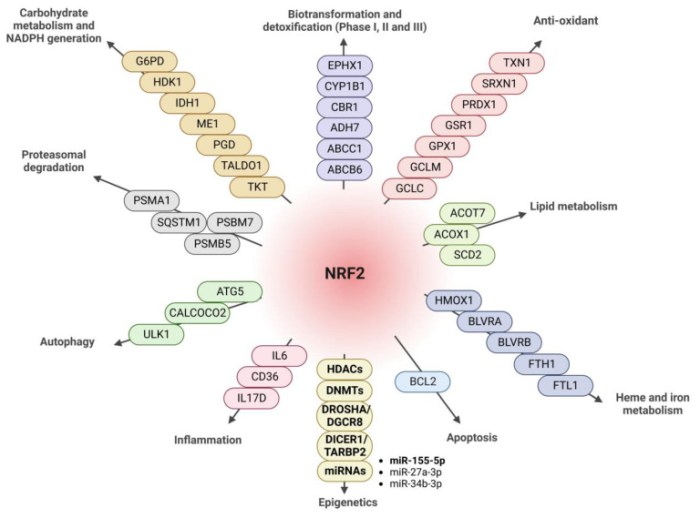
It would be incorrect to consider the effect of Nrf2 transcription factor at the organism level as exclusively antioxidant, anti-inflammatory, and, ultimately, anti-aging. Nrf2 controls many genes, products of which have complex, pleiotropic effects on the body:

  • No experiments that use Nrf2 chemical inducers as anti-aging drugs have been performed so far.
  • Nrf2 is not involved in life extension caused by caloric restriction.
  • Epigenetic clocks do not reveal transcription factors activity of which changes with aging.

Aging is accompanied by changes in gene expression profiles, which are tissue- and species-specific. These changes only to a small extent include genes controlled by Nrf2. At the moment, it cannot be concluded that Nrf2 is the master regulator of the aging process.”

https://link.springer.com/article/10.1134/S0006297922120045 “Does Nrf2 Play a Role of a Master Regulator of Mammalian Aging?”


PXL_20230520_182643299

Nrf2 Week #3: Epigenetics

To follow the Nrf2 Week #2 finding that chromatin accessibility parallels Nrf2 expression, this 2023 cell study explored how Nrf2 influences other epigenetic processes:

“We identified antioxidant response element sequences in promoter regions of genes encoding several epigenetic regulatory factors, such as histone deacetylases (HDACs), DNA methyltransferases (DNMTs), and proteins involved in microRNA biogenesis.

  • We treated cells with dimethyl fumarate (DMF), an activator of the NRF2 pathway through both the KEAP1 and GSK-3 pathways. NRF2 is able to modulate expression of HDAC1, HDAC2, HDAC3, and SIRT1 in different cell types.
  • DMF treatment induced DNMT1 and DNMT3b at both mRNA and protein levels. For DNMT3a, there was a slight induction of mRNA levels but not at the protein level.

antioxidants-12-00641-g007

  • Our data indicate that of all miRNAs analyzed, only miR-27a-3p, miR-27b-3p, miR-128-3p, and miR-155-5p associate with Nfe2l2 mRNA. NRF2 causes degradation of miR-155-5p, which is implicated in neuroinflammation and other pathologies, and is the main miRNA induced by LPS treatment in microglia. miR-155 alters expression of genes that regulate axon growth, supporting the bioinformatic prediction that miR-155 can regulate expression of genes involved in central nervous system development and neurogenesis.

Todate we only understand how epigenetic modifications affect expression and function of the NRF2 pathway. The fact that NRF2 can promote expression of type I HDACs, DNMTs, and proteins involved in miRNA biogenesis opens new perspectives on the spectrum of actions of NRF2 and its epigenetic influences.”

https://www.mdpi.com/2076-3921/12/3/641 “The Transcription Factor NRF2 Has Epigenetic Regulatory Functions Modulating HDACs, DNMTs, and miRNA Biogenesis”


PXL_20230515_185958612

Nrf2 Week #2: Neurons

To follow the Nrf2 Week #1 suggestion that Nrf2 target neurological disorders, this 2023 cell study investigated Nrf2 expression in neurons:

“Oxidative metabolism is inextricably linked to production of reactive oxygen species (ROS), which have the potential to damage all classes of macromolecules. Yet ROS are not invariably detrimental. Several properties make ROS useful signaling molecules, including their potential for rapid modification of proteins and close ties to cellular metabolism.

We used multiple single cell genomic datasets to explore Nrf2 expression and regulation in hundreds of neuronal and non-neuronal cell types in mouse and human. With few exceptions, Nrf2 is expressed at far lower levels in neurons than in non-neuronal support cells in both species.

This pattern is maintained in multiple disease states, and the chromatin accessibility landscape at the Nrf2 locus parallels these expression differences. These results imply that Nrf2 activity is limited in almost all neurons of the mouse and human central nervous system (CNS).

nrf2 expression

We separated cell types into neuron or non-neuronal ‘support’ cell categories. The general ‘support’ term is not meant to minimize the functional relevance of non-neuronal cells in the CNS, but is an umbrella term meant to cover everything from glial cell types (astrocytes, microglia, oligodendrocytes) to endothelial cells.

It is not clear why an important, near ubiquitous cytoprotective transcription factor like Nrf2 remains off in mature neurons, especially considering oxidative stress is a driver of many diseases. The simplest explanation is that Nrf2 activity also disrupts normal function of mature neurons.

ROS play a key role in controlling synaptic plasticity in mature neurons. These activity-dependent changes in synaptic transmission, which are important for learning and memory, are disrupted by antioxidants.

A subset of important Nrf2-targeted antioxidant genes (e.g., Slc3a2, Slc7a11, Nqo1, Prdx1) are also low in neurons. So it is likely that these and/or other Nrf2 targets must remain low or non-ROS-responsive in mature neurons. Future work exploring why this expression pattern persists in mature neurons will inform our models on roles of antioxidant genes in normal neuronal physiology and in neurological disorders.

https://www.biorxiv.org/content/10.1101/2023.05.09.540014v1.full “Limited Expression of Nrf2 in Neurons Across the Central Nervous System”


PXL_20230520_182827767

Don’t eat yourself into disease, Part 2

This blog’s 1000th curation is a 2023 rodent study associating gut microbiota, behavior, memory, and food reward:

“Energy intake and energy expenditure is regulated by the hypothalamus, and is referred to as homeostatic regulation of food intake. The reward system is the non-homeostatic regulation of food intake – pleasure-related consumption of foods enriched in fat and sugar. The pleasure is encoded by dopamine release from dopaminergic neurons projecting from the ventral tegmental area to the striatum, the nucleus accumbens, and the prefrontal cortex.

Food reward can be divided into three components – liking, wanting, and learning:

  • Liking refers to food hedonic value;
  • Wanting to the motivational process to seek out and consume certain foods; and
  • Learning to reinforcing conditioning behavior associated with food intake stimulus.

We confirmed that obese mice have a dysregulation of the learning and the wanting components of  food reward. Our previous data showed that the liking component was transferable through fecal material transplantation.

We demonstrated that gut microbes play a role in the regulation of food reward, and could be responsible for compulsive behavior and excessive motivation to obtain sucrose pellets. Moreover, obese gut microbes affected dopaminergic and opioid markers involved in reward system.

We identified 33HPP (produced exclusively by gut bacteria) as particularly increased in mice recipients of gut microbes from obese mice. We were able to demonstrate its effects as key mediator of the gut-brain axis controlling the reward response to palatable food.”

https://microbiomejournal.biomedcentral.com/articles/10.1186/s40168-023-01526-w “Obese-associated gut microbes and derived phenolic metabolite as mediators of excessive motivation for food reward”


PXL_20230415_195202937

Don’t eat yourself into disease, Part 1

Starting a sample of 2023 papers with a porcine review:

“Epigenetic programing predisposes pigs to insulin insensitivity, but pigs seem to sense this insensitivity and consequently eat less, preventing obesity. Pigs naturally prefer to eat small breakfasts and large dinners.

Deviating from this eating pattern or providing diets with a high glycemic burden can trigger obesity; however, pigs will restrict food intake to prevent serious obesity. In practice, problems with obesity are rarely seen, even when pigs are fed poorly timed diets similar to junk food, likely because swine diets are balanced for every nutrient.

Feeding pigs diets deficient in micronutrients does trigger obesity. For humans, several micronutrient requirements have not been set officially, and diets optimized for all micronutrients are rarely provided.

metabolites-13-00420-g015

Although we could debate whether this is a cause or effect, the above data on hyper-processed diets fed to pigs would indicate that it is causative. Pigs were fed a diet which included ‘human-targeted junk food’ but was adequate in phosphate, and they experienced no issues.

Controlled human studies are generally conducted with very small populations of subjects for very short durations, as emotions come into play. Humans are hard to persuade to follow a boring diet, especially over a longer period of time, and humans are easily tempted to deviate from a protocol if peer pressure or desires are high.

Even worse, in survey type experiments, people are asked what they ate for the past one or several days, and these data may well be subsequently extrapolated to patterns of behavior and then correlated with developments in health. Recalling what and especially how much a person ate yesterday is already a challenge for many, confounded even further by the desire not to include items that may be considered less acceptable.

On the swine side, knowledge on nutrient yield of foods and nutrient requirement appears further advanced, and controlled feeding trials are much easier to perform. Borrowing pig data is arguably much closer to the truth for humans than having no data at all.”

https://onlinelibrary.wiley.com/doi/10.1002/advs.202205346 “Eat like a Pig to Combat Obesity”


One fish in the gullet, another soon on its way

PXL_20230509_094500357

No exit

This 2023 rodent study investigated aging processes and gut microbiota in crowded conditions:

“Our study provides clear evidence that high-density crowding accelerates the aging process of Brandt’s voles. We also found that ‘high-density microbiota’ promote the aging-related phenotype in voles.

Because we minimized effects of direct fighting on mortality of voles, observed changes in lifespan in this study should mostly represent the natural aging processes of voles.

high-density survival

High density increased the level of stress hormone corticosterone, which disrupted gut microbiota composition by:

  • Decreasing abundance of anti-aging or anti-inflammatory bacterial species; and
  • Increasing the proportion of pathogenic bacteria.

This caused an increase in DNA oxidation and inflammation through upregulation of NF-kB and COX-2 pathways.

Although high-density relief and butyric acid administration interventions could reverse aging-related processes of adult voles, it remains unclear whether they could reverse the aging process in terms of lifespan.

Our results suggest that gut microbiota play a significant role in mediating aging-related processes of voles under high-density conditions, and can be used as a potential therapeutic target for treating stress-related diseases in humans.”

https://onlinelibrary.wiley.com/doi/10.1002/advs.202205346 “Gut Microbiota is Associated with Aging-Related Processes of a Small Mammal Species under High-Density Crowding Stress”


I came across this study by it citing Reversing hair greying for effects of stress interventions.

PXL_20230505_185253518.MP

Physical fitness and epigenetic clocks

This 2023 human study of 144 men average age 68 investigated relationships among physical fitness measurements and three epigenetic clocks:

“We investigated relationships between physical fitness and age-adjusted values from residuals of the regression of DNAm aging clocks to chronological age (DNAmAgeAcceleration: DNAmAgeAccel) and attempted to determine the relative contribution of physical fitness variables to DNAmAgeAccel in the presence of other lifestyle factors.

  • Volume of oxygen (VO2/kg) at ventilatory threshold and at Peak, fat free mass, calf circumference, serum HDL-C, daily intake of carbohydrates, iron, copper, vitamin C, and β-carotene were negatively related with DNAmAgeAccel.
  • Body fat, visceral fat area, and serum TG were positively related to DNAmAgeAccel.

figure 4

Frequent alcohol consumption and past- and current-smoking status were associated with accelerated DNAmAgeAccel, while a morning lifestyle was associated with deceleration of it. Multiple regression analysis suggested that – rather than physical fitness – serum triglycerides, carbohydrate intake, and smoking status were significantly associated with DNAmAgeAccel.

In conclusion, the contribution of cardiorespiratory fitness to DNAmAgeAccel was relatively low compared to lifestyle factors such as smoking. However, this study reveals a negative relationship between cardiorespiratory fitness and DNAmAgeAccel in older men.”

https://www.medrxiv.org/content/10.1101/2023.04.12.23288187v1.full-text “Associations between cardiorespiratory fitness and lifestyle-related factors with DNA methylation-based aging clocks in older men: WASEDA’S Health Study”


PXL_20230330_191228773

Remembering life before birth

This 2023 primate study investigated the body’s capability to remember prenatal experiences influencing later life:

“Maternal stressors and other environmental factors affect the developing embryo and fetus in ways that lead to increased susceptibility for chronic disease in later life. Developmental programming of chronic low-grade inflammation plays an important role in onset and progression of these diseases.

Establishing innate immune cell memory involves increased glycolysis, reduced oxidative phosphorylation, and expression of transcription factors which prime for pro-inflammatory activity. This memory relies on propagation of epigenetic modifications that develop in hematopoietic stem and progenitor cells (HSPCs), which can be passed on to progeny immune cells (i.e., macrophages).

These changes persist with altered epigenetic regulation for years after weaning – even when offspring are fed a conventional diet – predisposing offspring to inflammatory disease across their lifespans.

cell memory

Several factors may initiate metabolomic reprogramming in fetal HSPCs:

  • We found increased chromatin accessibility of gene regulatory regions and RNA signatures supporting activation of factors with a major role in regulating macrophage inflammatory activation, including FOS/JUN, NF-κB, C/EBPβ, and STAT6.
  • Our prior work demonstrated a persistently altered histone code in liver tissue from juvenile animals.
  • Maternal diet-supplied lipids, including oleic acid, in hematopoietic tissues may play an important role in priming inflammation and metabolism in fetal HSPCs and bone marrow-derived macrophages with postnatal persistence.

Striking changes in fetal bone marrow and liver HSPCs observed here suggest that the primary driver for developmental programming of inflammation takes place in utero. However, we cannot rule out that exposure to maternal diet during lactation postnatally triggers shifts in microbiome composition or function contributing to inflammation.

Components of maternal diet, rather than maternal obesity per se, are a modifiable risk factor with potential to alter developmental programming of offspring immune systems.”

https://www.cell.com/cell-reports/fulltext/S2211-1247(23)00404-7 “Maternal diet alters long-term innate immune cell memory in fetal and juvenile hematopoietic stem and progenitor cells in nonhuman primate offspring”


And there are other ways we remember everything that happened then and along the way. Big clues are in our out-of-context responses to present day events.

PXL_20230326_104753766

Year Three of Changing to a youthful phenotype with sprouts

1. I’ve continued daily practices from Year Two with microwaved 3-day-old broccoli, red cabbage, and mustard sprouts for 13 times longer now than any sulforaphane clinical trial. I haven’t had another three-year period in my life where I wasn’t sick even once!

The main difference over the past year is that I eat AGE-less chicken vegetable soup 3-4 times a week rather than twice a day. That was just too boring, plus I stopped eating lunch. My other dinners are often steamed vegetables and seafood.

2. I frame the above and following tactics as part of a strategy of delaying evolutionarily-determined programs from doing more to kill my body every year:

  • Eat 16-hour sprouted Avena nuda oats for breakfast to give my trillion+ gut microbiota what they need;
  • Eat 3-day-old hulled Avena sativa oat sprouts twice a day to provide antioxidants in another form, avenanthramides;
  • Eat AGE-less chicken vegetable soup 3-4 times a week to prevent diet-induced problems;
  • Take supplements that promote healthspan twice a day;
  • Exercise at least 30 minutes daily;
  • Take yeast cell wall β-glucan daily, with nothing else an hour before or after; and
  • Avoid undue stress by working from home 40 hours a week in my 26th year as a professional software developer.

These tactics’ main components activate AMPK, Nrf2, and associated signaling pathways, and inhibit pro-inflammatory pathways such as NF-κB.

But fixing inflammation doesn’t repair all existing damage. I wonder what could have been physically resolved if I had started thirty+ years earlier.

3. One place I take clues from are successful anti-aging animal research efforts such as a study reviewed here earlier this month. Last curated in Improve your internal environment, improve its constituents’ functions, it used plasma fraction treatment. Plasma fraction eclipsed a caloric restriction treatment’s previous record for maximum species lifespan by 5%.

This type of research clearly isn’t a priority for official sponsors to fund, though. Take responsibility for your own one precious life.

Eat broccoli sprouts to protect your lungs

This 2023 human cell study investigated sulforaphane’s effects on tuberculosis infections:

“Basic research efforts on tuberculosis (TB) immunotherapy are currently only the tip of the iceberg. This study highlights the association between autophagy-related genes and immune infiltration in TB, an infectious pathogen that has been around for tens of thousands of years.

Sulforaphane (SFN) is readily absorbed into the bloodstream by the intestine due to its lipophilic nature. Experiments in this study TB patient cells showed that SFN could promote autophagy in macrophages infected with Mycobacterium abscessus (Mab). Intracellular bacterial load of macrophages was associated with SFN-enhanced cellular autophagic processes.

1-s2.0-S156757692300276X-gr4

The relationship between autophagy and immune cells is complex, and recurrence of tuberculosis is significantly influenced by intracellular mycobacteria of macrophages. Macrophages have longer lifespans than neutrophils, and provide shelter for mycobacteria as they are better suited than neutrophils to establish strategies for targeting autophagy.

This is one of the reasons why autophagy in macrophages was the focus of this study. Appropriate autophagy is beneficial for the body and controls Mtb replication, but autophagic programmed cell death can activate tissues to produce an excessive inflammatory response, resulting in severe damage to lung tissues.

Autophagy-related genes regulated by SFN have good diagnostic potential, with FOXO1 potentially serving as a target for TB immunotherapy. Downstream targets of FOXO1 include important pro-inflammatory signaling molecules such as IL-1β and TNF-α, which are important for control of mycobacterium.”

https://www.sciencedirect.com/science/article/pii/S156757692300276X “Identifying autophagy-related genes as potential targets for immunotherapy in tuberculosis”

Take yeast cell wall β-glucan, too, and train your immune system.

Does eating broccoli sprouts influence biological age?

A 2023 review of 28 human clinical trials investigating broccoli sprout compounds brought up this post’s title by omitting discussion of it:

“In order to determine the effective reference dose of a broccoli sprouts beverage for detoxifying carcinogenic air pollutants (benzene), Chen et al. administrated a drink enriched with glucoraphanin (GR) and sulforaphane (SFN) from 3-day-old broccoli sprouts to healthy adults. Researchers focused on excretion of metabolites SFN-NAC, SFN-CYS, and non-esterified SFN, which represent 80–81%, 12–14%, and 5–7% of total SFN forms, respectively.

Excretion percentage did not change during the intervention, indicating that bioavailability remained constant.

Enhanced excretion of the urinary biomarker of benzene detoxification S-phenylmercapturic acid (SPMA) was measured in urine collected every 12 h during the 10-day intervention. Out of 132 samples analyzed, >95% had detectable concentrations of SPMA, being significantly increased after consumption of the high dose of beverage (600 and 40 μmol GR and SFN, correspondingly), suggesting that consumption of >10 μmol SFN per 24 h may represent the lowest effective dose of the BSE affecting this biomarker.

https://www.mdpi.com/2072-6643/15/6/1424 “Systematic Review on the Metabolic Interest of Glucosinolates and Their Bioactive Derivatives for Human Health”


These reviewers did much hand waving to draw their conclusions. They ignored that the only way randomized trials become better than non-randomized trials is in dealing with confounders.

The largest confounder with glucoraphanin is that an individual’s gut microbiota, not their human cells, metabolize it into isothiocyanates. A glucoraphanin randomized trial has to have sufficient numbers of subjects in each group to adequately deal with confounding individual differences in gut microbiota.

I highlighted the largest of the 28 trials:

Basic RGB

Sulforaphane studies have fewer confounders. Even so, Upgrade your brain’s switchboard with broccoli sprouts stated:

“Power analysis calculations suggest that a sample size of n = 50 would yield a significant result.”

An insufficient number of subjects in both the half dose and full dose groups caused that study’s researchers to frame their results as “suggesting that consumption of >10 μmol SFN per 24 h may..” rather than asserting significant results.


Addressing this post’s title, it’s been ten years since epigenetic clocks came into use. This review highlighted by omission that there still hasn’t been even one investigation of isothiocyanates’ effects on human biological age as measured by epigenetic clocks.

A 40 μmol ≈ 7 mg sulforaphane “high” dose of the cited study is easily achievable with microwaved 3-day-old broccoli sprouts. There’s little question that healthy people activating AMPK, Nrf2, and associated signaling pathways, and inhibiting pro-inflammatory pathways such as NF-κB with sulforaphane, will experience beneficial effects.

The cited study found no change in sulforaphane treatment bioavailability over ten days, and a predecessor study found the same over 12 weeks. I’ll guess those bioavailability findings will extend over longer time periods.

Where are the researchers who will take the next step to show isothiocyanate treatments cause positive changes in epigenetic clock / biological age measurements?

PXL_20230304_211250499

Eat broccoli sprouts for depression, Part 3

Here are two papers published after Part 2 that cited the Part 1 rodent study, starting with a 2023 rodent study performed by several Part 1 coauthors:

“We used a low-dose LPS-induced endotoxaemia model to mimic clinical characteristics of sepsis. We found that adolescent LPS treatment was sufficient to increase levels of inflammatory factor TNF-α in both the medial prefrontal cortex (mPFC) and hippocampus at post-natal day P22.

P21 LPS-treated mice were injected with sulforaphane (SFN) or saline intraperitoneally at P49 and then subjected to subthreshold social defeat stress (SSDS). We found that SFN preventative treatment significantly:

  • Decreased the social avoidance, anhedonia, and behavioural despair detected by the social interaction test, sucrose preference test, tail suspension test, and forced swim test, respectively.
  • Decreased anxiety-like behaviours without affecting locomotor activities.
  • Increased Nrf2 and brain-derived neurotrophic factor (BDNF) levels in the mPFC of P21 LPS-treated mice after SSDS compared with saline control mice.

The above results suggest that activation of the Nrf2-BDNF signalling pathway prevents the effect of adolescent LPS-induced endotoxaemia on stress vulnerability during adulthood.

sulforaphane and stress vulnerability

These results suggest that early adolescence is a critical period during which inflammation can promote stress vulnerability during adulthood. This might be due to increased inflammatory response in the mPFC, and mediated by decreased levels of Nrf2 and BDNF. These findings may shed light on the potential use of SFN as an alternative preventative intervention for inflammation-induced stress vulnerability.”

https://link.springer.com/article/10.1007/s00213-022-06285-4 “Lipopolysaccharide-induced endotoxaemia during adolescence promotes stress vulnerability in adult mice via deregulation of nuclear factor erythroid 2-related factor 2 in the medial prefrontal cortex” (not freely available)

This study demonstrated that adolescent diseases and stresses don’t necessarily develop into adult social problems. A timely intervention may even prevent future adult problems.

The one-time 10 mg/kg sulforaphane dose was the same as Part 1’s dose, a human equivalent of which is (10 mg x .081) x 70 kg = 57 mg.

I’d like to know more about how subjects’ memories of adverse events were retained, and subsequently affected their biology and behavior. Pretty sure limbic structures like the hypothalamus as well as lower brain structures played a part.


A 2022 review summarized what was known up to that time regarding Nrf2 and depression:

“Sulforaphane, an organosulfur compound isolated from Brassicaceae plants, is a potent natural NRF2 activator. Sulforaphane:

  • Exerts antidepressant- and anxiolytic-like activities and inhibits HPA axis and inflammatory response.
  • Has both therapeutic and prophylactic effects on inflammation-related depression.
  • Confers stress resilience.
  • Protects neurons via autophagy and promotes mitochondrial biogenesis by activating Nrf2.”

https://www.sciencedirect.com/science/article/pii/S2213231722002944 “Nrf2: An all-rounder in depression”


PXL_20230306_202857493

The goddess of destiny

A 2023 human study investigated exercise, klotho gene, and epigenetic clock relationships:

“Named after the spinner of the thread of life, klotho (KL) is involved in the aging process and may act as an anti-aging hormone in mammals. We hypothesize that circulating KL is correlated with age-associated methylation of the KL gene promoter region, and this is one reason for age-related decline in circulating KL.

202 subjects between ages 37 and 85 were included in the study. A great percentage of volunteers participated in the World Rowing Masters Regatta in Velence, Hungary. They were considered to be the trained group (TRND): n = 131; 80 males: age 59.14 ± 10.8; 51 females: age 57.24 ± 9.4. Results were compared to the sedentary group (SED): n = 71; 27 males: age 55.63 ± 13.4; 44 females: age 61.91 ± 10.5.

Circulating level of KL showed a negative correlation with chronological age in the TRND group, but not in the SED group.

klotho and exercise

Examining the relationship between circulating KL level and PhenoAge and GrimAge, KL is associated with PhenoAge acceleration in the TRND group only. It appears that higher KL can decelerate the DNA methylation-based aging process assessed by PhenoAge.

The present study revealed that circulating KL level is associated with exercise status level and general strength level, and is greatly dependent upon exercise-induced DNA methylation.”

https://www.mdpi.com/2073-4425/14/2/525 “The Circulating Level of Klotho Is Not Dependent upon Physical Fitness and Age-Associated Methylation Increases at the Promoter Region of the Klotho Gene”


PXL_20230216_200008595

Ancient parasite DNA within us

Two 2023 papers on endogenous retroviruses (ERVs) and aging relationships, starting with the Introduction section of a comprehensive study:

“Several causal determinants of aging-related molecular changes have been identified, such as epigenetic alterations and stimulation of senescence-associated secretory phenotype (SASP) factors. Although the majority of these studies describe aging determinants originating primarily from protein-coding genes, the non-coding part of the genome has started to garner attention as well.

ERVs belonging to long terminal repeat (LTR) retrotransposons are a relic of ancient retroviral infection, fixed in the genome during evolution, comprising about 8% of the human genome. As a result of evolutionary pressure, most human ERVs (HERVs) accumulate mutations and deletions that prevent their replication and transposition function. However, some evolutionarily young subfamilies of HERV proviruses, such as the recently integrated HERVK, maintain open reading frames encoding proteins required for viral particle formation.

In this study, using cross-species models and multiple techniques, we revealed an uncharacterized role of endogenous retrovirus resurrection as a biomarker and driver for aging. Specifically, we identified endogenous retrovirus expression associated with cellular and tissue aging and that the accumulation of HERVK retrovirus-like particles (RVLPs) mediates the aging-promoting effects in recipient cells. More importantly, we can inhibit endogenous retrovirus-mediated pro-senescence effects to alleviate cellular senescence and tissue degeneration in vivo, suggesting possibilities for developing therapeutic strategies to treat aging-related disorders.”

https://www.cell.com/cell/fulltext/S0092-8674(22)01530-6 “Resurrection of endogenous retroviruses during aging reinforces senescence”


This first paper’s foreword summarized their many experiments and findings:

“The study found that HERVK transcripts, viral proteins, and RVLPs were highly activated in prematurely aged human mesenchymal progenitor cells (hPMCs). This was similarly observed in aged human primary fibroblasts and hPMCs. They also discovered that decreasing silencing epigenetic marks DNA methylation and H3K9me3 while increasing H3K36me3 enabled HERVK expression.

erv aging mechanism

These observations also raise several intriguing questions:

  • HERVK is occasionally activated and eventually suppressed under physiological conditions, for example, in human embryonic cells. It would be fascinating to probe the possibility of mimicking physiological conditions in order to turn off the positive feedback between HERVK and senescence.
  • ERVs are hallmarks of aging in different species, including human, primate, and mouse. Future quantification of the absolute physiological level of ERVs across a broad population of various ages might provide further insights into the relationship between ERVs and organismal age.”

https://academic.oup.com/lifemedi/advance-article/doi/10.1093/lifemedi/lnad001/6982772 “Endogenous retroviruses make aging go viral”


Previously curated papers on these subjects include:

A study of our evolutionary remnants

“Repressive epigenetic marks associated with ERVs, particularly LTRs, show a remarkable switch in silencing mechanisms, depending on evolutionary age:

  • Young LTRs tend to be CpG-rich and are mainly suppressed by DNA methylation, whereas
  • Intermediate age LTRs are associated predominantly with histone modifications, particularly histone H3 lysine 9 (H3K9) methylation.
  • Evolutionarily old LTRs are more likely inactivated by accumulation of loss-of-function genetic mutations.”

Starving awakens ancient parasite DNA within us

Reality is sometimes stranger than what fiction writers dream up. 🙂

PXL_20230209_210243470

Environmental signaling rescues aging muscle stem cells

This 2023 rodent study applied An environmental signaling paradigm of aging concepts to muscle stem cells:

“The stem cell niche environment represents an important therapeutic target to enhance tissue regeneration in aging. We decoupled age-related cell-intrinsic effects, niche-mediated cell-extrinsic effects, and changes in population dynamics of muscle stem cells (MuSCs) and two key muscle-resident cells in young and aged mice.

in vivo model

We showed that:

  1. Age-related reduction in MuSCs is not stochastic.
  2. Despite differences in transcriptomes of MuSC clusters, the effect of age on gene expression is largely uniform, suggesting that the niche environment has a fundamental role in age-related changes in MuSC gene expression.
  3. A significant fraction of changes in the transcriptome of aging MuSCs can be reversed by exposure to the young muscle environment, i.e. are niche-responsive. Given the high percentage [46.6% at a stringent cutoff of s-value < 0.05] of reversibility in gene expression, our findings indicate that age-related changes in the niche are principal drivers of resulting alterations in the MuSC transcriptome.
  4. Aging is correlated with changes at the level of chromatin accessibility and DNA methylation in MuSCs.

Plasticity of the MuSC transcriptome suggests that modulating the niche environment can be a powerful tool to restore stem cell-mediated endogenous muscle regeneration in aging. Consequently, as opposed to focusing solely on MuSCs themselves to mitigate effects of aging on MuSCs, bioengineering of the niche in its entirety may be a viable therapeutic option.”

https://www.nature.com/articles/s41467-023-36265-x “Transcriptional reprogramming of skeletal muscle stem cells by the niche environment”


This study destroyed extremely well-funded directed research efforts that detract from science, especially those promoting irreversibility of epigenetic changes (but: Rockefeller) and randomness of pro-aging programming (but: Harvard).

These researchers showed they could do more with their ideas and careers than maintain an outdated and easily disproved status quo.