Don’t count on broccoli compounds bailing out a high-fat diet’s effects on gut microbiota

Two rodent studies of mature broccoli and broccoli sprouts’ effects on a high-fat diet, with the first from 2021 investigating broccoli florets and stalks:

“Addition of broccoli florets to a HFD ameliorated insulin sensitivity. Florets further promoted gut microbiota diversity and low-grade inflammatory-associated strains.

Stalk supplementation also altered gut microbiota, leading to increased Bacteroidetes/Firmicutes ratio and levels of communities that preserve mucus layer and gut integrity while simultaneously decreasing levels of potentially harmful species.

Addition of broccoli to a HFD did not ameliorate body and tissues weight gain or food intake. Both broccoli stalks and florets did not affect fat accumulation, carbohydrate, or lipid metabolism-related parameters.”

https://www.frontiersin.org/articles/10.3389/fnut.2021.680241/full “Broccoli Florets Supplementation Improves Insulin Sensitivity and Alters Gut Microbiome Population – A Steatosis Mice Model Induced by High-Fat Diet”


A 2020 study cited by this first study investigated compounds extracted from 1-day-old broccoli sprouts:

Bioaccessibility of aliphatic glucosinolates was shown to 76.2 ± 0.6%:

aliphatic glucosinolate bioavailability

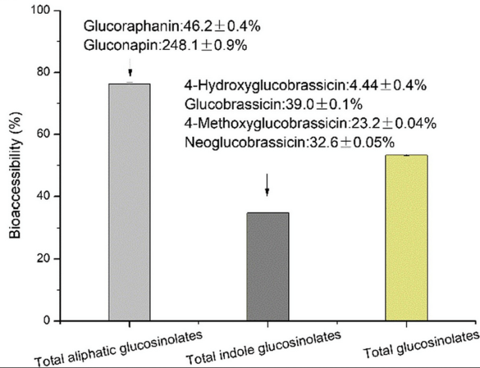
Glucoraphanin was the predominant glucosinolate with the highest bioaccessibility in broccoli, and could effectively prevent HFD-induced body weight gain in mice, especially increases in liver weight and the accumulation of lipids in adipocytes. Furthermore, supplementation with glucoraphanin reduced the level of oxidative stress, regulated genes of FAS, PPARα, CPT1 and ACOX associated with lipid metabolism, and might be associated with changes in composition of gut microbiota.”

https://www.frontiersin.org/articles/10.3389/fnut.2021.680241/full “Effect of glucoraphanin from broccoli seeds on lipid levels and gut microbiota in high-fat diet-fed mice”

This study’s title was “Effect of glucoraphanin from broccoli seeds..” although its Materials and methods section disclosed:

“1 day after germination from broccoli seeds, sprouts were boiled in water for 30 min. The resulting aqueous extract was processed by liquid solid separation and condensation and was subsequently spray-dried to yield an extract powder containing 249 mg glucoraphanin.”


Eat broccoli sprouts every day and its predecessor study demonstrated that broccoli intake every day had beneficial effects during shorter periods than either of these studies.

Both studies had many “may”, “could”, and “might” statements. Not sure that broccoli compounds / gut microbiota relationships are adequately investigated by choosing a few out of tens of thousands of gut microbiota species as both studies attempted to do.

There are too many additive / antagonistic / synergistic combinations to analyze even before reaching twenty gut microbiota species. But researchers aren’t often sponsored for studies unless they conform to existing research.


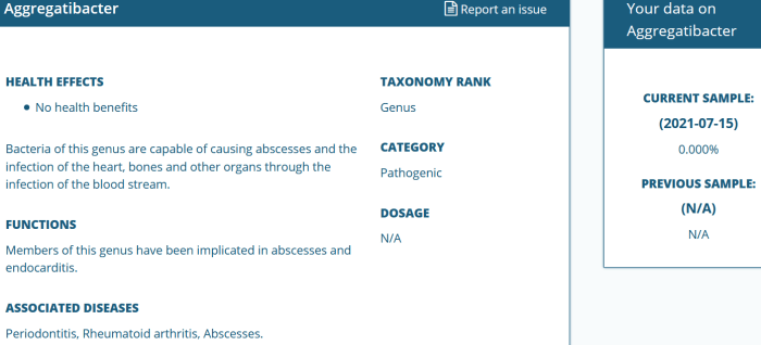
I haven’t made headway in understanding my top 10 of 42,156 gut microbiota species’ exact causes, effects, and interactions. The top three by themselves are considered beneficial:

top 1-10 species

Uncertainty is fine for now, though, with a 40-hour work week interfering. Finding out what my gut microbiota generally want and giving that to them has been a productive approach this year.

Part 3 of Make your gut microbiota happy

Continuing from Part 2, my 7/15/2021 sample found that no bad bacteria needed work. Top three reasons why this may be are:

1. I’ve eaten microwaved broccoli sprouts every day for 68 weeks now. Relevant research:

helicobacter 0

2. This is the 17th year of training my immune system every day with yeast cell wall β-glucan.

acinetobacter

3. Basic hygiene practices such as brushing my teeth twice a day.

aggregatibacter 0


PXL_20210731_094258419.NIGHT

Blood pressure and pain

A trio of papers, with the second and third citing a 2013 review:

“The relationship between pain and hypertension is potentially of great pathophysiological and clinical interest, but is poorly understood. Perception of acute pain initially plays an adaptive role, which results in prevention of tissue damage.

The consequence of ascending nociception is recruitment of segmental spinal reflexes through physiological neuronal connections:

  • In proportion to magnitude and duration of the stimulus, these spinal reflexes cause sympathetic nervous system activation, which increases peripheral resistances, heart rate, and stroke volume; and
  • The response also involves the neuroendocrine system, in particular, the hypothalamic-pituitary-adrenal axis, in addition to further activation of the sympathetic system by adrenal glands.

Persistent pain tends to become chronic and to increase BP values. After a long time, dysfunction of release of endogenous opioids results in a reduction of their analgesic effect. A vicious circle is established, where further pain leads to a reduction in pain tolerance, associated with decreased analgesia mediated by baroreceptors, in a kind of process of exhaustion.”

https://onlinelibrary.wiley.com/doi/epdf/10.1111/jch.12145 “The Relationship Between Blood Pressure and Pain”


A second paper was a 2021 human experimental pain study:

“We investigated the effectiveness of physiological signals for automatic pain intensity estimation that can either substitute for, or complement patients’ self-reported information. Results indicate that for both subject-independent and subject-dependent scenarios, electrodermal activity (EDA) – which is also referred to as skin conductance (SC) or galvanic skin response – was the best signal for pain intensity estimation.

EDA gave mean absolute error (MAE) = 0.93 using only 3 time-series features:

  1. Time intervals between successive extreme events above the mean;
  2. Time intervals between successive extreme events below the mean; and
  3. Exponential fit to successive distances in 2-dimensional embedding space.

Although we obtained good results using 22 EDA features, we further explored to see if we could reach similar or better results with fewer EDA features. This plot highlights that by considering only the top 3 features, we obtained the same level of performance given by all 22 features together.

journal.pone.0254108.g002

This is the first study that achieved less than 1-unit error for continuous pain intensity estimation using only one physiological sensor’s 3 time-series feature, and a Support Vector Regression machine learning model. Considering that this is an encouraging result, we can estimate objective pain using only the EDA sensor, which needs neither a complex setup nor a complex computationally intense machine learning algorithm.

This study paves the way for developing a smart pain measurement wearable device that can change the quality of pain management significantly.”

https://doi.org/10.1371/journal.pone.0254108 “Exploration of physiological sensors, features, and machine learning models for pain intensity estimation”


A third paper was a 2020 human rotator cuff surgery study:

“Results of our study demonstrated that:

  • Pain during the early postoperative period;
  • Time until occurrence of a retear; and
  • Existence of hypertension

were correlated with severity of pain in patients with a retorn rotator cuff.

Pain was selected as the sole outcome parameter of this study because:

  • Pain is an important factor that compels patients to seek treatment for rotator cuff tears, along with functional disability;
  • Pain and subjective functional deficits are important factors that influence a surgeon’s decision to continue with treatment in cases of retearing; and
  • Analyzing pain severity can be a good way to determine patients’ overall satisfaction after rotator cuff repair.

However, pain is not always correlated with disease severity or tear size and vice versa. A lack of pain does not necessarily depend on integrity of the repaired tendon or constitute a good prognosis. In fact, patients with partial-thickness rotator cuff tears showed more pain than did those with full-thickness tears.

Existence of hypertension had a proportional relationship with pain at 12 months postoperatively in patients with retears. This can be interpreted as a suggestion that pain in patients with retears is not acute, but rather chronic, and may be connected to pain in the early postoperative period at 3 months. However, results of this study cannot explain benefits of controlling hypertension in alleviating pain in patients with retears.”

https://journals.sagepub.com/doi/10.1177/2325967120947414 “Factors Related to Pain in Patients With Retorn Rotator Cuffs: Early Postoperative Pain Predicts Pain at 12 Months Postoperatively”


PXL_20210722_100353787

Back pain and advanced glycation end products (AGEs)

Two 2020 rodent studies investigated intervertebral disk degeneration, with the first on AGEs’ role:

“This study evaluated the role of AGEs and RAGE in driving early intervertebral disk (IVD) degeneration processes in mice. Aging and diabetes are associated with increased low-back pain and IVD degeneration, yet causal mechanisms remain uncertain. AGEs:

  • Accumulate in IVDs from aging;
  • Are implicated in diabetes-related disorders;
  • Alter collagen; and
  • Induce proinflammatory conditions.

A mixed population of 23 male and female wild type AC57BL/6J mice were each assigned to two isocaloric diet groups after weaning. They received either low-AGE chow containing 7.6 μg/mg AGE, or high-AGE chow containing 40.9 μg/mg AGE generated via high-temperature heating (NIH-31 open formula chow autoclaved for 30 minutes at 120°C [248° F]). This in vivo dietary model was previously shown to increase IVD AGE accumulation without systemic obesity or diabetes.

disc AGE damage

AGE accumulation leads to RAGE-dependent collagen disruption in the annulus fibrosus, and can initiate molecular and tissue level collagen disruption. Second harmonic generation (SHG) and collagen-hybridizing peptide (CHP) analyzes were sensitive to collagenous alterations at multiple hierarchical levels due to AGE.

These methods may be useful in identifying additional contributors to collagen damage in IVD degeneration processes.”

https://onlinelibrary.wiley.com/doi/10.1002/jsp2.1126 “Advanced glycation end products cause RAGE-dependent annulus fibrosus collagen disruption and loss identified using in situ second harmonic generation imaging in mice intervertebral disk in vivo and in organ culture models”

Other human studies found degenerative spine disorders start at detectable levels during adolescence. Those study designs didn’t trace disc degeneration to diet, though.


A second study was summarized in a conference I’m sure researchers would like to reconvene:

“Kritschil et al investigated the role of insulin-like growth factor 1 (IGF-1) signaling on progression of disc degeneration in aging mice. They showed that diminished IGF-1 bioavailability confers both beneficial effects of decreased disc cell senescence and extracellular matrix catabolism, whilst at the same time negatively impacting proteoglycan production.”

jsp21134-fig-0001-m

https://onlinelibrary.wiley.com/doi/10.1002/jsp2.1134 “Advancing basic and preclinical spine research: Highlights from the ORS PSRS 5th International Spine Research Symposium”

https://onlinelibrary.wiley.com/doi/10.1002/jsp2.1112 “Effects of suppressing bioavailability of insulin-like growth factor on age-associated intervertebral disc degeneration”

This study asserted:

“Despite some inconsistent findings on the role of IGF-1 among human centenarian and animal model studies, there is overwhelming evidence to support that disruptions to the IGF-1 signaling pathway promotes healthy longevity.”

See Take responsibility for your one precious life – DHEA for other evidence on IGF-1.


Spent a large part of this weekend reading abstracts and studies concerning diet interactions with spinal disc degeneration. This AGE study provided more evidence than others on these relationships.

I’ve eaten AGE-less chicken vegetable soup almost every day for two years:

  • 237 g chicken breast cubes, 179 g celery, and 262 g carrots in 1 cup Savignon Blanc get up to 100° C around 9 minutes initially, then again about 6 minutes after I add 1 quart chicken broth, then I turn off the Instant Pot.
  • I stir in 340 g mushrooms, 31 g garlic, and 387 g Roma tomatoes five minutes later at about 85° C, and they cool the soup down to around 70° C. I let it stew for another 15 minutes before eating half (1.5 quarts).
  • A 1.5 quart leftover heated the next day for six minutes in a 1000W microwave reaches 55° C.

I do stretches every day to accommodate a L5-S1 disc replacement with a titanium-cage-and-rods apparatus done ten years ago, and a C5-C6-C7 similar operation done eleven years ago. Can’t say whether recent diet, last decades’ disc replacement surgeries, daily stretches and exercises, or other factors are responsible for absence of spine pain.

PXL_20210717_093614850.NIGHT

ω-6 to ω-3 PUFA ratio

Three human-evidenced publications on omega-6 and omega-3 polyunsaturated fatty acids, with the first a 2021 blog post that cited 72 references:

“In the area of heart health, which is why most consumers swallow fish oil, the data is hopelessly conflicted:

  • One meta-analyses found that protective effects were dose-related, which is always persuasive;
  • In marked contrast, three recent powerful clinical trials found fish oil to have no effects on cardiovascular pathology in either primary or secondary prevention; and
  • Yet another meta-analysis found null results, except for a slight degree of protection in subjects who had gallantly taken fish oil supplements for over ten years.

Can these all be right? I think they can, based on secondary bioavailability.

Levels of omega 3s in the bloodstream are irrelevant, except in terms of their calorie content. That is not where they do their anti-inflammatory thing. They become precursors for resolvins, maresins, protectins, and anti-inflammatory eicosanoids only after they have been incorporated into the host’s cell membranes.

Getting them into cell membranes is secondary bioavailability (or bio-efficacy), and this is a much more complicated procedure. Seafood does it, but fish oil doesn’t.

Specifically, there is something in oily fish which enables secondary bioavailability, but which is missing in commercial fish oils. That something is a lipophillic polyphenol called phlorotannin.”

https://drpaulclayton.eu/blog/fish-oil-upgrade-to-snake-oil/ “Fish Oil? Upgrade to Snake Oil!”


A second paper was a 2021 review that focused on ratios of ω-6 to ω-3 PUFAs:

“Chronic diseases including obesity, type 2 diabetes, cardiovascular disease, cancer, and Alzheimer’s disease are rising exponentially in the modern world. Though these diseases are multifactorial in nature, their prevalence is mostly associated with an unbalanced increase in dietary n-6 PUFAs and decrease in n-3 PUFAs.

Mostly, these diseases escalate on the fact that inflammation in conjunction with obesity is the basis of every chronic disease.

Considering antagonistic effects of n-3 and n-6 PUFAs, both n-3 and n-6 SC-PUFAs and LC-PUFAs in their proportional ratio with each other, which is close to 4:1, play a significant role in regulating body homeostasis of inflammation and anti-inflammation, vasodilation and vasoconstriction, bronchoconstriction and bronchodilation, and platelet aggregation and antiaggregation.”

https://www.hindawi.com/journals/jl/2021/8848161/ “Overconsumption of Omega-6 Polyunsaturated Fatty Acids (PUFAs) versus Deficiency of Omega-3 PUFAs in Modern-Day Diets: The Disturbing Factor for Their ‘Balanced Antagonistic Metabolic Functions’ in the Human Body”


A third paper was a 2020 human adolescent study:

“Obese youth 9–19 y of age with nonalcoholic fatty liver disease were treated to see whether 12 wk of a low n–6:n–3 PUFA ratio (4:1) normocaloric diet mitigated fatty liver.

Independent of weight loss, a low n–6:n–3 PUFA diet ameliorated the metabolic phenotype of adolescents with fatty liver disease. This trial was registered at clinicaltrials.gov as NCT01556113.”

https://academic.oup.com/jn/article/150/9/2314/5870325 “A Low ω-6 to ω-3 PUFA Ratio (n–6:n–3 PUFA) Diet to Treat Fatty Liver Disease in Obese Youth”


My ω-6 to ω-3 PUFA 4 : 1 (1400 / 350) intake at breakfast and dinner via Balance Oil:

PXL_20210704_161714382

At lunch I eat an ounce of walnuts with a ω-6 to ω-3 PUFA 4.4 : 1 ratio:

walnuts 1 oz


PXL_20210710_093234225.NIGHT

PTSD susceptibility?

This 2021 rodent study investigated post-traumatic stress disorder (PTSD) susceptibility:

“PTSD is an incapacitating trauma-related disorder, with no reliable therapy. We show distinct DNA methylation profiles of PTSD susceptibility in the nucleus accumbens (NAc). Data analysis revealed overall hypomethylation of different genomic CpG sites in susceptible animals.

Is it possible to treat PTSD by targeting epigenetic processes? Such an approach might reverse genomic underpinning of PTSD and serve as a cure.

To test plausibility of such an approach, a reliable animal (rat) model with high construct validity is needed. Previously, we reported one such model, which uses predator-associated trauma, and cue reminders to evoke recurring trauma. This simulates clinical PTSD symptoms including re-experiencing, avoidance, and hyperarousal.

Individual PTSD-like (susceptible) behavior is analyzed, enabling identification of susceptible animals separately from those that are non-PTSD-like (resilient). This model captures salient features of this disorder in humans, in which only a fraction of trauma victims develop PTSD, while others are resilient.

experimental model

Sprague–Dawley rats were exposed to trauma and to three subsequent trauma-associated reminders. Freezing behavior was measured under conditions of:

  • Exploration;
  • Social interaction (with a companion); and
  • Hyperarousal.

Controls were exposed to identical conditions except for the traumatic event.

PTSD-like behavior of each animal was compared with baseline and with the population. Two unambiguous sub-populations were identified, resilient and susceptible.

After exposure to trauma and its reminders, susceptible animals showed an increase from baseline in freezing behavior, and over time in all three behavioral tests, as opposed to resilient and control groups.

DMRs

Differentially methylated sites in susceptible and resilient animals compared to control group.

Although we focused in this study on DNA methylation changes that associate with susceptibility, we also report unique changes in DNA methylation that occur in resilient animals. Inhibition of critical genes that are downregulated in susceptible animals convert resilient animals to become susceptible.”

https://www.researchgate.net/publication/353192082_Reduction_of_DNMT3a_and_RORA_in_the_nucleus_accumbens_plays_a_causal_role_in_post-traumatic_stress_disorder-like_behavior_reversal_by_combinatorial_epigenetic_therapy “Reduction of DNMT3a and RORA in the nucleus accumbens plays a causal role in post-traumatic stress disorder-like behavior: reversal by combinatorial epigenetic therapy” (registration required)


Rodents with the same genetics and environment displayed individual differences in their responses to traumatic events. Researchers, provide evidence for that before venturing elsewhere.

Not sure why it took 3+ years for this study received in November 2017 to finally be published in July 2021. Sites other than https://doi.org/10.1038/s41380-021-01178-y are more transparent about their peer review and publication processes.

No causes for PTSD susceptibility were investigated. PTSD effects and symptoms aren’t causes, notwithstanding this study’s finding that:

“Our results support a causal role for the NAc as a critical brain region for expression of PTSD-like behaviors, and a role for programming genes by DNA methylation in the NAc in development of PTSD-like behaviors.”

Can’t say that I understand more about causes for PTSD susceptibility now than before I read this study. Researchers attaching significance to gene functional groups seemed like hypothesis-seeking efforts to overcome limited findings.

Will this study’s combination of a methyl donor with a Vitamin A metabolite address PTSD causes in humans? If it only temporarily alleviates symptoms, what lasting value will it have?


Several brain and body areas that store traumatic memories other than the nucleus accumbens were mentioned in The role of recall neurons in traumatic memories. A wide range of epigenetic memory storage vehicles is one reason why effective human therapies need to address each individual, their whole body, and their entire history.

PXL_20210714_095056317

Osprey breakfast

Improving gut barriers

Three papers on gut barriers, with the first a 2020 review of four intestinal barrier layers:

“The epithelial cell layer and outer/inner mucin layer constitute the physical barrier. Intestinal alkaline phosphatase (IAP) produced by epithelial cells and antibacterial proteins secreted by Paneth cells represent the functional barrier.

Multiple layers of this barrier, from intestinal lumen to systemic circulation, include:

  1. Luminal intestinal alkaline phosphatase (IAP) that dephosphorylates bacterial endotoxin lipopolysaccharide (LPS) to detoxify it;
  2. Mucus layer that provides a physical barrier preventing interactions between gut bacteria and intestinal epithelial cells;
  3. Tight junctions between epithelial cells that limit paracellular transport of bacteria and/or bacterial products to systemic circulation; and
  4. Antibacterial proteins secreted by specialized intestinal epithelial cells or Paneth cells, and IgA [immunoglobulin A] secreted by immune cells present in lamina propria underlying the epithelial cell layer.

m_bvz039f0001

The presence of LPS in systemic circulation is identified as a causal or complicating factor in diverse diseases such as:

  • Diet-induced metabolic diseases;
  • Autism;
  • Alzheimer’s disease;
  • Parkinson’s disease;
  • Arthritis;
  • Obesity-induced osteoarthritis;
  • Asthma; and
  • Several autoimmune diseases.

Causal relationships between circulating LPS levels and development of multiple diseases underscore the importance of changes in intestinal barrier layers associated with disease development.

Correcting intestinal barrier dysfunction to modulate multiple diseases can be envisioned as a viable therapeutic option. Identifying precise defects by use of specific biomarkers would facilitate targeted interventions.”

https://academic.oup.com/jes/article/4/2/bvz039/5741771 “Intestinal Barrier Dysfunction, LPS Translocation, and Disease Development”


A second 2020 review focused on IAP:

“IAP plays a vital role in intestinal barrier function, affecting bicarbonate secretion, duodenal surface pH, nutrient resorption, local intestinal inflammation, and gut microbiota. Disturbances of IAP functions are associated with persistent inflammatory diseases associated with aging (i.e.,inflammageing), inflammatory bowel diseases, type 2 diabetes mellitus, obesity, metabolic syndrome, and chronic kidney disease (CKD).

Expression and activity of IAP are directly affected by food intake, i.e., quantity and type of macro- and micronutrients including vitamins and other bioactive nutrients, or by absence of food, as well as indirectly by composition of gut microbiota that in turn are highly dependent on food intake. Increased IAP gene expression and activity promoting detoxification of LPS may lead to improvement of both intestinal and systemic inflammation, reduced bacteria translocation, and maintaining gut barrier function.

IAP could be used as an inflammatory marker together with other markers, such as interleukins, to predict inflammation and diseases that are based on chronic inflammatory processes.”

https://doi.org/10.1007/s13167-020-00228-9 “Intestinal alkaline phosphatase modulation by food components: predictive, preventive, and personalized strategies for novel treatment options in chronic kidney disease” (not freely available)


A third paper was a 2021 rodent study by coauthors of the first paper:

“We developed intestine-specific IAP transgenic mice (IAPTg) overexpressing human chimeric IAP to examine direct effects of increased IAP expression on barrier function and development of metabolic diseases. We evaluated effects of intestine-specific IAP overexpression in hyperlipidemic Ldlr−/− mice. The data presented demonstrated significant attenuation of Western-type diet (WD)-induced LPS translocation in Ldlr−/−IAPTg mice, with significant reduction in intestinal lipid absorption, hyperlipidemia, hepatic lipids, and development of atherosclerotic lesions.

circresaha.120.317144.fig09

IAP is produced by enterocytes, and catalyzes removal of 1 of the 2 phosphate groups from the toxic lipid A moiety of LPS. This produces monophosphoryl-LPS, and results in attenuation of the downstream TLR (Toll-like receptor)-4–dependent inflammatory cascade.

IAP also:

  • Dephosphorylates other proinflammatory molecules such as flagellin and ATP, resulting in their detoxification;
  • Regulates expression of key gap junction proteins (zonula occludens, claudin, and occludin) and their cellular localization, which directly modulates intestinal barrier function;
  • Promotes growth of various commensal bacteria in the gut by decreasing luminal concentrations of nucleotide triphosphates via dephosphorylation; and
  • Translocates from the apical surface of enterocytes during fat absorption. Increased serum IAP accompanies fat absorption, which is consistent with observed increased levels of circulating LPS in WD-fed mice, providing one more likely mechanism by which WD affects intestinal barrier function via IAP.

Nutrients and food components/supplements that increase IAP include galacto- or chito- oligosaccharides, glucomannan, and vitamin D3. These provide a novel opportunity to develop simple strategies for modulation of diet/nutrition to target metabolic diseases including diabetes, fatty liver disease, atherosclerosis, and heart disease.”

https://www.ahajournals.org/doi/10.1161/CIRCRESAHA.120.317144 “Over-Expression of Intestinal Alkaline Phosphatase Attenuates Atherosclerosis”


Previously curated IAP studies were:

PXL_20210710_093310161.NIGHT

Berry polyphenols

This 2021 review subject was berries and health:

“Phenolic compounds present in different berries (raspberry, blueberry, goji berry, black currant, strawberry, cranberry, and blackberry) were summarized based on up-to-date information and their beneficial health effects. Compounds such as anthocyanins, flavonols, and phenolic acids occur in different concentrations depending on berry type.

1-s2.0-S2214799321001028-fx1_lrg

Polyphenols are the ‘new’ prebiotics. A more recent definition of prebiotics is ‘a substrate that is selectively utilised by host microorganisms and conferring a health benefit.’

Only 5–10% of total intake is absorbed in the small intestine. Remainders can reach the large intestinal lumen, where they may be subjected to gut microbial community enzymatic activities. Microbiota can catabolize flavonoids that have not been absorbed into smaller molecules, such as phenolic and aromatic acids, which can then be absorbed by intestinal villi.

Increase of beneficial bacteria such as Bacteroidetes, decrease of Firmicutes, and production of short-chain fatty acids is almost consensus among studies. More in vivo data are required to understand mechanisms of action, while clinical trials using different characteristics (i.e., gender, age, existence of diseases) should be performed so new information on bioactivity of berries can be unveiled.”

https://www.sciencedirect.com/science/article/abs/pii/S2214799321001028 “Berry polyphenols and human health: evidence of antioxidant, anti-inflammatory, microbiota modulation, and cell-protecting effects (not freely available) Thanks to Dr. Anderson S. Sant’Ana for providing a copy.


It’s summer, and time to gorge on berries! We’ll deal with overindulgences later.

IMG_20190705_100825

Gut and brain health

This 2021 human review subject was interactions of gut health and disease with brain health and disease:

“Actions of microbial metabolites are key for appropriate gut-brain communication in humans. Among these metabolites, short-chain fatty acids (SCFAs), tryptophan, and bile acid metabolites / pathways show strong preclinical evidence for involvement in various aspects of brain function and behaviour.

Dietary fibres, proteins, and fats ingested by the host contain components which are metabolized by microbiota. SCFAs are produced from fermentation of fibres, and tryptophan-kynurenine (TRP-KYN) metabolites from dietary proteins. Primary bile acids derived from liver metabolism aid in lipid digestion, but can be deconjugated and bio-transformed into secondary bile acids.

1-s2.0-S0149763421001032-gr1

One of the greatest challenges with human microbiota studies is making inferences about composition of colonic microbiota from faeces. There are known differences between faecal and caecal microbiota composition in humans along with spatial variation across the gastrointestinal tract.

It is difficult to interpret microbiome-host associations without identifying the driving influence in such an interaction. Large cohort studies may require thousands of participants on order to reach 20 % explanatory power for a certain host-trait with specific microbiota-associated metrics (Shannon diversity, relative microbial abundance). Collection of metadata is important to allow for a better comparison between studies, and to identify differentially abundant microbes arising from confounding variables.”

https://www.sciencedirect.com/science/article/pii/S0149763421001032 “Mining Microbes for Mental Health: Determining the Role of Microbial Metabolic Pathways in Human Brain Health and Disease”


Don’t understand why these researchers handcuffed themselves by only using PubMed searches. For example, two papers were cited for:

“Conjugated and unconjugated bile acids, as well as taurine or glycine alone, are potential neuroactive ligands in humans.”

Compare scientific coverage of PubMed with Scopus:

  • 2017 paper: PubMed citations 39; Scopus citations 69.
  • 2019 paper: PubMed citations 69; Scopus citations 102.

Large numbers of papers intentionally missing from PubMed probably influenced this review’s findings, such as:

  1. “There are too few fibromyalgia and migraine microbiome-related studies to make definitive conclusions. However, one fibromyalgia study found altered microbial species associated with SCFA and tryptophan metabolism, as well as changes in serum levels of SCFAs. Similarly, the sole migraine-microbiota study reported an increased abundance of the kynurenine synthesis GBM (gut-brain module).
  2. Due to heterogeneity of stroke and vascular disease conditions, it is difficult to make substantial comparisons between studies. There is convincing evidence for involvement of specific microbial genera / species and a neurovascular condition in humans. However, taxa were linked to LPS biosynthesis rather than SCFA production.
  3. Several studies suggest lasting microbial changes in response to prenatal or postnatal stress, though these do not provide evidence for involvement of SCFA, tryptophan, or bile-acid modifying bacteria. Similar to stress, there are very few studies assessing impact of post-traumatic stress disorder on microbiota.”

These researchers took on a difficult task. Their study design could have been better.


PXL_20210628_095746132

Wildlife

PXL_20210710_100826663

Improving epigenetic clocks’ signal-to-noise ratio

This 2021 computational study investigated several methods of improving epigenetic clock reliability:

“Epigenetic clocks are widely used aging biomarkers calculated from DNA methylation data. Unfortunately, measurements for individual CpGs can be surprisingly unreliable due to technical noise, and this may limit the utility of epigenetic clocks.

Noise produces deviations up to 3 to 9 years between technical replicates for six major epigenetic clocks. Elimination of low-reliability CpGs does not ameliorate this issue.

We present a novel computational multi-step solution to address this noise, involving performing principal component analysis (PCA) on the CpG-level data followed by biological age prediction using principal components as input. This method extracts shared systematic variation in DNAm while minimizing random noise from individual CpGs.

Our novel principal-component versions of six clocks show agreement between most technical replicates within 0 to 1.5 years, equivalent or improved prediction of outcomes, and more stable trajectories in longitudinal studies and cell culture. This method entails only one additional step compared to traditional clocks, does not require prior knowledge of CpG reliabilities, and can improve the reliability of any existing or future epigenetic biomarker.

PC-based clocks showed greatly improved agreement between technical replicates, with 90+% agreeing within 1-1.5 years. The median deviation ranged from 0.3 to 0.8 years, whereas CpG clocks ranged from 0.9-2.4 years.

PCPhenoAge vs. PhenoAge

The most dramatic improvement was in PhenoAge. CpG-trained PhenoAge has a median deviation of 2.4 years, 3rd quartile of 5 years, and maximum of 8.6 years. In contrast, PCPhenoAge has a median deviation of 0.6 years, 3rd quartile of 0.9 years, and maximum of 1.6 years. PCPhenoAge was trained directly on phenotypic age based on clinical biomarkers rather than DNAm.

Correlations between different PC clocks was stronger than between CpG clocks. This may be partly due to the shared set of CpGs used to train PCs, or due to the reduction of noise that would have biased correlations towards the null. Correlations between PC clocks and CpG clocks tended to be stronger compared to correlations between CpG clocks and CpG clocks, consistent with a reduction of noise.

PC clocks preserve relevant aging signals unique to each of their CpG counterparts. They reduce technical variance but maintain relevant biological variance.

PCA is a commonly used tool and does not require specialized knowledge. High reliability of principal component-based epigenetic clocks will make them particularly useful for applications in personalized medicine and clinical trials evaluating novel aging interventions.”

https://doi.org/10.1093/geroni/igab046.015 “A Computational Solution to Bolster Epigenetic Clock Reliability for Clinical Trials and Longitudinal Tracking”


I appreciate that a coauthor – who is the originator of PhenoAge – is open to evidence and improvements. There’s a fun do-it-yourself demo of PCA at https://setosa.io/ev/principal-component-analysis/.

I found this study from it citing a 2021 review:

https://www.sciencedirect.com/science/article/abs/pii/S1084952121000094 “Aging biomarkers and the brain” (not freely available)

I found that review from it citing a 2020 study:

https://www.cell.com/iscience/fulltext/S2589-0042(20)30384-9 “Human Gut Microbiome Aging Clock Based on Taxonomic Profiling and Deep Learning”

Maybe this last study could be improved from its “mean absolute error of 5.91 years” with PCA? See Part 2 for another view.


PXL_20210704_092829847

Take acetyl-L-carnitine for early-life trauma

This 2021 rodent study traumatized female mice during their last 20% of pregnancy, with effects that included:

  • Prenatally stressed pups raised by stressed mothers had normal cognitive function, but depressive-like behavior and social impairment;
  • Prenatally stressed pups raised by control mothers did not reverse behavioral deficits; and
  • Control pups raised by stressed mothers displayed prenatally stressed pups’ behavioral phenotypes.

Acetyl-L-carnitine (ALCAR) protected against and reversed depressive-like behavior induced by prenatal trauma:

alcar regime

ALCAR was supplemented in drinking water of s → S mice either from weaning to adulthood (3–8 weeks), or for one week in adulthood (7–8 weeks). ALCAR supplementation from weaning rendered s → S mice resistant to developing depressive-like behavior.

ALCAR supplementation for 1 week during adulthood rescued depressive-like behavior. One week after ALCAR cessation, however, the anti-depressant effect of ALCAR was diminished.

Intergenerational trauma induces social deficits and depressive-like behavior through divergent and convergent mechanisms of both in utero and early-life parenting environments:

  • We establish 2-HG [2-hydroxyglutaric acid, a hypoxia and mitochondrial dysfunction marker, and an epigenetic modifier] as an early predictive biomarker for trauma-induced behavioral deficits; and
  • Demonstrate that early pharmacological correction of mitochondria metabolism dysfunction by ALCAR can permanently reverse behavioral deficits.”

https://www.nature.com/articles/s42003-021-02255-2 “Intergenerational trauma transmission is associated with brain metabotranscriptome remodeling and mitochondrial dysfunction”


This study had an effusive endorsement of acetyl-L-carnitine in its Discussion section, ending with:

“This has the potential to change lives of millions of people who suffer from major depression or have risk of developing this disabling disorder, particularly those in which depression arose from prenatal traumatic stress.”

I take a gram daily. Don’t know about prenatal trauma, but I’m certain what happened during my early childhood.

I asked both these researchers and those of Reference 70 for their estimates of a human equivalent to “0.3% ALCAR in drinking water.” Will update with their replies.


No word from those researchers, so here’s what I calculate:

  • (.003 x .081) x 70 kg = 1.7% human equivalent dose.
  • 1 liter water = 1 kg, so .017 x 1000 g = 17 g per liter of water.

We all drink at least a liter of water every day. A 17 gram/liter dose is way too high for humans, considering:

I downgraded this study to Required further work. It’s likely these researchers overdosed mice to ensure their treatment produced an effect. That’s counterproductive to the purpose of animal studies: to help humans.


PXL_20210704_095621886

Eat broccoli sprouts every day

This 2020 rodent study demonstrated benefits from daily cooked broccoli intake, even when it contained no myrosinase enzyme and no sulforaphane:

“Broccoli consumption by rats influenced several metabolic pathways that impact liver health. Plasma metabolite changes are potential biomarkers of liver health, and also monitor broccoli benefits.

Rats fed a broccoli diet exhibited an enhanced Nrf2-Nqo1 pathway by day 4:

nrf2-nq01 pathway activation

Amino acid synthesis and glutathione (GSH) synthesis pathways were upregulated by Day 7. Fatty acid synthesis pathways, specifically α-linoleic acid synthesis pathways, were downregulated by Day 14.

Glucosinolate (GSL) metabolite sulforaphane alters liver GSH metabolism. It might be that consumption of any brassica, since all have GSLs, may lead to plasma glutamine and S-methyl-L-cysteine (SMC) as biomarkers. Future studies are needed to confirm whether glutamine and SMC are broccoli-specific or GSL-specific biomarkers.

Dietary broccoli caused plasma metabolite changes that correlate with:

  • Improved GSH status, suggesting protection from oxidative stress; and
  • Diversity and abundance of gut microbiota, suggesting that changes in gut microbiome may contribute to health benefits caused by dietary broccoli.”

https://www.mdpi.com/2072-6643/12/9/2514/htm “Biomarkers of Broccoli Consumption: Implications for Glutathione Metabolism and Liver Health”


I came across this study as a result of it citing the second study of A pair of broccoli sprout studies:

“A human clinical study reported changes in plasma fatty acids and GSH/GSH component levels after even a single meal of broccoli sprouts, similar to pathways we report here for rat plasma. We saw levels drop initially, then rise.

In that 2-day study, levels dropped like ours, but it was not sufficiently long to see the recovery and overshoot that we saw by 14 days when glutamine abundance and liver Nrf2 and NQO1 expression were all increased, suggesting increased GSH production, which might provide protection of liver from reactive oxygen species.”

Maybe a better comparison would have been against 0, 1-day, and 2-day rodent measurements, since the human study sampled at 0, 3, 6, 12, 24, and 48-hour intervals? People ate fresh broccoli sprouts only at time 0, though, whereas rodents ate a 10% cooked broccoli diet (0.11 mg/g glucoraphanin) ad libitum.


PXL_20210628_092632681

Vitamin K-dependent proteins

This 2020 review focused on three Vitamin K-dependent proteins (VKDPs):

“We summarize three important emerging VKDPs: Growth arrest‑specific protein 6 (Gas 6), Gla‑rich protein (GRP) and periostin in terms of their functions in physiological and pathological conditions. As examples:

  • Carboxylated Gas 6 and GRP effectively protect blood vessels from calcification;
  • Gas 6 protects from acute kidney injury and is involved in chronic kidney disease;
  • GRP contributes to bone homeostasis and delays progression of osteoarthritis; and
  • Periostin is involved in all phases of fracture healing and assists myocardial regeneration in the early stages of myocardial infarction.

IJMM-47-03-4835-g00

The ‘+’ refers to promotion and ‘-‘ refers to inhibition. Green represents Gas 6 physiological effects and red represents its pathological effects.

  • Gas 6 resists vascular calcification: i) Gas 6 promotes proliferation and migration of endothelial progenitor cells (EPCs); ii) Gas 6 inhibits apoptosis and senescence of vascular smooth muscle cells (VSMCs) by binding Tyro3, Axl and Mer (TAM) receptors; iii) Gas 6 decreases expression of inflammatory factors, including TNF-α and ICAM-1.
  • Gas 6 protects from acute kidney injury: i) Gas 6 significantly reduces creatinine and blood urea nitrogen; ii) Gas 6 enhances macrophages to uptake apoptotic cells; iii) Gas 6 reduces the expression of pro-inflammatory cytokines, such as IL-1β.
  • Gas 6 assists tumor progression: i) Gas 6 is necessary for survival, proliferation and growth of tumor cells; ii) Gas 6 contributes to drug resistance and tumor angiogenesis; iii) Gas 6 negatively regulates tumor immunity.

Numerous physiological benefits of vitamin K2 have been identified, such as anti-vascular calcification, glycemic control, and lipid-lowering effects. However, some questions about relationships between vitamin K2 and cancers remain unsolved. VKDPs are expected to be biomarkers for many diseases.”

https://www.spandidos-publications.com/10.3892/ijmm.2020.4835?text=fulltext “Role of emerging vitamin K‑dependent proteins: Growth arrest‑specific protein 6, Gla‑rich protein and periostin (Review)”


This review’s VKPD biomarkers included:

  • Vascular calcification;
  • Asthma;
  • Bronchial obstruction;
  • Diabetic nephropathy; and
  • Fracture risk.

Elaborating on this last item:

“In a cohort of 607 postmenopausal women from France that were followed up for 7 years, a positive correlation between serum periostin and fracture risk was observed. The association was independent of bone mineral density and prior fractures, indicating that periostin is an independent predictive marker of fracture risk.”

As pointed out in Chronological age by itself is an outdated clinical measurement, bone mineral density is one of several historical measurements that were selected for their relative convenience instead of chosen for their efficacy. We’re in a different century now.

PXL_20210628_094545802

Part 2 of Week 63 of Changing to a youthful phenotype with sprouts

To follow up Part 1, received Thursday’s lab results yesterday. Downloaded the workbook at https://michaellustgarten.com/2019/09/09/quantifying-biological-age/ and filled it in. Went to http://aging.ai/, selected 3.0, and entered values.

My starting point’s calculated values were:

biological age 1

A biological age snapshot from a year ago‘s video included optimal ranges:

optimizing biological age

Values in these optimal ranges were:

  • Albumin: 46;
  • Creatinine: 1.07;
  • high-sensitivity C-reactive protein: 0.24;
  • Red cell distribution width: 11.8; and
  • White blood cell count: 4.6.

I have some work to do on the other four. Good health while aging seldom happens on its own.

Reading more about Phenotypic age and its biological relationships. It definitely doesn’t mean I can do things I did 9.5 years ago like play golf and Frisbee football on the weekends.

I’d probably use DNAm PhenoAge to compare with other epigenetic clocks. Not sure how to use Aging.ai 3.0 calculations.

Sometime over the past year, Labcorp changed their adult alkaline phosphatase reference range from 39-117 to 48-121. Don’t know whether alkaline phosphatase’s optimal range will change with Labcorp’s new range, since < 48 was based on all-cause-mortality data.

PXL_20210622_093759263