Beneficially stressing sprouts with light

This 2021 study investigated effects of light on red cabbage sprouts and microgreens. I’ll highlight its 3-day-old sprout findings:

“Periodic ultraviolet UV-B (280–320 nm) pulses at low doses improved morphological development of red cabbage sprouts, and probably will have the same effect on other sprouts. Similarly, total phenolic content, total flavonoid content, and total antioxidant capacity presented a UV-B dose-dependence response.

Although UV-B radiation may cause damage to plant tissues, an optimum dose can promote accumulation of antioxidant and UV-protective molecules that enhance nutraceutical biosynthesis in plant foods without altering sensory quality. Such an increase in concentration of bioactive compounds is mainly due to environmental stress generated by UV-B light, which leads to changes in morphology, physiology, and molecular conformation of DNA, RNA, and proteins.

red cabbage sprout phenolics

Total phenolic content of control (0, CTRL) or UV-B-treated (5, 10, and 15 kJ m−2) red cabbage sprouts after 10 days growth at 20 °C. Different capital letters indicate significant differences among treatments at < 0.05. Different lowercase letters indicate significant differences among time of analysis of the same treatment at p < 0.05.

Our results demonstrated that application of UV-B light during germination induces a positive effect on growth of red cabbage sprouts, as well as on secondary metabolite content related to nutritional quality. Analysed bioactive compounds (phenolics, flavonoids, and carotenoids) increased during germination, and tended to remain constant throughout a refrigerated shelf life.”

https://www.mdpi.com/2311-7524/7/12/567/htm “UV-B Radiation as Abiotic Elicitor to Enhance Phytochemicals and Development of Red Cabbage Sprouts”


I came across this study after lead author Dr. Lorena Martínez-Zamora provided an earlier study, Postharvest UV-B and UV-C radiation enhanced the biosynthesis of glucosinolates and isothiocyanates in Brassicaceae sprouts (not freely available). Its focus was also a worthwhile commercialization of cruciferous microgreens.

Nearby researchers also published studies such as Red cabbage effects on gut microbiota and Sprout bioaccessibility last year. Let’s hope a push for cruciferous sprouts and microgreens continues, although consumer acceptance is limited by products not being sweet.

I think these research efforts will succeed. Take a look at oat milk’s quick rise, for example. I had trouble getting delivery of Avena sativa seeds last month because oat milk producers bought up last year’s supplies and futures on this year’s crops.

For me, it’s been 98 weeks of spending at least 45 minutes a day growing 3-day-old broccoli, red cabbage, mustard, and oat sprouts at home. I’m satisfied with results, and won’t turn my kitchen into a laboratory to eke out extra effects with light and other elicitors.

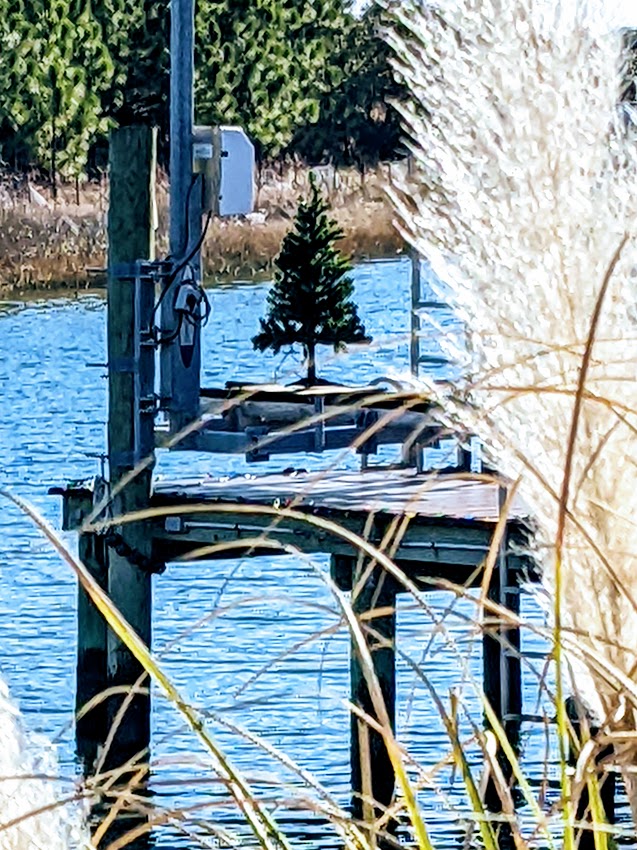
Here’s a photo of this study’s sponsoring institution’s harbor from the latest of two visits:

IMG_20170611_125837

I’d like to return whenever we individually stop being herded and recover our sanity.

Your thymus and calories

This 2022 paper studied caloric restriction in humans followed up by rodent experiments:

“Extension of lifespan driven by 40% caloric restriction (CR) in rodents causes trade-offs in growth, reproduction, and immune defense that make it difficult to identify therapeutically relevant CR-mimetic targets. We report that about 14% CR for 2 years in healthy humans improved thymopoiesis.

thymus cr

Expression of the gene PLA2G7 is inhibited in humans undergoing CR. Deletion of Pla2g7 in mice showed decreased thymic lipoatrophy, protection against age-related inflammation, lowered NLRP3 inflammasome activation, and improved metabolic health.

Twenty-four-month-old PLA2G7-deficient mice (analogous to ~70-year-old humans) had larger thymi and higher thymocyte abundance, and were protected from age-related thymic involution.

thymus mice

We propose that reduction of PLA2G7 caused by CR in humans might contribute to better adipose tissue metabolism, lower inflammation, and reduced thymic lipoatrophy.”

https://www.science.org/doi/10.1126/science.abg7292 “Caloric restriction in humans reveals immunometabolic regulators of health span” (not freely available). Thanks to Dr. Alexander Predeus for providing a copy.


I would have liked rodent experiments to continue another year or so to determine the control group’s and PLA2G7-deficient group’s healthspans and lifespans. These researchers could have strengthened their findings if increased healthspans also increased lifespans.

CD38 and balance

I’ll highlight this 2022 review’s relationships between inflammation and cluster of differentiation 38:

“We review the nicotinamide adenine dinucleotide (NAD) catabolizing enzyme CD38, which plays critical roles in pathogenesis of diseases related to infection, inflammation, fibrosis, metabolism, and aging.

NAD is a cofactor of paramount importance for an array of cellular processes related to mitochondrial function and metabolism, redox reactions, signaling, cell division, inflammation, and DNA repair. Dysregulation of NAD is associated with multiple diseases. Since CD38 is the main NADase in mammalian tissues, its contribution to pathological processes has been explored in multiple disease models.

CD38 is upregulated in a cell-dependent manner by several stimuli in the presence of pro-inflammatory or secreted senescence factors or in response to a bacterial infection, retinoic acid, or gonadal steroids. CD38 is stimulated in a cell-specific manner by lipopolysaccharide, tumor necrosis factor alpha, interleukin-6, and interferon-γ.

dysregulated inflammation

CD38 plays a critical role in inflammation, migration, and immunometabolism, but equally important is resolution of the inflammatory response which left unchecked leads to loss of self-tolerance, tissue infiltration of lymphocytes, and circulation of autoantibodies.

  • Depending upon context, CD38 can either promote or protect against an autoimmune response.
  • Chronic mucosal inflammation and tissue damage characteristic of inflammatory bowel disease predisposes IBD patients to development of colorectal cancer, and the risks increase with duration, extent, and severity of inflammation.
  • Pulmonary fibrosis occurs in the presence of unresolved inflammation and dysregulated tissue repair, and results from an array of injurious stimuli including infection, toxicant exposure, adverse effects of drugs, and autoimmune response.
  • Modulating CD38 and NAD levels in kidney disease may provide therapeutic approaches for prevention of inflammatory conditions of the kidney.
  • Inflammation as well as evidence of senescence are present in pathophysiology of chronic liver diseases that progress to cirrhosis.
  • Inflammation-associated metabolic diseases impair vascular function. Chronic inflammation can lead to vascular senescence and dysfunction.

One cause of NAD decline during aging is due to increase of NAD breakdown in the presence of increased CD38 expression and activity on immune cells, thus linking inflammaging with tissue NAD decline. Other sources of NAD decline include increased DNA-damage requiring PARP1 activation, and decreased NAMPT levels leading to diminished NAD synthesis through the salvage pathway.

Inflammation is among the major risk factors that predispose organisms to age-associated diseases. During aging, accumulation of senescent cells creates an environment rich in proinflammatory signals, leading to ‘inflammaging.’ Metabolically active cells lose their replicative capacity by entering an irreversible quiescent state, and are considered both a cause and a consequence of inflammaging.

Recent findings uncover a major role of CD38 in inflammation and senescence, showing that age-related NAD+ decline and the sterile inflammation of aging are partially mediated by a senescence / senescence associated secretory phenotype (SASP)-induced accumulation of CD38+ inflammatory cells in tissues. Given the clear association between the phenomenon of inflammaging, senescence, and CD38, as well as the impact of CD38 on degradation of NAD and the NAD precursor NMN, future studies should focus on CD38 as a druggable target in viral illnesses.”

https://journals.physiology.org/doi/abs/10.1152/ajpcell.00451.2021 “The CD38 glycohydrolase and the NAD sink: implications for pathological conditions”


We extend good-vs.-bad thinking to nature. Does that paradigm explain much, though?

All pieces of a puzzle are important. Otherwise, evolution would have eliminated what wasn’t necessary for its purposes.

Restoring balance to an earlier phenotype suits my purposes. Don’t want to eliminate inflammatory responses, but instead, calm them down so that they’re evoked appropriately.

Studying AGEs and neurodegeneration

This 2022 review suggested more effective ways to conduct in vitro studies of advanced glycation end products (AGEs) and neurodegenerative diseases:

“The main goal of this review was to present and discuss in vitro models that were applied or have the potential to be used in research on AGEs and ND.

  • We introduced and explained current knowledge on AGEs regarding their formation and accumulation in humans.
  • We presented existing evidence linking involvement of AGEs in ND and explained basic concepts of brain physiology and immunology affected by AGEs.
  • We presented and discussed available in vitro models to study AGE-mediated neurodegeneration by dividing them into sections from simple models. These have been applied to more complex models that have not been yet applied in the field of AGEs, but offer opportunities.
  • We gathered advisable in vitro tools based on their relevance to three primary endpoints that AGEs can impact brain pathophysiology and their characteristics and suitability to mimic ND pathophysiology.

nutrients-14-00363-g002

Several studies have indicated intracellular formation of AGEs by microglia or neurons, but identification of intracellular AGEs in those cases is made by immunoassays, which have received much criticism regarding their reliability to identify and quantify AGEs. Concerns about these techniques are mostly related to undefined specificity and affinity of anti-AGE antibodies.

The source of observed AGE accumulation in the brain of patients (dietary or endogenous) is not yet fully understood. For that reason, studies on AGE digestion and absorption (i.e., in vitro digestion models) are crucial to understanding the type of dietary AGEs that will circulate and cross the BBB to reach the brain.

On the other hand, endogenous AGEs can also be formed due to increased glucose levels derived from a high glycemic diet. Highly reactive molecules in the brain can contribute to locally produced AGEs extracellularly or intracellularly.

Clinical studies mainly focus on the fate and metabolism of dietary AGEs. Exposure based on consumption of certain foods is difficult to translate to a concentration that cells are going to be exposed to. The complexity and multiple sources of protein glycation require application of in vitro models to understand potential contribution to neurodegeneration.”

https://www.mdpi.com/2072-6643/14/2/363/htmIn Vitro Methodologies to Study the Role of Advanced Glycation End Products (AGEs) in Neurodegeneration”


While we’re waiting for research to catch up, we can hedge neurodegenerative disease bets by:

  • Not spiking our blood glucose levels;
  • Avoiding foods with medium and high levels of AGEs;
  • Giving our gut microbiota the intake they need instead of what our unconscious programming dictates; and
  • Maintaining youthful activities.

PXL_20220212_194256506

An epigenetic regulator of vascular aging

This 2022 rodent and human cell study investigated the smooth muscle cell mineralocorticoid receptor:

“Vascular stiffness increases with age and independently predicts cardiovascular disease risk. Epigenetic changes, including histone modifications, accumulate with age, but the global pattern has not been elucidated nor are the regulators known.

Rising mineralocorticoid receptor (MR) in aging vascular smooth muscle cells downregulates EZH2 to globally shift to a more open chromatin thereby allowing MR to be recruited to promoters to transcriptionally upregulate target genes involved in vascular stiffness. This mechanism provides multiple potential targets to prevent vascular stiffness in aging humans.

cvac007f8

We demonstrate for the first time that:

  1. MR expression increases with age in primary, low passage, human aortic smooth muscle cell (SMC) and correlates with age in whole aortic tissue from aging humans;
  2. The global proteomic profile of histone modifications in mouse vessels changes profoundly with aging with a significant overall decrease in H3K27 methylation;
  3. Expression of H3K27 methyltransferase EZH2 decreases with age in mouse vessels and in human SMCs in a MR-dependent manner and negatively correlates with MR expression in whole human aortic tissue;
  4. The aging-induced decline in EZH2 associates with reduced H3K27 methylation and increased H3K27 acetylation in vitro and in vivo;
  5. These epigenetic changes in aging human SMC and mouse vessels correspond with increased expression of the vascular stiffness genes, CTGF and integrin-α5, previously identified vascular MR target genes;
  6. Induction of an aging phenotype in human SMC associates with increased MR enrichment and H3K27 acetylation at these stiffness gene promoters; and
  7. Inhibition of MR in aged mice and aged human aortic SMCs reverses the entire process; increasing EZH2 and H3K27 methylation, increasing locus-specific EZH2 enrichment and decreasing H3K27 acetylation at stiffness gene promoters, decreasing vascular expression of CTGF and integrin-α5, and decreasing the stiffness and adhesiveness of aged human SMC in vitro and mouse aortic stiffness and fibrosis in vivo.”

https://academic.oup.com/cardiovascres/advance-article-abstract/doi/10.1093/cvr/cvac007/6502304 “Smooth muscle mineralocorticoid receptor as an epigenetic regulator of vascular ageing” (not freely available) Thanks to Dr. Seung Kyum Kim for providing a copy.

Intergenerational epigenetic inheritance of trained immunity, Part 2

A 2022 McGill University rodent study couldn’t replicate Part 1 findings:

“We find that using similar mouse models of trained immunity induced by:

  1. Live vaccination (BCG);
  2. PAMPs (β-glucan); or
  3. Infection (C. albicans),

protection against:

  1. Viral (influenza virus);
  2. Bacterial (Mycobacterium tuberculosis (Mtb)); or
  3. Fungal (C. albicans)

infections was the same between offspring of trained and non-trained parents.

BCG-vaccination-in-the-offspring-of-vaccinated-parents-does-not-enhance-trained-immunity_W640

BCG vaccination in the offspring of vaccinated parents does not enhance trained immunity in macrophages.

a) Mice were vaccinated with BCG-iv (1 × 10⁶ CFU) for one month and mated with vaccinated or naive counterparts. 6–8 week-old F1.1 and F1.3 offspring were then vaccinated or not with BCG-iv (1 × 10⁶ CFU).

b), c) At 1 month post BCG vaccination, protective capacities of BMDM from BCG-iv vaccinated and nonvaccinated F1.1 (b), or F1.3 (c) offspring from naïve or BCG-iv vaccinated parents were assessed against M. tuberculosis (H37Rv, MOI 1) infection. * p < 0.05.”

https://www.nature.com/articles/s41590-021-01102-0 “Lack of evidence for intergenerational inheritance of immune resistance to infections” (not freely available)


Part 1 coauthors replied:

“We are very encouraged that this topic is gaining increased interest. The reason for the discrepancy between findings in the two studies is unclear. It likely involves local differences in mouse substrains, housing, diet, microbiome, infection models, or other factors.

These findings underscore the effect of environment on intergenerational inheritance of infection resistance. What these environmental factors are and how these factors are integrated with regards to intergenerational inheritance remains largely elusive at this time.

One intriguing possibility that needs to be tested in future studies is whether such effects may be more robust in outbred wild mice, in which subtle environmental changes may have less strong impact.”

https://www.nature.com/articles/s41590-021-01103-z “Reply to: ‘Lack of evidence for intergenerational inheritance of immune resistance to infections'”


PXL_20211031_112819792

Gut microbiota’s positive epigenetic effects

Three papers with the first a 2021 review:

“Gut microbiota along with their metabolites are involved in health and disease through multiple epigenetic mechanisms including:

  • Affecting transporter activities, e.g. DNA methyltransferases (DNMTs), histone methyltransferases (HMTs), histone acetyltransferases (HATs), and histone deacetylases (HDACs);
  • Providing methyl donors to participate in DNA methylation and histone modifications; and
  • miRNAs that can lead to gene transcriptional modifications.

ijms-22-06933-g003

These mechanisms can participate in a variety of biological processes such as:

  • Maturation of intestinal epithelial cells (IECs);
  • Maintenance of intestinal homeostasis;
  • Inflammatory response;
  • Development of metabolic disorders; and
  • Prevention of colon cancer.”

https://www.mdpi.com/1422-0067/22/13/6933/htm “Dissecting the Interplay Mechanism between Epigenetics and Gut Microbiota: Health Maintenance and Disease Prevention”


A second 2022 review added subjects such as crotonate (aka unsaturated butyrate):

“Studies are carving out potential roles for additional histone modifications, such as crotonylation and ethylation, in facilitating crosstalk between microbiota and host. Lysine crotonylation is a relatively less studied histone modification that is often enriched at active promoters and enhancers in mammalian cells.

While addition or removal of crotonyl motifs can be catalyzed by specialized histone crotonyltransferases and decrotonylases, HATs and HDACs have also been reported to exhibit histone crotonyl-modifying activity. Microbiota stimulate multiple types of histone modifications and regulate activity of histone-modifying enzymes to calibrate local and extra-intestinal chromatin landscapes.”

https://www.tandfonline.com/doi/full/10.1080/19490976.2021.2022407 “Epigenetic regulation by gut microbiota”


A third 2021 review added subjects such as broccoli sprout compounds’ epigenetic effects:

“Glucosinolates are converted into isothiocyanates (ITCs) by bacteria that regulate host epigenetics. Levels of ITCs produced following broccoli consumption are highly dependent on the functional capacity of individual microbiomes, as much interindividual variability exists in gut microbiota composition and function in humans.

Sulforaphane inhibits HDAC activity both in vitro and in vivo, and protects against tumor development. Microbial-mediated production of ITCs represents a strong diet-microbe interaction that has a direct impact on host epigenome and health.”

https://www.sciencedirect.com/science/article/pii/S0955286321000516 “The interplay between diet, gut microbes, and host epigenetics in health and disease”


Clearing the channel

PXL_20220118_203446833

The aryl hydrocarbon signaling pathway

I’ll emphasize this densely packed 2021 review’s broccoli sprout compounds / gut microbiota / health interactions:

“The aryl hydrocarbon receptor (AhR) senses cues from environmental toxicants and physiologically relevant dietary/microbiota-derived ligands. AhR signaling mediates bidirectional host-microbiome interactions in a wide range of cellular functions in a ligand-, cell type-, species-, and context-specific manner.

Brassicaceae family plants are rich sources of glucobrassicin, the glucosinolate precursor of indole-3-carbinol (I3C). Glucobrassicin can be enzymatically hydrolyzed and converted into I3C by myrosinase, which is present in intact plant cells and gut microbiota.

I3C activates AhR but exhibits low binding affinity. However, in acidic conditions found in the stomach, I3C undergoes acid condensation reaction to generate a variety of more potent AhR ligands, such as 3,3′-diindolylmethane (DIM).

AhR activation by natural AhR ligands (e.g., I3C) has been shown to prevent pathogenic gut microbial dysbiosis by altering gut microbiome composition in mice with colitis. Depletion of AhR ligands in the diet decreased α diversity of gut microbiota, while I3C supplementation restored microbiota composition.

I3C treatment is effective for treating IBD patients, partly by upregulating IL-22. Targeting AhR could modulate the amplitude and duration of IL-22 signaling to treat IBD patients.

Administration of I3C or DIM significantly reduced the number of tumors in the cecum and small intestine. Supplementation of I3C reduces the number of colorectal tumors in WT, but not in AhR null mice.

nihms-1759454-f0003

Gut microbiota and diet are major sources of AhR ligands that influence the whole body, including gut, liver, brain, and the immune system. Many human diseases are associated with decreased circulating levels of AhR ligands, partly due to dysbiosis.

The ability of AhR signaling to regulate self-renewal and differentiation of intestinal stem cells intrinsically or extrinsically has recently been brought into the spotlight.”

https://www.ncbi.nlm.nih.gov/labs/pmc/articles/PMC8667662/ “Diet–Host–Microbiota Interactions Shape Aryl Hydrocarbon Receptor Ligand Production to Modulate Intestinal Homeostasis”


Young hawk

PXL_20220109_195611152

Nrf2 and circadian rhythm

This 2021 rodent study investigated aging’s effects:

“We investigated aging consequences on temporal patterns of antioxidant defenses, molecular clock machinery, and blood pressure.

We observed circadian rhythms of catalase (CAT) and glutathione peroxidase (GPx) mRNA expression, as well as ultradian rhythms of Nrf2 mRNA levels, in the hearts of young adult rats. We also found circadian oscillations of CAT and GPx enzymatic activities, reduced glutathione (GSH), and BMAL1 protein.

Aging abolished rhythms of CAT and GPx enzymatic activities, phase-shifted rhythm acrophases of GSH and BMAL1 protein levels, and turned circadian the ultradian oscillation of Nrf2 expression.

aging changes Nrf2 oscillation

Moreover, aging phase-shifted the circadian pattern of systolic blood pressure. In conclusion, aging modifies temporal organization of antioxidant defenses and blood pressure, probably as a consequence of disruption in the circadian rhythm of the clock’s transcriptional regulator, BMAL1, in heart.”

https://doi.org/10.1007/s10522-021-09938-7 “Aging disrupts the temporal organization of antioxidant defenses in the heart of male rats and phase shifts circadian rhythms of systolic blood pressure” (not freely available)


A human equivalent to this study’s 3-month-old young adult group is around 19 years. The older group’s 22-month age is roughly equivalent to a 68-year-old human.

Couldn’t say whether Nrf2 oscillations flattening out with age is specific to heart tissue, or is a more general trend. I’m pretty sure that humans have to make good things happen while aging, because bad things are pre-programmed.

I came across this study from a citation trail of a comment to Eat broccoli sprouts for your workouts. I didn’t curate the mentioned study because one of its coauthors tainted it by designing and supervising Problematic rodent sulforaphane studies.

How would you answer the comment’s question?


Repairs needed: The story of 2021

PXL_20220104_194204601

Sulforaphane vs. too much oxygen

This 2021 rodent study investigated perinatal effects of hyperoxia and sulforaphane:

“We demonstrated that early-life oxidant-induced acute lung injury had significant consequences later in life on NRF2-dependent respiratory syncytial virus (RSV) susceptibility in mice. We also determined that increased antioxidant conditions in utero potentially contribute to a decreased risk of postnatal airway disease as we found that prenatal antioxidant sulforaphane (SFN) protected developing lungs from bronchopulmonary dysplasia (BPD)-like oxidative pathogenesis in mice.

Unexpectedly, our results indicated that prenatal SFN-mediated postnatal protection against BPD-like phenotypes are not NRF2-dependent. Prenatal SFN markedly improved hyperoxia-caused severe BPD-like lung injury parameters in Nrf2−/− pups while we observed relatively marginal protection by in utero SFN in hyperoxia-resistant Nrf2+/+ pups.

SFN is a strong NRF2 and ARE gene inducer for cytoprotection by NRF2 stabilization. However, SFN also acts through other mechanisms, including NF-κB inhibition, MAPK activation, and histone deacetylase inhibition for anti-inflammation, chemoprevention, apoptosis, and autophagy.

Our study provided new insights into infant oxidant lung injury severity influence on persistence of pulmonary morbidity and therapeutic intervention for NRF2 agonists. Our results also provided justification for further studies on feto–placental barrier crossing of SFN metabolites and SFN-triggered molecular and epigenetic aspects of maternal cues for barrier and fetal lung signaling.”

https://www.mdpi.com/2076-3921/10/12/1874/htm “Murine Neonatal Oxidant Lung Injury: NRF2-Dependent Predisposition to Adulthood Respiratory Viral Infection and Protection by Maternal Antioxidant”


This study’s oral human-equivalent dose for treatment dams was 9 mg sulforaphane (1.67 mg x .081 x 70 kg) every other day during the last half of pregnancy. A small dose per How much sulforaphane is suitable for healthy people?

“The daily SFN dose found to achieve beneficial outcomes in most of the available clinical trials is around 20-40 mg.”

PXL_20211226_120528134

Eat broccoli sprouts for your workouts

This 2021 human study investigated effects of pre- and post-workout glucoraphanin intake:

“The tablets used in this study contained 30 mg/3 tablets of sulforaphane glucosinolate [properly termed glucoraphanin], a precursor of sulforaphane (SFN), which is converted to SFN in the intestinal lumen by intestinal microflora. Subjects took one tablet of SFN supplement per meal, three times a day. Healthy men without exercise habits, smoking, or medication were included in the experiment:

eccentric exercise subjects

Pain on palpation reached its peak 1–2 days after exercise and recovered to baseline 5 days after exercise. Muscle soreness on palpation and range of motion were significantly lower 2 days after exercise in the sulforaphane group:

range of motion

Serum malondialdehyde, an indicator of exercise-induced oxidative stress, showed significantly lower levels 2 days after exercise in the sulforaphane group. SFN intake may protect the balance of antioxidant capacity and suppress excessive oxidative stress caused by exercise.

Continuation of SFN intake – from 2 weeks before and up to 4 days after eccentric exercise – suppressed exercise-induced oxidative stress and inhibited muscle soreness and muscle damage. To our knowledge, this study is the first to analyze these effects of SFN in humans.”

https://physoc.onlinelibrary.wiley.com/doi/10.14814/phy2.15130 “Effect of a sulforaphane supplement on muscle soreness and damage induced by eccentric exercise in young adults: A pilot study”


This study found that four days wasn’t enough time for 19-to-23-year-old men to fully recover from bicep eccentric exercise, regardless of glucoraphanin treatment or control group status. What’s an appropriate exercise recovery time? found a similar result using taurine as treatment with 20-to-33-year-old recreationally fit men who didn’t fully recover from bicep eccentric exercise after three days.

These researchers referenced Autism biomarkers and sulforaphane to acknowledge that this study’s daily 30 mg of glucoraphanin wasn’t sufficient to fully activate Nrf2 signaling pathways:

When SFN was added to PBMCs of healthy subjects in ex vivo experiments, NQO1 expression was increased, while HO-1 was not increased at a low SFN concentration (0.5 µM). However, when 2 or 5 µM of SFN was added to PBMCs, both NQO1 and HO-1 gene expression were increased. Concentration of the SFN supplement may be a reason why the amount of supplementation used in our protocol did not increase HO-1 expression.”


I create isothiocyanates by microwaving 3-day-old broccoli / red cabbage / mustard sprouts at 1000 W to 60°C (140°F) shortly before eating them. Unlike this study, I don’t depend on metabolism after the stomach to produce isothiocyanates from glucosinolates:

  • Less dependence on these subjects’ gut microbiota for sulforaphane production would have reduced a source of dose variability. Broccoli sprout compounds and gut microbiota first paper reviewed that subject.
  • Glucoraphanin intake with nothing else an hour before and after would have also reduced chances of sulforaphane loss by reacting with food. See Week 19 item 2 for two studies that found eating protein, fats, and fiber along with broccoli sprouts lowered isothiocyanates’ bioavailability.

Still, this was a step forward in research. Have fun with New Year’s resolutions.

PXL_20211226_123100412

Defend yourself with taurine

This densely packed 2021 review subject was taurine:

“Taurine (Tau), a sulphur-containing non-proteinogenic β-amino acid, has a special place as an important natural modulator of antioxidant defence networks:

  • Direct antioxidant effect of Tau due to scavenging free radicals is limited, and could be expected only in a few tissues (heart and eye) with comparatively high concentrations.
  • Maintaining optimal Tau status of mitochondria controls free radical production.
  • Indirect antioxidant activities of Tau due to modulating transcription factors leading to upregulation of the antioxidant defence network are likely to be major molecular mechanisms of Tau’s antioxidant and anti-inflammatory activities.
  • A range of toxicological models clearly show protective antioxidant-related effects of Tau.”

antioxidants-10-01876-g001-550

https://www.mdpi.com/2076-3921/10/12/1876/htm “Taurine as a Natural Antioxidant: From Direct Antioxidant Effects to Protective Action in Various Toxicological Models”


PXL_20211226_120547077

Your lungs and Nrf2 activity

Two 2021 papers of Nrf2 activation effects on lung diseases, with the first a McGill University review:

“Oxidative stress and subsequent activation of Nrf2 have been demonstrated in many human respiratory diseases. The purpose of this review is to summarize involvement of Nrf2 and its inducers in acute respiratory distress syndrome, chronic obstructive pulmonary disease (COPD), asthma, and lung fibrosis in both human and experimental models.

fphys-12-727806-g004

These inducers have proven particularly effective at reducing severity of oxidative stress-driven lung injury in various animal models. In humans, these compounds offer promise as potential therapeutic strategies for management of respiratory pathologies associated with oxidative stress, but there is thus far little evidence of efficacy through human trials.

Perhaps, by analogy with biologics, patients with demonstrated deficient antioxidant responses to their disease should be selected for study in future clinical trials.”

https://www.frontiersin.org/articles/10.3389/fphys.2021.727806/full “Role of Nrf2 in Disease: Novel Molecular Mechanisms and Therapeutic Approaches – Pulmonary Disease/Asthma”


A second paper was a human/rodent study of COPD:

“We investigated Nrf2 expression and epigenetic regulation, and mechanisms by which the Nrf2 signaling pathway in ferroptosis is related to COPD. These findings elucidated pathways of ferroptosis in bronchial epithelial cells in COPD, and revealed Nrf2 as a potential target for COPD treatment.

COPD_A_340113_t0001

DNA hypermethylation at specific CpG sites of the Nrf2 promoter in primary epithelial cells and in clinical lung tissues is correlated with decreased Nrf2 expression, which is related to COPD occurrence and development.”

https://www.ncbi.nlm.nih.gov/pmc/articles/PMC8684379/ “Hypermethylation of the Nrf2 Promoter Induces Ferroptosis by Inhibiting the Nrf2-GPX4 Axis in COPD”


Similar to this second paper’s CpG findings, Eat broccoli sprouts for your heart found:

“Sulforaphane (SFN) reduced Ang II‐induced CpG hypermethylation and promoted Ac‐H3 [histone H3 acetylation] accumulation in the Nrf2 promoter region, accompanied by inhibition of global DNMT [DNA methyltransferase] and HDAC [histone deacetylase] activity, and a decreased protein expression of key DNMT and HDAC enzymes. Overall, DNA methylation and histone deacetylation are considered to inhibit gene transcription with a synergistic effect.

Nrf2 can also be regulated independently of Keap1. Evidence indicates that SFN may indirectly activate Nrf2 by affecting activity of several upstream kinases.”

However, this second paper didn’t measure DNMT and HDAC inhibition, although their therapeutic effects in reducing oxidative injury and inflammation may have been present.

PXL_20211225_193439146

Gut microbiota vs. disease risks

This 2021 review subject was risk relationships between diseases from the perspective of gut microbiota:

“There is a significant inverse relationship between the onset of Alzheimer’s disease/Parkinson’s disease (AD/PD) and cancer, but the mechanism is still unclear. Considering that intestinal flora can connect them, we briefly introduced the relationship among AD/PD, cancer, and intestinal flora, studied metabolites or components of the intestinal flora, and the role of intestinal barriers and intestinal hormones in AD/PD and cancer.

According to existing evidence:

  • Bifidobacterium and Lactobacillus positively affect AD/PD and cancer;
  • Ruminococcaceae, Prevotellaceae, and Prevotella significantly improve on AD/PD but harm cancer; and
  • Blautia has universal anticancer ability, but it may aggravate AD pathology.

1-s2.0-S0753332221011276-gr1_lrg

This may partially explain the antagonistic relationship between neurodegenerative diseases and cancer. When some individuals suffer from one disease, their intestinal flora change to obtain a stronger resistance to the other disease than healthy individuals, which is consistent with statistical data.”

https://www.sciencedirect.com/science/article/pii/S0753332221011276 “Composition of intestinal flora affects the risk relationship between Alzheimer’s disease/Parkinson’s disease and cancer”


PXL_20211224_180111266

Inevitable individual differences

This 2021 review subject was individual differences:

“We will focus on recent findings that try to shed light on the emergence of individuality, with a particular interest in Drosophila melanogaster.

fphys-12-719038-g001

Another possible source of potential behavioral variability might come from the interaction of individuals with environmental microbes, from Wolbachia infections to changes in the gut microbiome. In this particular case, no genetic variation or neural circuit alteration would be responsible for the change in behavior.

Finally, from an evolutionary point of view, individuality might play an essential role in providing an adaptive advantage. For example, we have described that animals might use diversified bet-hedging as a mechanism to produce high levels of variation within a population to ensure that at least some individuals will be well-adapted when facing unpredictable environments.”

https://www.frontiersin.org/articles/10.3389/fphys.2021.719038/full “Behavior Individuality: A Focus on Drosophila melanogaster


Other papers on this subject include:

PXL_20211218_192020643