Eat broccoli sprouts for your heart, Part 2

A 2025 rodent study investigated synergistic effects of sulforaphane (SFN) and nicotinamide mononucleotide (NMN) on diabetic cardiomyopathy:

“Diabetic cardiomyopathy (DCM) as a significant diabetes complication remains a major human challenge. In this study, we provide evidence that the fat mass and obesity-associated protein (FTO) plays a pivotal role in DCM pathogenesis.

Downregulation of FTO in DCM acts as a critical inducer of ferroptosis by increasing expression of acyl-CoA synthetase long-chain family 4 (ACSL4), a key positive mediator of ferroptosis. FTO-mediated mitigation of ferroptosis occurs in an ACSL4-dependent manner which leads to increased methylation of Acsl4 transcripts.

  1. Ferroptosis plays an essential role in the pathogenesis of DCM.
  2. As the most widespread mRNA modification, N6-methyladenosine (m6A) is globally downregulated and implicated in diabetes and its complications.
  3. FTO, which is an m6A demethylase, was found to be downregulated in diabetes and its cardiovascular complications.
  4. NAD+ enhances the demethylase activity of FTO. Dietary supplementation with NMN, a critical intermediate in the NAD+ biosynthetic pathway, has been shown to efficiently elevate endogenous NAD+ levels.

Enhancing the demethylase activity of FTO with NMN combined with SFN targeting NRF2 could synergistically reduce the level of lipid peroxides to inhibit ferroptosis, providing an effective avenue for alleviating DCM.

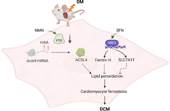
We found that NMN could alleviate ferroptosis and improve heart function through enhancing FTO. SFN could prevent ferroptosis and partly rescue heart function via AMPK-mediated NRF2 activation.

We demonstrated that SFN combined with NMN treatment could significantly inhibit lipid peroxidation and rescue cardiac function in DCM compared to SFN or NMN treatment alone.

Although the combined regimen further suppressed ferroptosis and improved cardiac performance, it fell short of complete remission, underscoring that additional pathways also contribute substantially to the pathogenesis of DCM.”

https://link.springer.com/article/10.1007/s12012-025-10080-w “FTO-Mediated Mitigation of Ferroptosis Occurs in an ACSL4-Dependent Manner in Diabetic Cardiomyopathy”


The epigenetic mechanism involved with this study’s dietary dissolved-in-water 100mM NMN dose was Non-CpG methylation. This study used the same very low sulforaphane dose intraperitoneally injected as Eat broccoli sprouts for your heart. Discussion of that study provided an example that if a person waited until a diabetes-related disease condition became a problem, capabilities to adequately address causes and prevent the problem may be lost.

Notice in the last bar of the second graphic above taken from Figure 7 that the combined treatment was also provided to non-diabetic mice. These researchers provided over a dozen other measurements in Figure 7 to show similar short-term non-effects of the combined treatment, i.e. that it neither benefited nor harmed non-diabetic subjects. Grok interpreted this study’s 3-month-long intervention to be a 1-to-5 year human equivalent, depending on the measured effect (shorter for metabolic effects like MDA, longer for structural cardiac changes like reduced ferroptosis.)

The male subjects began at 2-months old, a human-equivalent 15-20 years old. These researchers gave them diabetes by feeding them a “high-fat diet for 3 months to induce insulin resistance, followed by a single intraperitoneal injection of streptozotocin (STZ) (in 0.1 mol/L of citrate acid buffer, 60 mg/kg) to induce partial insulin deficiency.” A 5-months old mouse is a 25-30 years old human equivalent.

Grok considered this study’s NMN human equivalent dose to be extremely high if provided in drinking water, but not if injected, depending on volume. However, the study didn’t state that its NMN dose was injected, and there was no dose volume indicated.

Human studies of astaxanthin – Part 1

Here are three 2025 clinical trials of the Nrf2 activator astaxanthin’s effects. Let’s start with a clinical trial of inflammation-related diabetic complications and insulin resistance:

“We investigated effects of 10 mg/day astaxanthin (ASX) supplementation for 12 weeks on microRNAs (miRNAs), lysophosphatidylcholine (LPC), and α-hydroxybutyrate (α-HB) as novel factors in development of a variety of diabetes-related complications.

  • LPC is believed to play a significant role in atherosclerosis and inflammatory diseases by modifying functions of multiple cell types, including smooth muscle cells, endothelial cells, monocytes, macrophages, and T cells. LPC can interfere with glucose-stimulated insulin secretion by impairing calcium homeostasis and other signaling pathways that are crucial for the proper functioning of beta cells. This impairment exacerbates hyperglycemia in diabetic patients. LPCs may impede insulin signaling pathways, thereby contributing to insulin resistance (IR).
  • α-HB is also an indicator of IR and impaired glucose regulation, both of which appear to result from excessive lipid oxidation and oxidative stress. The European population cohorts in 2016 identified α-HB as a selective biomarker for decreased glucose tolerance and prediabetes, which was independent of age, sex, BMI, and fasting glucose.
  • A number of studies have established a link between miR-21, miR-34a, and miR-155 and diabetic complications such as retinopathy and nephropathy.

In the ASX group, participants were divided into 2 subgroups according to the urinary albumin-to-creatinine ratio (ACR) (< 30 mg/g or ≥ 30 mg/g, an indicator of diabetic kidney disease).

  • The level of fasting plasma glucose before and after 12 weeks of treatment with ASX was 139.27 ± 21.18 vs. 126.43 ± 18.97 (p = 0.002), demonstrating a significant reduction compared to the placebo group.
  • In the ASX group, the mean HbA1c level at baseline was 7.89 ± 0.79 and declined to 7.05 ± 0.35 after the supplementation period, which was statistically significant.
  • Supplementation with ASX resulted in a statistically significant drop in HOMA-IR levels, whereas this parameter was not altered significantly in the placebo group.
  • The ASX group, in comparison with the placebo group, demonstrated marked changes in lipid profile factors such as TC, TG, and LDL (p = 0.011, p = 0.043, and p = 0.022, respectively).

Clinical studies indicate that rigorous diabetes management does not substantially diminish appearance of complications. Modifications in oxidative stress and IR markers, as well as miRNA expression, must be analyzed to identify biological markers with sufficient predictive power for development of complications in diabetic patients.

Supplementation with ASX substantially diminished the levels of α-HB, LPC, and inflammation-related miRNAs in diabetic patients with and without complications.”

https://onlinelibrary.wiley.com/doi/10.1155/ije/5878361 “Astaxanthin Modulates Inflammation in Type 2 Diabetes via Regulation of microRNAs, Lysophosphatidylcholine, and α-Hydroxybutyrate”


Another clinical trial investigated astaxanthin’s effects in heart failure patients:

“Chronic heart failure (HF) is often linked to increased oxidative stress and metabolic issues like high uric acid, which can worsen outcomes.This study aimed to investigate the effects of ASX supplementation on oxidative stress markers as the primary outcome and clinical symptoms in patients with HF.

80 patients with HF were enrolled and randomly assigned to receive either ASX (20 mg/day) or a placebo (20 mg/day of maltodextrin) for 8 weeks. Biomarkers including total antioxidant capacity (TAC), malondialdehyde (MDA), superoxide dismutase (SOD), serum uric acid (UA), and clinical symptoms (dyspnea, fatigue, appetite) were assessed pre-and post-intervention.

Daily supplementation with 20 mg of ASX for eight weeks in patients with HF resulted in significantly greater improvements in oxidative stress biomarkers compared to placebo group. This improvement included reductions in uric acid and MDA, along increases in TAC and SOD.

In our study, participants received the cis-isomer form of ASX. The cis-isomer of ASX demonstrates greater anti-inflammatory and antioxidant properties than the trans-isomer, along with enhanced bioavailability. Inconsistencies among studies may be attributed to differences in participants’ baseline antioxidant status, underlying medical conditions, dosage, isomeric form and formulation of ASX used, and the duration of intervention.

One of the strengths of this study is that it represents the first randomized clinical trial to evaluate the effects of ASX supplementation on oxidative stress markers, UA levels, and clinical symptoms in patients with HF. Additionally, potential confounding factors were controlled as much as possible. However, several limitations were identified, including the relatively short intervention duration, limited sample size, limited generalizability of the findings due to the single-center design, absence of blood ASX level measurements, and lack of long-term follow-up.”

https://link.springer.com/article/10.1186/s12872-025-05260-zImpact of astaxanthin on oxidative markers, uric acid, and clinical symptoms in heart failure: a randomized clinical trial”


A third clinical trial evaluated astaxanthin’s effects as an adjunct to standard treatment of community-acquired pneumonia:

“Adult patients diagnosed with community-acquired pneumonia (CAP) were enrolled and assigned to receive either 12 mg/day ASX or a placebo in addition to standard antibiotic therapy for 7 days. Inflammatory markers, including interleukin-6 (IL-6), tumor necrosis factor-alpha (TNF-α), and interleukin-10 (IL-10), were measured at baseline and post-treatment. Secondary outcomes included Sequential Organ Failure Assessment (SOFA) and Acute Physiology and Chronic Health Evaluation II (APACHE II) scores.

A total of 80 patients (40 per group) completed the study. Patients receiving ASX exhibited significant reductions in pro-inflammatory cytokines compared to the placebo group. IL-6 and TNF-α levels were significantly lower in the ASX group at the end of the study (P < 0.05). Additionally, SOFA and APACHE II scores showed greater improvements in ASX-treated patients, suggesting a potential role in mitigating disease severity.

These findings suggest that ASX may help preserve organ function, limit the progression of inflammatory injury, and reduce overall disease severity in hospitalized patients with CAP.

ASX is widely regarded as the most potent carotenoid, owing to its unique molecular structure. Its polar-nonpolar-polar configuration enables it to span lipid bilayers and neutralize ROS both within and outside cellular membranes—an advantage not shared by other carotenoids that tend to localize at the membrane surface.

Despite the positive findings of this study, some limitations should nevertheless be considered.

  • The relatively small sample size may have limited the statistical power to detect differences in some outcomes and affects the generalizability of the findings.
  • Microbiological data on CAP pathogens were not collected. As different microorganisms can trigger distinct inflammatory responses, this limits our ability to assess pathogen-specific variations in ASX efficacy.
  • A notable limitation of this study is the short follow-up duration, with outcomes assessed only over a 7-day period. While this timeframe offers insight into the acute effects of ASX on inflammatory and OS markers, it does not clarify whether these benefits are sustained beyond the immediate treatment window.
  • The fixed dose of 12 mg once daily may not have maintained optimal therapeutic levels throughout the day. Dose-ranging studies and evaluations of alternative regimens are needed to determine the most effective strategy.”

https://www.frontiersin.org/journals/pharmacology/articles/10.3389/fphar.2025.1621308/full “The anti-inflammatory and antioxidant effects of astaxanthin as an adjunctive therapy in community-acquired pneumonia: a randomized controlled trial”


Part 2 continues with four more 2025 human studies of astaxanthin.


Ancient DNA fragments enable adult neurogenesis

A 2025 rodent study investigated mechanisms by which erythropoietin (EPO) enables adult neurogenesis and cognitive function:

“We mapped epigenomic and transcriptional landscapes of adult mouse hippocampus under recombinant human EPO (rhEPO) treatment. We discovered significant lineage-specific remodelling of chromatin accessibility predominantly in newly formed pyramidal neurons, highlighting a robust EPO-driven neurogenic response. Notably, transposable elements (TEs), particularly ancient LINEs and SINEs, emerged as critical cis-regulatory elements (cCREs).

EPO is known to be upregulated in the brain under hypoxic or injury conditions, and it has been considered a natural neuroprotective agent. We demonstrated that EPO, a traditionally hematopoietic hormone, can profoundly reprogram the adult neural epigenome to drive neurogenesis.

EPO may activate a specific subclass of dormant regulatory elements to drive nearby genes. Such a mechanism would represent a previously unappreciated mode of gene regulation: the de novo recruitment of ancient genomic elements to drive a contemporary cellular response.

Our data support the model that EPO drives differentiation of progenitors rather than inducing widespread cell division. The net effect is an enrichment of pyramidal neurons at the cost of interneurons. Pyramidal neurons integrate in the hippocampal circuitry, leading to potential implications for mood, memory, cognitive enhancement, and recovery from brain injury.

We propose a conserved evolutionary mechanism at play: ancient TEs embedded in the genome have been repurposed as cCREs in neural cells, and during an EPO-induced neurogenic stimulus, the brain taps into this reservoir of regulatory elements to rapidly reshape gene expression. In evolutionary terms, this represents an efficient strategy.”

https://www.biorxiv.org/content/10.1101/2025.10.13.682070v1.full “Transposable Element-Mediated Epigenomic Remodeling Drives Erythropoietin-Induced Neurogenesis in the Adult Hippocampus”


Plasmapheresis doesn’t reduce biological age

A 2025 clinical trial investigated effects of plasmapheresis as measured with epigenetic clocks:

“This study aimed to assess whether plasmapheresis without volume replacement with young plasma or albumin affects epigenetic age and other biomarkers in healthy adults. No significant epigenetic rejuvenation was observed based on epigenetic clock measurements. Instead, plasmapheresis was associated with increases in DNAmGrimAge, the Hannum clock, and the Dunedin Pace of Aging.

  1. The relatively small sample size of 34 finishing participants comprising of first-time plasma donors limits the statistical power and generalizability of our findings.
  2. Our cohort was restricted to individuals aged 40 to 60 years in accordance with Czech regulatory guidelines, which, although intentional to focus on an older population where rejuvenating effects might be most apparent, constrains evaluation of age-related differences across a broader demographic.
  3. The 18-week duration of the study, while sufficient to detect rapid alterations in key biomarkers under an intensive plasmapheresis protocol, may not fully capture the long-term implications of these changes.
  4. Due to our trial taking place during spring and summer months, we cannot fully separate the effects of increased sunlight exposure, outdoor physical activity, and dietary changes from the observed rises in Vitamin D and concurrent shifts in DNAm-based aging metrics. We did not collect objective measures of activity or diet, so these factors remain potential confounders.

The protocol of donating plasma every two weeks, although deemed safe by many countries around the world, is not yet well researched and cannot therefore be marked as benefiting to the donor right now. Further refinement to balance clearance of pro-aging factors with maintenance of systemic homeostasis is needed.”

https://www.nature.com/articles/s41598-025-05396-0 “Human clinical trial of plasmapheresis effects on biomarkers of aging (efficacy and safety trial)”


Betaine as an exercise mimetic

A 2025 human study investigated effects of long-term exercise:

“Exercise has well-established health benefits, yet its molecular underpinnings remain incompletely understood. We conducted an integrated multi-omics analysis to compare effects of acute vs. long-term exercise in healthy males.

Acute exercise induced transient responses, whereas repeated exercise triggered adaptive changes, notably reducing cellular senescence and inflammation and enhancing betaine metabolism. Exercise-driven betaine enrichment, partly mediated by renal biosynthesis, exerts geroprotective effects and rescues age-related health decline in mice.

Betaine binds to and inhibits TANK-binding kinase 1 (TBK1), retarding the kinetics of aging.

Betaine effectively alleviated senescence phenotypes by reduced senescence-associated β-galactosidase (SA-β-Gal)-positive cells, decreased p21 expression, lowered DNA damage indicator γ-H2A.X, and elevated heterochromatin mark H3K9me3. Betaine treatment also enhanced cellular antioxidant capacity, as evidenced by increased NRF2 phosphorylation and reduced ROS accumulation.

These findings systematically elucidate the molecular benefits of exercise, and position betaine as an exercise mimetic for healthy aging.”

https://doi.org/10.1016/j.cell.2025.06.001 “Systematic profiling reveals betaine as an exercise mimetic for geroprotection” (not freely available) Thanks to Dr. Weimin Ci for providing a copy.


Nrf2 activators and transcriptomic clocks

Two preprint studies looked at making transcriptional aging clocks using Nrf2 activators. Let’s start with a 2025 nematode study that used constant exposure to sulforaphane at different concentrations:

“To explore the potential of sulforaphane as a candidate natural compound for promoting longevity more generally, we tested the dose and age-specific effects of sulforaphane on C. elegans longevity, finding that it can extend lifespan by more than 50% at the most efficacious doses, but that treatment must be initiated early in life to be effective. We then created a novel, gene-specific, transcriptional aging clock, which demonstrated that sulforaphane-treated individuals exhibited a “transcriptional age” that was approximately four days younger than age-matched controls, representing a nearly 20% reduction in biological age.

The clearest transcriptional responses were detoxification pathways, which, together with the shape of the dose-response curve, indicates a likely hormetic response to sulforaphane. The hormetic, stress-pathway inducing properties of sulforaphane may indicate that many beneficial dietary supplements work in a fairly generic fashion as mild toxins rather than being driven by the biochemical properties of the compounds themselves (e.g., as antioxidants).

These results support the idea that robust longevity-extending interventions can act via global effects across the organism, as revealed by systems level changes in gene expression.”

https://www.biorxiv.org/content/10.1101/2025.05.11.653363v1 “The broccoli derivative sulforaphane extends lifespan by slowing the transcriptional aging clock”

There are difficulties in researchers translating nematode studies to mammals and humans. Nematodes lack a homolog to the Keap1 protein, which is sulforaphane’s main mammalian target to activate Nrf2.


A 2024 study developed various mammalian epigenetic clocks:

“A unified transcriptomic model of mortality that encompasses both aging and various models of lifespan-shortening and longevity interventions (i.e., mortality clocks) has been lacking. We conducted an RNA-seq analysis of mice subjected to 20 compound treatments in the Interventions Testing Program (ITP).

We sequenced the transcriptomes of a large cohort of ITP mice subjected to various neutral and longevity interventions, expanded the dataset with publicly available gene expression data representing organs of mice and rats across various strains and lifespan-regulating interventions, connected these models with survival data, and performed a meta-analysis of aggregated 4,539 rodent samples, which allowed us to identify multi-tissue transcriptomic signatures of aging, mortality rate, and maximum lifespan.

Aging and mortality were characterized by upregulation of genes involved in inflammation, complement cascade, apoptosis, and p53 pathway, while oxidative phosphorylation, fatty acid metabolism, and mitochondrial translation were negatively associated with mortality, both before and after adjustment for age.

Utilizing the aggregated dataset, we developed rodent multi-tissue transcriptomic clocks of chronological age, lifespan-adjusted age, and mortality. While the chronological clock could distinguish the effect of detrimental genetic and dietary models, it did not show a decrease in biological age in response to longevity interventions. In contrast, clocks of lifespan-adjusted age and mortality both captured aging-associated dynamics and correctly predicted the effect of lifespan-shortening and extending interventions.

Transcriptomic biomarkers developed in this study provide an opportunity to identify interventions promoting or counteracting molecular mechanisms of mortality, and characterize specific targets associated with their effects at the level of cell types, intracellular functional components, and individual genes. Our study underscores the complexity of aging and mortality mechanisms, the interplay between various processes involved, and the clear potential for developing therapies to extend healthspan and lifespan.”

https://www.biorxiv.org/content/10.1101/2024.07.04.601982v1.full “Transcriptomic Hallmarks of Mortality Reveal Universal and Specific Mechanisms of Aging, Chronic Disease, and Rejuvenation”


This second study’s references included an ITP study curated in Astaxanthin and aging, which stated:

“Despite the fact that the average diet contained 1840 ppm astaxanthin (only 46% of the target), median lifespans of male UM-HET3 mice were significantly improved. Amounts of dimethyl fumarate (DMF) in the diet averaged 35% of the target dose, which may explain the absence of lifespan effects.”

So screw-ups in making both astaxanthin and DMF mouse chows ended up with study data that didn’t measure the full lifespan impacts of activating transcription factor Nrf2. I’ll assert that such faulty data may have deviated this second study by downplaying Nrf2 activation’s impact on aging, chronic disease, and rejuvenation.

Sponsors may be less likely to be presented sulforaphane and other Nrf2 activator candidates for future aging and chronic disease studies as this first study suggests, thinking that these have already been studied in mammals. Well, maybe these compounds haven’t been accurately studied. There’s no effective way to fix a rodent study’s missing DMF Nrf2 data and faulty astaxanthin Nrf2 data to train an epigenetic clock in this second study.

I could be wrong about this second study using faulty astaxanthin Nrf2 data. It was cited as Reference 27 in the Introduction as an ITP study, but not specifically cited in the Method section. I don’t know how findings such as one of Nrf2’s target genes (“Remarkably, one of the top genes positively associated with maximum lifespan and negatively associated with chronological age and expected mortality was Gpx1, encoding the selenoprotein glutathione peroxidase 1″) and a Nrf2 specific pathway (Phase II) (“Pathways positively associated with lifespan and negatively with mortality, both before and after adjustment for age, included..xenobiotic metabolism..”) were made without Reference 27. Neither of the above studies has been peer reviewed yet.


The third phase of reversing aging and immunosenescent trends

Here’s a 2025 interview with Dr. Greg Fahy:

“We found that we could statistically demonstrate thymic regeneration morphologically on single individuals at single time points. MRI changes really are detecting shifts from the fatty tissue infiltration state of the involuted thymus to the regenerated thymus with functional thymic epithelial cells.

When you go through puberty your thymus involutes so you don’t have much left even when you’re 40. Essentially the process consists of loss of functional thymic mass and replacement of that functional thymic mass with adipose tissue, that’s what thymic involution is. It continues throughout life, but you retain a small amount of functional thymic mass all the way out to the age of 107.

The function of the thymus is to essentially manufacture half of your immune system. You have precursor cells arise from the bone marrow. They either go into the meiotic lineage and turn into the innate immune system, or you have the lymphocytic cells for what turns into T cells that enter the thymus and are educated in the thymus to grow up into newborn T cells and they’re released into the bloodstream.

The thymus has two jobs. It manufactures these lovely T cells without which you die but it also has a secondary finishing school. In the thymus cortex you manufacture all these lovely T cells but in the thymus medulla the T cells go to the medulla and if they don’t pass the second examination that they have to pass before they release into the body they’re all killed off. That second examination is: Do you reject self? As we get older, the thymus weakens in both the functions of making the T cells and screening out the ones that attack self. It stands to reason as we get older and the thymus’ influence wanes, we’re going to get more autoimmune disorders.

It took people a while to catch on to the fact that this involution problem is really a significant issue because the T cells that you made when you were 12, and even 20 and 40, they’re probably lasting until you’re 60. But at some point they don’t get replaced as fast as they’re going out of existence, and then your immune system goes off the cliff. Between the ages of 62 and 78 you lose 98% of your ability to recognize foreign antigens, and you still have a lot of capacity left.

We had nine guys in the first trial. Second trial we had 18 men 6 women and 2 controls that happen to be contemporaneous with that group. We have some more controls now that are either finished or or nearing completion. The second population was older than the first population by about nine years, but based on the epigenetic clocks that we looked at, they were starting off biologically younger.

On this last data analysis for Triim XA we looked at 21 different aging clocks. One aspect of the noise that we’re talking about is that biological aging as measured by some of these clocks is circadian. If you measure your age at 4:00 a.m. versus 11:00 a.m. you’re going to get a different result. It’s dynamic and there’s a trend and over time you change in a certain direction, but over any short period of time you can bounce around a little bit. The clocks predict your probability of cognitive dysfunction, they predict your probability of having impairments in your daily life, and they also predict your mortality.

We’re pretty much wrapping up that second clinical trial and going into the third. As we look at more data we understand more and more things and we see more and more things that we previously were not aware of. We began to look at a phenomena that may be responsible for limiting the magnitude of responses that we’re seeing limiting the aging reversal.

Triim-XD which is the next flavor of Triim-X is going to be looking at shifting biochemical pathways in such a way that it optimizes effects of these three medications that we’re giving people [human growth hormone, DHEA, and metformin] and prevents contradictions between them and prevents side effects of each one of these things. That’s about all I can tell you right now.”


Charts regarding the discussed item of how long effects may last are covered in The next phase of reversing aging and immunosenescent trends which was the last time I curated this research effort.


2025 α-ketoglutarate research

I haven’t mentioned α-ketoglutarate for a while, although I’ve taken it twice a day for several years. Here are four 2025 papers on α-ketoglutarate, starting with a review of its role in bone health:

“α-Ketoglutarate (α-KG) serves as a pivotal intermediate in various metabolic pathways in mammals, significantly contributing to cellular energy metabolism, amino acid metabolism, and other physiological processes. α-KG may be a therapeutic target for a variety of bone-related diseases, such as osteoporosis, osteoarthritis, and rheumatoid arthritis, because of its role in maintaining metabolic balance of bone.

α-KG, as a rate-determining mitochondrial intermediate, is crucial in cell energy metabolism because it connects intracellular carbon and nitrogen metabolism between isocitrate and succinyl coenzyme A. Additionally, α-KG is closely involved in the amino acid cycle. As a precursor of amino acids such as glutamine and glutamic acid, α-KG plays a direct role in energy production and a wide range of cellular chemical reactions. α-KG provides an energy source, stimulating protein synthesis, inhibiting protein degradation in muscle, and serving as a significant metabolic fuel for gastrointestinal cells.

α-KG promotes osteogenic differentiation of stem cells, increases activity of osteoblasts to promote osteogenesis, and inhibits bone resorption activity of osteoclasts. α-KG in articular cartilage promotes differentiation and maturation of chondrocytes and formation of a cartilage matrix. The protective effect of α-KG on bone has practical value in treatment of abnormal bone loss symptoms in various bone tissue diseases.”

https://www.sciengine.com/ABBS/doi/10.3724/abbs.2025020 “Essential role of the metabolite α-ketoglutarate in bone tissue and bone-related diseases”


A rodent study explored adding α-KG to osteoarthritis treatment:

“Mesenchymal stem cell (MSC) therapy represents a promising treatment strategy for osteoarthritis (OA). Nevertheless, the therapeutic efficacy of MSCs may be attenuated under conditions of cellular senescence or when the available clinical quantity is insufficient. α-Ketoglutarate (AKG) exerts beneficial effects on skeletal tissues and activity of stem cells. The present study was designed to explore the potential of AKG in augmenting viability of MSCs and the potential of their combined utilization in treatment of OA.

AKG plays a crucial role in multiple biological processes. It is involved in regulating stem cell differentiation, exerts anti-apoptotic effects, modulates the body’s immune and inflammatory responses, contributes to muscle and bone development, and is essential for maintaining stability of the cartilage matrix.

Platelet-rich plasma (PRP) has been demonstrated to have protective effects on chondrocytes and can effectively repair damaged cartilage in OA. However, PRP has intractable problems in terms of product quality control and allogeneic application, and its long-term therapeutic effect gradually weakens.

Combining AKG’s regulation of cellular metabolism with the multi-directional differentiation and immunomodulatory functions of MSCs is likely to generate a synergistic effect. This combined treatment modality targets the complex pathological processes of OA, including cartilage damage, inflammatory responses, and extracellular matrix imbalance, in a more comprehensive manner than a single therapy.”

https://www.sciencedirect.com/science/article/pii/S2707368825000032 “The repair effect of α-ketoglutarate combined with mesenchymal stem cells on osteoarthritis via the hedgehog protein pathway”


A rodent study investigated whether α-KG has a role in determining frailty:

“Frailty is an age-related geriatric syndrome, for which the mechanisms remain largely unknown. We performed a longitudinal study of aging female (n = 40) and male (n = 47) C57BL/6NIA mice, measured frailty index, and derived metabolomics data from plasma samples.

We find that frailty related metabolites are enriched for amino acid metabolism and metabolism of cofactors and vitamins, include ergothioneine, tryptophan, and alpha-ketoglutarate, and present sex dimorphism. We identify B vitamin metabolism related flavin adenine dinucleotide and pyridoxate as female-specific frailty biomarkers, and lipid metabolism related sphingomyelins, glycerophosphoethanolamine and glycerophosphocholine as male-specific frailty biomarkers.

We were interested to observe whether metabolite abundance at any specific timepoint was associated with frailty at a future timepoint. Unfortunately, we didn’t observe any metabolites that showed an overall significant association with future FI (FIf) or future devFI (devFIf). When focusing only on the abundance of metabolites at the baseline time point (∼400 days), we found a single metabolite, alpha-ketoglutarate, was negatively associated with both FIf and devFIf.”

https://www.biorxiv.org/content/10.1101/2025.01.22.634160v1.full “Metabolomics biomarkers of frailty: a longitudinal study of aging female and male mice”


Wrapping up with a rodent study adding α-KG to exercise for its effects on depression and learning:

“aKG acts as a prophylactic and antidepressant to effectively counteract social avoidance behaviors by modulating BDNF levels in the hippocampus and nucleus accumbens. Exercise increases aKG levels in the circulation.

In mice, aKG supplementation prolongs lifespan and reduces aging-associated frailty. aKG supplementation also reverses aging in humans as measured by DNA methylation patterns.

aKG functions as a co-factor for epigenetic enzymes. Changes in the intracellular αKG/succinate ratio regulates chromatin modifications, including H3K27me3 and ten-eleven translocation (Tet)-dependent DNA demethylation. The ability of aKG to influence epigenetic status of cells may explain both its prophylactic and anti-depressant effects since transcriptional dysregulation and aberrant epigenetic regulation are unifying themes in psychiatric disorders. This may also explain its ability to differentially regulate BDNF expression in the hippocampus and NAc.

If exercise mediates its effects through aKG, aKG may be a pivotal component of an exercise pill along with lactate and BHB that can serve as both a prophylactic and antidepressant treatment for depression.”

https://www.sciencedirect.com/science/article/pii/S266717432500031X “α-ketoglutarate (aKG) is a circulatory exercise factor that promotes learning and memory recall and has antidepressant properties


Epigenetic clock analysis of a clinical trial

A 2025 paper performed post-hoc epigenetic clock analyses of a supplement and exercise clinical trial completed earlier this decade:

“We report results of a post hoc analysis among 777 participants of the DO-HEALTH trial on the effect of vitamin D (2,000 IU per day) and/or omega-3 (1 g (330 mg EPA plus 660 mg DHA from marine algae) per day) and/or a home exercise program (a strength-training exercise program performed for 30 min three times per week) on four next-generation DNA methylation (DNAm) measures of biological aging (PhenoAge, GrimAge, GrimAge2 and DunedinPACE) over 3 years. Omega-3 alone slowed the DNAm clocks PhenoAge, GrimAge2 and DunedinPACE, and all three treatments had additive benefits on PhenoAge.

Inclusion criteria were age 70 years and older, living at home, having no major health events (no cancer or myocardial infarction) in the 5 years before enrollment, having sufficient mobility to visit the study centers without help and having good cognitive function with a Mini-Mental State Examination score of at least 24. 777 provided consent for these analyses and had samples available after the application of the exclusion criteria. This group of individuals formed our analysis sample, which had the following characteristics: 59% were women; the mean age at baseline was 75 years; 30% had 25-hydroxyvitamin D (25(OH)D) levels of <20 ng ml−1; 53% were healthy agers as defined in the Nurses’ Health Study (free of major chronic diseases, disabilities, cognitive impairments and mental health limitations); and 88% were physically active (29% were active one to three times per week, and 59% were active more than three times per week). The Swiss participant subgroup represents a healthier and more active subgroup within the total DO-HEALTH population.

Overall, from baseline to year 3, standardized effects ranged from 0.16 to 0.32 units (2.9–3.8 months). In summary, our trial indicates a small protective effect of omega-3 treatment on slowing biological aging over 3 years across several clocks, with an additive protective effect of omega-3, vitamin D, and exercise based on PhenoAge.”

https://www.nature.com/articles/s43587-024-00793-y “Individual and additive effects of vitamin D, omega-3 and exercise on DNA methylation clocks of biological aging in older adults from the DO-HEALTH trial”

These epigenetic clock measurements of a subset of trial subjects was interesting, although I didn’t find it particularly relevant to what I do. I take twice as much Vitamin D and omega-3s everyday, do resistance exercises once or twice a week whenever I’ve recovered from the previous session, walk a few miles on the beach if the weather is nice, and other things.

I don’t bother with epigenetic clock measurements anymore because the free one (PhenoAge) is too variable to be personally accurate. For other clocks, it would be meaningless if all I got was a 2-3 month improvement over a three year period like this trial. Studies usually find that the most deficient subjects at the beginning are the ones that show the greatest improvements with effective treatments. Unhealthiness on any epigenetic clock parameter probably wouldn’t be my starting point, so I may not show even a one-month improvement over three years.


Dr. Goodenowe offered his opinion on the paper:

“DHA is a polyunsaturated fatty acid that is essential for maintaining youthful fluidity of the body’s membranes. While our bodies can make DHA from the essential omega-3 dietary fatty acid, as we get older, our ability to make DHA decreases and oxidative stress on our bodies increases. These two factors contribute to our membranes becoming stiffer and less pliable as we age, in other words, ‘older.’

Because getting older and losing function appear to go hand in hand, we equate aging with a loss of function. As such, we think that aging causes this loss of function, like a disease. Instead, the opposite is true, and it’s the loss of function that causes aging. To slow aging you need to focus on maintaining function.”

https://www.prevention.com/health/a63850396/vitamin-exercise-boost-longeivty-study/ “Scientists Find Taking This Vitamin Boosts Longevity, Add Years to Your Life”

Prevention magazine’s editors need to better proof their writers’ work before it gets published. Unlike the headline, the trial had nothing to do with adding years to human lifespan.

Failed aging paradigms

A 2024 paper with 81 coauthors presented different views of aging:

“This article highlights the lack of consensus among aging researchers on fundamental questions such as the definition, causes, and onset of aging as well as the nature of rejuvenation. Our survey revealed broad disagreement and no majority opinion on these issues.

We obtained 103 responses (∼20% of which were submitted anonymously). The respondents included 29.8% professors, 25% postdoctoral fellows, 22.1% graduate students, 13.5% industry professionals, and 9.6% representing other categories (a total of eight additional groups).

When does aging begin? At 20 years (22%), gastrulation (18%), conception (16.5%), gametogenesis (13%), 25 years (11%), birth (8%), 13 years (5%), and 9 years (4%). Nobody chose the only remaining option (30 years).

m_pgae499f3

It is clear from responses that aging remains an unsolved problem in biology. While most scientists think they understand the nature of aging, apparently their understanding differs. Where some may stress the importance of targeting underlying mechanisms, others focus on ameliorating the phenotypes.”

https://academic.oup.com/pnasnexus/article/3/12/pgae499/7913315?login=false “Disagreement on foundational principles of biological aging”


I’ll assert that these researchers were unable to incorporate information outside of their chosen paradigm. This would explain why only 18% understood the embryonic stage of gastrulation as aging’s start, although the 2022 paper Epigenetic profiling and incidence of disrupted development point to gastrulation as aging ground zero in Xenopus laevis provided epigenetic clock evidence that:

“It is not birth, marriage, or death, but gastrulation which is truly the most important time in your life.”


I’ve cited Josh Mitteldorf’s work about aging a few times. His paradigm of aging is in his 2017 book Cracking the Aging Code: The New Science of Growing Old – And What It Means for Staying Young that:

“Aging has an evolutionary purpose: to stabilize populations and ecosystems.”

However, there isn’t evidence of such causal inheritance mechanisms that would begin an organism’s aging during embryogenesis, i.e., that an embryo’s development of aging elements at gastrulation is causally affected by population and ecosystem factors.


Dr. Goodenowe recently had a casual conversation Episode 8 – Perpetual Health, Exploring The Science Behind Immortality where he asserted items such as:

“What we’re all fighting is entropy. Entropy is the tendency of all things to reach a level of randomness. Aging is not a disease. It’s just apathy and entropy. The body just doesn’t care – people don’t pay attention.

This notion that we are programmed for death is wrong. We’re not programmed to die. We actually teach ourselves to die. The body learns how to die, so as your function decreases, it adjusts. It appears to be programmed because of the association with chronological age.”

I haven’t seen any of his papers that put these and his other assertions up for review. For example, I doubt the entropy-caused randomness assertion would survive peer review per Stochastic methylation clocks?:

“Entropic theories of aging have never been coherent, but they are nevertheless experiencing a resurgence in recent years, primarily because neo-Darwinist theories of aging are all failing. I find this ironic, because the neo-Darwinist theories arose precisely because scientists realized that the Second Law of Thermodynamics does not apply to living systems.”


The funny thing about failed aging paradigms is that quite a few of their treatments improve healthspan, but not lifespan. If they don’t “target aging underlying mechanisms” they “ameliorate aging phenotypes.” None so far have positively affected both human healthspan and lifespan.

PXL_20241129_174732711.MP~2

Activate Nrf2 to reduce biological age

A 2024 primate study investigated effects of an off-patent drug on age-related changes:

“We evaluated geroprotective effects of metformin on adult male cynomolgus monkeys. The study encompassed a comprehensive suite of physiological, imaging, histological, and molecular evaluations, substantiating metformin’s influence on delaying age-related phenotypes at the organismal level.

monkey nrf2

Results highlighted a significant slowing of aging indicators, notably a roughly 6-year regression in brain aging. Metformin exerts a substantial neuroprotective effect, preserving brain structure and enhancing cognitive ability.

Geroprotective effects on primate neurons were partially mediated by activation of Nrf2, a transcription factor with anti-oxidative capabilities.”

https://www.cell.com/cell/abstract/S0092-8674(24)00914-0 “Metformin decelerates aging clock in male monkeys” (not freely available). Thanks to Dr. Pradeep Reddy for providing a copy.


From this study’s Nrf2 activation findings:

“Metformin treatment resulted in increased nuclear phosphorylated Nrf2, accompanied by up-regulation of Nrf2 target genes like HO-1, NQO-1, SOD3, GPX2, and GPX1, which were generally suppressed and typically down-regulated during human neuron senescence.

Genes pivotal for neuronal function, such as dendrite morphogenesis/extension and synapse assembly (e.g., GSK3B, GRID2, and NRG3), were down-regulated during aging in excitatory neurons (ExN), inhibitory neurons (InN), oligodendrocytes (OL), oligodendrocyte progenitor cells (OPC), microglia, and astrocyte but were restored by metformin treatment. By contrast, pathways that were up-regulated during aging, including activation of the immune response, complement activation, and regulation of the TGF-b receptor signaling pathway, were reset to lower levels by metformin treatment.

metformin neuronal gene pathways

We verified that markers associated with brain aging and progression of neurodegenerative diseases were restored by metformin treatment to levels similar to those observed in young monkeys. Additionally, we observed that reduced myelin sheath thickness, a characteristic of aged monkeys, was rebuilt to a younger state following metformin treatment.

These findings align with the levels of nuclear-localized phosphorylated Nrf2, suggesting that Nrf2 pathway activation is a key mechanism in metformin’s role in delaying human neuronal aging and, by extension, brain aging. Consistent with our in vitro findings, Nrf2 pathway activation was also detected across multiple tissues in metformin-treated monkeys, including frontal lobe neurons.


At last count, I’ve curated 250+ papers this decade on cruciferous vegetables, and many of these explored relationships with Nrf2 activation. Basically, eating a clinically-relevant daily dose of 3-day-old cruciferous sprouts and taking off-patent metformin both induce Nrf2 activation effects.

Don’t expect to see many researchers highlighting this equivalency. They’d rather wait another decade to nitpick other studies with not-enough-subjects / not-exactly replicated / other nitpicks before expressing opinions urging caution from their nursing home beds.

But even then, they won’t get their facts straight. For example, a contemporaneous opinion article https://www.nature.com/articles/d41586-024-02938-w “The brain aged more slowly in monkeys given a cheap diabetes drug” attempted to summarize this study, and flubbed two points:

1. The study said: “We conducted a proof-of-concept study involving male cynomolgus monkeys (Macaca fascicularis) aged between 13 and 16 years, roughly equivalent to approximately 40–50 years in humans. Monkeys adhered to this regimen for a period of 1,200 days, approximately 3.3 years, which corresponds to about 10 years in humans.”

The opinion claimed: “Animals took the drug for 40 months, which is equivalent to about 13 years for humans.”

2. The opinion quoted a New York City researcher involved in a separate metformin study and employed at a medical school for:

“Research into metformin and other anti-ageing candidates could one day mean that doctors will be able to focus more on keeping people healthy for as long as possible rather than on treating diseases.”

This statement is a big break from the realities of medical personnel daily actions at least so far this decade, which is when I started to pay close attention:

  • Doctors have very little diet and exercise training in medical school. There’s no way they can give health advice. There’s no way that a “keeping people healthy” paradigm will emerge from the current medical system.
  • Fixing a disease doesn’t restore a patient’s health. Dr. (PhD) Goodenowe cites several examples in his talks, such as a study that compared colorectal cancer therapy with post-operation patient health.
  • If you listen to yesterday’s two-hour-long podcast, the currently injured person in the first hour gave plenty of contrary evidence of doctors’ focuses: behaviors of trying to blame and gaslight the patient, thinly-disguised punitive actions, CYA etc., all of which they will be sued for one day. The doctor in the second hour provided an example of the quoted researcher in her explanation of how doctors higher in the hierarchy either can’t see or can’t admit realities of doctor/patient interactions, and what therapies have actually benefited or harmed a patient.

A heterochromatin loss theory of aging? Or just an unhealthy system?

A 2024 rodent study investigated epigenetic effects of loosening compacted chromatin:

“We show using a novel mouse strain, (TKOc), carrying a triple knockout of three methyltransferases responsible for H3K9me3 deposition, that the inducible loss of H3K9me3 in adulthood results in premature aging. TKOc mice exhibit:

  • Reduced lifespan;
  • Lower body weight;
  • Increased frailty index;
  • Multi-organ degeneration;
  • Transcriptional changes with significant upregulation of transposable elements; and
  • Accelerated epigenetic age.

TKOc survival

Through simultaneous depletion of Setdb1 and Suv39h1/2 methyltransferases, crucial to formation of constitutive heterochromatin, our model analyzes consequential transcription changes including a potential source of genomic instability by activation of endogenous mobile genetic elements, specifically transposable elements.

These findings reveal the importance of epigenetic regulation in aging, and suggest that interventions targeting epigenetic modifications could potentially slow down or reverse age-related decline.”

https://www.biorxiv.org/content/10.1101/2024.07.24.604929v1.full “Loss of H3K9 trimethylation leads to premature aging”


Many of these findings could be restated without viewing them as age-related, i.e.: failure to maintain an adult’s methyltransferase system results in a loss of health. For example, an unhealthy methyltransferase system indicated by parameters like homocysteine levels (not mentioned) can be reversed to healthy function regardless of age. Healthy vs. unhealthy system function wasn’t the paradigm these researchers operated under, though.


PXL_20240726_101120498

Eat broccoli sprouts to reverse or prevent glucose-induced metabolic memories

A 2024 human cell study investigated endothelial cell memories of hyperglycemia:

“Transient exposure to high glucose induces enduring transcriptional and chromatin alterations in endothelial cells (ECs). Activation of the NRF2 pathway with sulforaphane can mitigate these cellular memories, offering valuable insight into mechanisms and management of diabetes-associated complications.

LSA-2023-02382_GA

Remarkably, sulforaphane not only prevents most of the aforementioned alterations caused by high glucose (HG), but it can also revert them once established. Although NRF2-independent chemoprotective mechanisms for sulforaphane have been described, our data showing that NRF2 gene overexpression resulted in a similar outcome suggest that beneficial effects conferred by sulforaphane in our HG and memory treatments occur mainly through activation of the NRF2 pathway.

We hypothesize that transient hyperglycemia impacts the epigenetic and functional states of enhancers, priming them to amplify or sustain the transcriptional changes. This mechanism mirrors how inflammation can imprint an enhancer’s epigenetic memory in immune cells and ECs. Ergo, in diabetes patients, repetitive cycles of pathological hyperglycemia could set enhancers into a pathological memory state.

The metabolic memory phenomenon has been studied for over three decades, yet currently, there are no specific treatments to ameliorate diabetes-associated vascular complications, which comprise the leading causes of morbidity and mortality in patients with this disease. Our study highlights the potential use of sulforaphane to revert high-glucose–induced transcriptional and epigenetic memories in human ECs.”

https://www.life-science-alliance.org/content/7/8/e202302382 “Reversal of high-glucose–induced transcriptional and epigenetic memories through NRF2 pathway activation”


A seven-month-long back-and-forth official correspondence history among these researchers and peer reviewers was also published in the Reviewer Comments pdf file, which was informative as to what was and wasn’t included in this study. For examples, in response to peer review comments, the researchers performed an unplanned in vivo rodent study that wasn’t added because it didn’t continue long enough to confirm in vitro human cell primary results. A five-item limitation section was added to this study, though.

PXL_20240516_164820343

Taurine’s effects on healthspan and lifespan, Part 2

Four 2023 papers that cited Part 1, starting with a review of hypothetical parameters for taurine clinical trials that aren’t going to happen because:

  • Drug companies can’t make money from a research area that’s cheap, not patentable, and readily accessible.
  • Government sponsors are likewise not incentivized to act in the public’s interest per their recent behavior.

“We propose the rationale that an adequately powered randomized-controlled-trial (RCT) is needed to confirm whether taurine can meaningfully improve metabolic and microbiome health, and biological age.

taurine hypothetical trial

Using long-term survival as a primary outcome is desirable but difficult; any demonstrable difference in this outcome will require a substantial sample size with prolonged follow-up (e.g., 5 years or longer) if the effect size is relatively small (or modest at best). Biological age based on DNA methylation biomarkers according to the Levine PhenoAge or newer biological age models is increasingly being recognized as an important dynamic health parameter, and hence it can also be used as a surrogate outcome in assessing benefits of taurine supplementation.

The recent taurine trial on nonhuman primates used an equivalent dose that was between 3 and 6 g per day for an 80-kg person, and this could represent a reasonable dose range for any human RCTs. We believe that a 6-month or longer interventional period matching what was successfully done on nonhuman primates will be an acceptable time frame in assessing potential efficacy of taurine on human metabolic health in a RCT.”

https://www.sciopen.com/article/10.26599/1671-5411.2023.11.004 “Flattening the biological age curve by improving metabolic health: to taurine or not to taurine, that’s the question”

A six-month duration and a 6 grams per day dose were in the above table’s desirable features column, but epigenetic clock measurements weren’t included as an outcome. I’d guess that its omission reflected disagreements among coauthors, because the desirability of using epigenetic clocks as surrogate measures of human healthspan and lifespan was mentioned several times.


Another review:

“As described in the first half of this review, recent advances in omics analysis technology have led to research to detect the causative gene of dilated cardiomyopathy. It has been found that rare mutations in the taurine transporter gene contribute to the development of dilated cardiomyopathy in humans. It is unlikely that a taurine-deficient diet is a factor in dilated cardiomyopathy, but taurine intake may have positive cardiovascular effects.

The second half summarizes the relationship between taurine and healthspan and lifespan. It is difficult to summarize the effect of age in whole body taurine content, which may vary in species, strain, sex, and age of animal models. Future human studies will clarify the relationship between dietary taurine intake and healthy life expectancy.”

https://www.sciencedirect.com/science/article/pii/S1347861323000749 “Taurine deficiency associated with dilated cardiomyopathy and aging”


A human study investigated brain chemicals that fluctuate with our circadian rhythm:

“We conducted a MRS study at 7 T, where occipital NAD content, lactate, and other metabolites were assessed in two different morning and afternoon diurnal states in healthy participants. Salivary cortisol levels were determined to confirm that the experiment was done in two circadian different physiological conditions.

Although no significant differences in NAD+, NADH, and NAD+/NADH were detected between the morning and afternoon sessions, there was a significant variance difference in NAD+/NADH, with a higher variance of NAD+/NADH redox ratio in the morning.

None of the over 30 measured brain metabolites were significantly affected by the circadian rhythm (CR) except for taurine, which decreased in the afternoon. Further CR studies should consider the prospective measurement of taurine levels in different regions of the human brain, and explore how taurine supplements could impact brain CR metabolism in health and diseases.”

https://www.frontiersin.org/articles/10.3389/fphys.2023.1285776/full “Effect of circadian rhythm on NAD and other metabolites in human brain”

I omitted findings regarding this study’s pathetic Balloon Analogue Risk Task (BART) test. Older studies that drew spurious findings from this video game include:


A rodent study modeled human childhood cataracts:

“Our analysis identified targets that are required for early normal differentiation steps and altered in cataractous lenses, particularly metabolic pathways involving glutathione and amino acids. Glutathione and taurine were spatially altered, and both taurine and the ratio of reduced glutathione to oxidized glutathione, two indicators of redox status, were differentially compromised in lens biology.

1-s2.0-S2213231723002707-ga1_lrg

Dietary amino acid supplementation has been shown to prevent cataract development, and dietary intake of taurine was protective in a glutathione depletion-derived opacity model. This opens up the possibility that dietary supplementation of taurine could be used as a strategy to prevent human congenital cataracts.

Our findings shed light on molecular mechanisms associated with congenital cataracts, and point out that unbalanced redox status due to reduced levels of taurine and glutathione, metabolites already linked to age-related cataracts, could be a major underlying mechanism behind lens opacities that appear early in life.”

https://www.sciencedirect.com/science/article/pii/S2213231723002707 “Unbalanced redox status network as an early pathological event in congenital cataracts”


PXL_20240103_191340418

What is the health utility of white blood cell type count ratios?

This post explores CBC ratios of neutrophils-to-lymphocytes (NLR) and lymphocytes-to-monocytes (LMR) as healthy biomarkers.

Uses of the neutrophils-to-lymphocytes ratio asserted:

A normal range of NLR is between 1–2, and values higher than 3.0 and below 0.7 in adults are pathological.

I saw only one study out of 151 references, reference 61 from 2017, that demonstrated a “below 0.7” range. All of its sampled subjects had sepsis or septic shock, though. Not exactly people from which to derive healthy parameters for the general population.

I looked through all 2023 papers of the 223 papers that cited this review. I didn’t see any that questioned a healthy status of a low neutrophils-to-lymphocytes ratio in humans. Almost all 2023 papers focused on diseases, not health, as if that’s what we want from researchers and medical professionals.

So if a review gets enough citations, its assertions become a fait accompli, elevated to an indisputable fact. Nevermind that unlike study researchers, reviewers aren’t bound to demonstrate evidence from tested hypotheses. Citing paper writers also aren’t obligated to actually read and understand what they cite.


I looked at papers that cited any of the four papers in Uses of the lymphocytes-to-monocytes ratio. There were hundreds of citations over the years, but I didn’t see any 2023 papers that related the LMR to health rather than disease.

The reciprocal monocytes-to-lymphocytes ratio may have prognostic value or “association with” other disease conditions. But do patients care about abstract values such as area under the curve?

Are hormone ratios useful in explaining health? Behavior? Neurobiology? Anything? had a similar situation:

“Analysis of individual variables offers more information and a more accurate picture of underlying relationships.”

This paper suggested by analogy that researching treatments to increase lymphocyte and/or decrease monocyte absolute counts rather than change ratios should be emphasized for health.


Labcorp blood tests from earlier this week came back yesterday. I’ll repeat a paragraph from another blog post that illustrates my viewpoint on them:

“Every explanation of those reference ranges, and optimal ranges built from all-cause mortality statistics, requires a suffix “of people who didn’t positively change their healthspan and lifespan.”

  • What value is there in optimizing (pick a measurement) against those outcomes?
  • Why compare my efforts, or results, or any other aspect of my life, to people who didn’t actionably care about their one precious life?”

Relevant white blood cell type counts and ratios from the current and three previous blood tests are:

nlr lmr

I’ve trained my innate immune system every day for the past 19 years with yeast cell wall β-glucan because every disease is connected to the immune system. I also haven’t been sick even one day this decade.

Let’s start with high-specificity C-reactive protein (hsCRP). Bookend values show very low inflammation over the past 2.5 years. Missing and regular CRP <1 values were due to medical professionals ignoring my written instructions.

Next are innate immune system monocytes, counts of which haven’t changed over the past 2.5 years. There have been no viruses, bacteria, fungi, or parasites that survived initial defenses. So monocyte-derived dendritic cell and macrophage activity hasn’t changed.

Next are adaptive immune system lymphocyte (T cells, natural killer cells, and B cells) counts that had a steady 31% increase from 1.6 to 2.1. It would be interesting to see which lymphocyte subtypes responded to the various therapeutic regimes I’ve implemented over these past 2.5 years. But I won’t become a lab rat to find out.

Last are innate immune system neutrophil counts, which our bone marrow makes copious amounts every day to fight infections. Mine have undergone a 43% decrease over the past 2.5 years. My bone marrow apparently doesn’t have metabolic imperatives to produce more short-lived neutrophils, probably because there isn’t a health emergency to immediately defend against.

To summarize, focusing on white blood cell type counts rather than their ratios better serves health purposes. The CBC test has coarse measures, though, so more refinement could be achieved.


Here’s an epigenetic clock summary of this week’s metabolomic results:

2023 epigenetic age

This summary from 2.5 years ago used the same calculations:

2021 epigenetic age

Maybe the additional 5.6 year difference in this first measurement instead of an opposite 2.5 year change along with chronological age is a signal that I’m getting more healthy. Maybe it’s noise. Recent memories argue against phenotypic age having anywhere near the impact of chronological age.

Comparing my two results against people who didn’t positively change their healthspan and lifespan has limited value, although this could reduce their denominator’s influence. We each have our life at stake, and bad things will happen on their own. If we want good things to happen, we have to make them happen.


PXL_20231127_180605329