DNA damage to fat cells may cause obesity and insulin resistance

This 2015 Indiana rodent study found:

“DNA damage is a root cause of adipocyte senescence [fat cells that can no longer replicate], which plays a determining role in the development of obesity and insulin resistance.”

The researchers removed the capability for the subject mice to produce a protein that “plays an essential role in preventing cutaneous cancer caused by UV radiation-induced DNA damage.” They showed that this genetic deficiency:

“Causes obesity with visceral fat accumulation, hepatic steatosis, hyperleptinemia, hyperinsulinemia, and glucose intolerance.”

These researchers – in contrast with the Pulling on the chain of causes and effects with insulin resistance study – investigated causes for the various effects that included insulin resistance. However, the study’s applicability to humans wasn’t clear, since we most often develop symptoms such as insulin resistance due to causes other than genetics.

The study also demonstrated that treatment with a common dietary supplement – N-acetyl cysteine (NAC) – or metformin (Met):

“Reduce[d] adipose DNA damage.

Ameliorated cellular senescence and metabolic abnormalities.”

Body fat

High-fat and high-fructose diets caused the opposite effects in the subject genetic-deficient mice.

http://www.pnas.org/content/112/33/E4556.full “Ablation of XP-V gene causes adipose tissue senescence and metabolic abnormalities”

A study of how “age” itself wasn’t a causal factor for wound-healing differences

This 2015 California rodent study found:

“A surprising beneficial effect of mitochondrial dysfunction at young age (accelerated wound closure), and a potential mechanism for the reduced epidermal regeneration at older ages (stem cell depletion).”

The researchers generated mitochondrial oxidative stress by deleting:

“A nuclear gene that encodes the mitochondrial antioxidant enzyme superoxide dismutase 2 (Sod2). Epidermal Sod2 loss induced cellular senescence, which irreversibly arrested proliferation in a fraction of keratinocytes.

Surprisingly, in young mice, Sod2 deficiency accelerated wound closure, increasing epidermal differentiation and reepithelialization, despite the reduced proliferation.

In contrast, at older ages, Sod2 deficiency delayed wound closure and reduced epidermal thickness, accompanied by epidermal stem cell exhaustion.”

The term “cellular senescence” used above is defined as: a cell can no longer replicate. Although the word “senescence” implies that chronological age is a factor, “cellular senescence” by definition isn’t about age.


This study’s etiologic findings weren’t “age” itself, but:

  1. Sod2 deficiency – the subjects’ genetic condition – which increased free radicals;
  2. The interplay of Sod2 deficiency with varying keratinocyte and epidermal stem cell levels; and
  3. Sod2 deficiency’s influence on other items shown in the supplementary material, to include varying mRNA levels of wound healing-related growth factors.”

I guess the “age was the cause” meme is hard to stop repeating, though. The researchers said they could “identify a previously unidentified age-dependent role for mitochondria in quality and wound closure,” and repeated the “age-dependent” phrase in the study title.

Is pitching this meme an organizational imperative for the Buck Institute for Research on Aging, no matter what their researchers find?

http://www.pnas.org/content/112/33/10407.full “Pleiotropic age-dependent effects of mitochondrial dysfunction on epidermal stem cells”

Are a child’s genes the causes for their anxiety?

This 2015 Wisconsin macaque study was another attempt to justify the school’s continuing captivity of thousands of monkeys. The researchers performed a study that – if its experimental design was truly informative for helping humans – could have been done with humans.

A problem I saw in the news coverage was that the finding of:

“35 percent of variation in anxiety-like tendencies is explained by family history”

was attributed to genetics, with headlines such as “Anxious Brains Are Inherited, Study Finds.” The lead researcher encouraged this misinterpretation with statements such as:

“Over-activity of these three brain regions are inherited brain alterations that are directly linked to the later life risk to develop anxiety and depression.”

However, the researchers produced this finding by running numbers on family trees, not by studying genetic samples to assess the contributions of genetic and epigenetic factors!

The study’s “family history” correlation was different than finding an inherited genetic causation that wasn’t influenced by the subjects’ caged environments!

The study found:

“Metabolism within a tripartite prefrontal-limbic-midbrain circuit mediates some of the inborn risk for developing anxiety and depression.

The brain circuit that was genetically correlated with individual differences in early-life anxiety involved three survival-related brain regions. These regions were located in the brain stem, the most primitive part of the brain; the amygdala, the limbic brain fear center; and the prefrontal cortex, which is responsible for higher-level reasoning and is fully developed only in humans and their primate cousins.”


The 592 subjects were the human-equivalent ages of 3 to 12 years old. Primate brainstems and limbic systems are fully-developed BEFORE these ages.

The researchers skipped over potential evidence for the important contributions of epigenetic factors to “the later life risk to develop anxiety and depression” that change the studied brain areas during womb-life, infancy, and early childhood. Studies such as:

show:

  1. A developing fetus adapts to being constantly stressed by an anxious mother.
  2. When these adaptations persist after birth, they may present as physiological and behavioral maladaptations of the infant and young child to a non-stressful environment.
  3. Later in life, these enduring changes may be among the causes of symptoms such as the anxious overreactions the current study found.

http://www.pnas.org/content/112/29/9118.full “Intergenerational neural mediators of early-life anxious temperament”

Perpetuating the meme that rodent PTSD experiments necessarily apply to humans

This 2015 Texas A&M rodent study found:

“Propranolol administration dampened the stress-induced impairment in extinction observed when extinction training is delivered shortly after fear conditioning.”

The researchers were way off base in extrapolating this study to humans:

“Propranolol may be a helpful adjunct to behavioral therapy for PTSD, particularly in patients who have recently experienced trauma.”

Would National Institutes of Health Grant R01MH065961 money have been available without perpetuating the meme that rodent PTSD experiments necessarily apply to humans? Or are a priori findings necessary in order to get research funded?

In rodent studies such as this one, the origins of both the disease and the “cure” are all exerted externally. But humans aren’t lab rats. We can perform effective therapy that doesn’t involve some outside action being done to us.

Studies such as Fear extinction is the learned inhibition of retrieval of previously acquired responses make clear that extinction is equivalent to suppression. “Behavioral therapy for PTSD” that suppresses symptoms can’t be a “cure” for humans since the original causes for the symptoms aren’t treated.

Even if this study’s recommendation to administer a drug applied to humans, neither drugs nor “behavioral therapy for PTSD” address the underlying causes.

http://www.pnas.org/content/112/28/E3729.full “Noradrenergic blockade stabilizes prefrontal activity and enables fear extinction under stress”

Using epigenetic DNA methylation markers to estimate biological age

I curated this 2015 Georgia human study only for its use of two methods of estimating biological age. The researchers misguidedly used these techniques to help paint a scientific patina on an agenda.

One of the methods was originated by a coauthor of The degree of epigenetic DNA methylation may be used as a proxy to measure biological age study. He compared his epigenetic clock technique with the other technique here:

  • His technique used the same 353 DNA regions (CpGs, cytosine and guanine separated by only one phosphate link) across different tissues to compare tissue/organ ages;
  • “The DNA methylation levels of 193 of these markers increase with age but the remaining 160 markers show the opposite behavior.”

  • His technique had a Pearson correlation coefficient of r=0.96 with chronological age in this 2013 study;
  • The other technique:

    “Works poorly for blood samples from subjects who are younger than 20.”

That such methods were available calls into question why the researchers of A study of biological aging in young adults with limited findings didn’t avail themselves of these techniques. They used techniques that were less informative such as telomere length. As an example of how that study’s methods were known to be limited, this 2009 study found that the correlation between chronological age and telomere length was r = −0.51 in women and r = −0.55 in men.

http://www.pnas.org/content/112/33/10325.full “Self-control forecasts better psychosocial outcomes but faster epigenetic aging in low-SES youth”

Interruptions to the circadian cycle negatively affect memory consolidation

This 2015 German rodent study found:

“The control of sleep and memory consolidation may share common molecular mechanisms.”

Somewhat counter to the “Enhanced memory consolidation” in the study’s title, the researchers also found:

“Elevated IGF2 [insulin-related growth factor 2] signaling in the long term, however, has a negative impact on cognitive processing.”

The IGF2 finding was in genetically altered mice that had their circadian rhythm permanently disturbed, however. The study didn’t clearly determine the contribution of other factors that could have contributed to the cognitive decline.


The study traced fear memories induced by stress through the cerebrum to the anterior cingulate cortex and hippocampus parts of the limbic system.

Researchers have no problems studying emotional memories in these brain areas with rodents. In human memory experiments, however, emotional content is consistently excluded, as if none of our memories had anything to do with our feelings.

http://www.pnas.org/content/112/27/E3582.full “Enhanced memory consolidation in mice lacking the circadian modulators Sharp1 and -2 caused by elevated Igf2 signaling in the cortex”

The effects of inescapable, uncontrollable, repeated stress on the hippocampus

This 2015 MIT rodent study found:

Behavioral stress impairs cognitive function via activation of a specific direct neural circuit from the basolateral amygdala to the dorsal hippocampus. Moreover, we delineate a molecular mechanism by which behavioral stress is translated to hippocampal dysfunction via a p25/Cdk5 (cyclin-dependent kinase 5)-dependent pathway and epigenetic alterations of neuroplasticity-related gene expression.”

The researchers made several intermediate findings to develop their main finding:

1. “Repeated stress is accompanied by

  • generation of p25,
  • up-regulation and phosphorylation of glucocorticoid receptors,
  • increased HDAC2 [the gene encoding the histone deacetylase 2 enzyme] expression, and
  • reduced expression of memory-related genes [most, but not all that were tested] in the hippocampus.”

2. “BLA [basolateral amygdala] activation is both necessary and sufficient for stress-associated molecular changes and memory impairments.”

3. “This effect [2. above] relies on direct glutamatergic projections from the BLA to the dorsal hippocampus.”

4. “p25 generation is necessary for the stress-induced memory dysfunction.”

From the Results section:

“Control mice showed a significant preference for the novel over the familiar object or location, whereas RFS [repetitive foot shock]-treated mice performed no better than chance.”

The subject adult mice underwent:

“Inescapable, uncontrollable repeated stress.”

Do humans also experience impaired “cognitive function” and “hippocampal dysfunction” and “epigenetic alterations of neuroplasticity-related gene expression” caused by “inescapable, uncontrollable repeated stress”?

And what are the real histories of people who aren’t curious, who don’t show “a significant preference for the novel over the familiar object or location”?

http://www.pnas.org/content/112/23/7291.full “Basolateral amygdala bidirectionally modulates stress-induced hippocampal learning and memory deficits through a p25/Cdk5-dependent pathway”

Epigenetic changes in the developing brain change behavior

This 2015 review cited 143 studies to tie together findings in epigenetic chemistry and behavioral neuroscience.

In addition to studies I’ve previously curated, other research included:

  • a 2012 study which completely abolished mouse maternal behavior by silencing a gene encoding an estrogen receptor;
  • a 2012 study which found that stress-induced changes in the rat hippocampus were heritable;
  • a 2014 study that distinguished between transgenerational and intergenerational epigenetic effects such as:

    in utero exposure to nutritional status, stress, or toxic environmental factors that act on the developing embryo and its germ line”

  • a 2013 study that showed how genomic imprinting coordinated:

    “Genetic coadaptation where beneficially interacting alleles evolve to become coinherited.”

The current status of research incorporating both epigenetic chemistry and behavioral neuroscience was summed up as:

“A large number of behavioral epigenetic studies attempt to correlate epigenetic marker changes at global levels and in mixed populations of cells with phenotypic changes.

Specific changes at specific gene levels and at single cell levels correlating with behavioral changes remain largely unknown.”

http://www.pnas.org/content/112/22/6789.full “Epigenetic changes in the developing brain: Effects on behavior”

Stress in early life can alter physiology and behavior across the entire lifespan

I’ll quote a few sections of this 2014 summary of 111 studies concerning stress, including the authors’ research:

“The brain is the central organ of stress and adaptation to stressors because:

  • It not only perceives what is threatening or potentially threatening and initiates behavioral and physiological responses to those challenges,
  • But also is a target of the stressful experiences and the hormones and other mediators of the stress response.

The stress history of parents is a significant factor in the resilience of their offspring.

Environmental stress transduces its effects into lasting changes on physiology and behavior, which can vary even among genetically identical individuals.

Stress in early life can alter physiology and behavior across the entire lifespan.

Structural stress memory is even more apparent with regard to gene expression in stress-sensitive brain regions like the hippocampus.

Individual history is important and that there is a memory of stress history retained by neurons at the cellular level in regions like the hippocampus.

Stress has a number of known effects on epigenetic marks in the brain, producing alterations in DNA methylation and histone modifications in most of the stress-sensitive brain regions examined, including the hippocampus, amygdala, and prefrontal cortex.”


It seemed to be taboo to note that most of – and the largest of – detrimental effects of stress occurred during womb-life in the mother’s environment. The authors instead opted for a politically correct “the stress history of parents” phrase.

Referenced studies had findings relevant to the earliest periods of life, including Figure 1:

interactions

“Those organs that show the highest levels of retrotransposon [a repeat element (mobile DNA sequences often involved in mutations) type formed by copy-and-paste mechanisms] activity, such as the brain and placenta, also seem to be both steroidogenic and steroid-sensitive.”

However, Figure 1 was given a beneficial context, and other studies’ findings weren’t mentioned in their contexts of detrimental effects on fetuses of mothers who were stressed while pregnant.

http://www.pnas.org/content/112/22/6828.full “Stress and the dynamic genome: Steroids, epigenetics, and the transposome”

Running a marathon, cortisol, depression, causes, effects, and agendas

Let’s imagine that you decide you want to run a marathon. You haven’t run in six months, and you know you’ll have to train.

On the first day of training, as you run your first mile a friend pops out of nowhere and says, “You’re sweating! That means you’re going up to Mile 14 today! Good job, you’re on your way!”

You may appreciate the encouragement, but would a friend’s assessment have anything to do with your physical reality? Before you’ve run one mile, can an observer of your sweat say with certainty that you’ll run 14 miles on your first day of training?

Yeah. That’s how I felt when reading this 2014 UK study that found:

“Adolescent boys who have high levels of stress hormone ‘cortisol’ along with some symptoms of depression are at a 14 times higher risk of the condition than their peers.”

The researchers latched onto teenagers (12-16 years old, mean 13.7) to assess a psychiatric condition. They stated that a physical effect as common as visible sweat was a biomarker that predicted where some of the teenagers were going with their lives.


The study’s only physical measurements were cortisol from saliva samples at 8:00 a.m. on four consecutive days, then repeated a year later. For comparison, a standard lab test is to measure cortisol from saliva taken four times in one day at 9:00 a.m., 1:00 p.m., 5:00 p.m., and 9:00 p.m.

Cortisol is an effect of multiple potential causes, including stress, which itself is often an effect of multiple potential causes. One common cause of stress and its cortisol byproduct is diet, for example, when a person consumes caffeine.

“Mean time between waking and morning-cortisol collection was 50 min.”

I found it hard to believe that teenagers who:

  • woke up at 7:10 a.m.,
  • gulped down who knows what for breakfast,
  • got ready for, and then
  • went to school for an 8:00 a.m. cortisol test

wouldn’t have relatively “elevated morning cortisol” from the resultant stress.

Subjects self-reported depressive symptoms via a 33-item questionnaire initially and again every four months. They were interviewed for psychiatric diagnoses.


The largest separator used for stratification within subjects was an autobiographic memory test. Without this test, the study wouldn’t have made its main finding, so let’s look at the test’s details:

Anxious and depressed adolescent patients report significantly elevated levels of over-general categoric memories compared with well controls. Six positive and six negative words are presented on flashcards in pseudorandom order, and participants are instructed to recall a particular memory of an event in their life after each word. Sixty seconds were allowed for each response.

Responses were categorized as specific if they referred to an event with a specific time and place, lasting no longer than 1 d[ay]. Responses were considered overgeneral if they formed a general class of repeated events.”

We can see that the autobiographical memory test only considered the subjects’ verbal expressions – within a short time period – of their recalls of emotionally triggered memories. As informed by the principles described in Agenda-driven research on emotional memories, the recall of an emotional memory is a product of the cerebrum responding to input from limbic system and lower brain areas. When someone describes their recall of an emotionally triggered memory, it’s yet another level further removed from the brain areas that store emotional memories.

We can also see that test scores of the subjects’ verbal expressions aren’t capable of providing any etiologic evidence for an effect of high cortisol. A correlation is the best that could ever be shown by an autobiographic memory test. Again, the study’s main finding hinged on this third-order observational method of trying to figure out what’s going on inside subjects’ brains.


The researchers developed a control group, and made only a token attempt to trace the control group teenagers’ histories:

“The primary caregiver was interviewed about the quality of the family environment in three epochs (0–5, 6–11, and 12–14 y of age).

Four classes were found: optimal class, aberrant parenting, discordant, and hazardous.”

Were we supposed to believe that any primary caregiver would tell the truth about anything in a teenager’s history that indicated they had damaged their child? Good luck with that.

Anyway, the researchers didn’t act as though teenagers’ histories had any significant relationships with any present or future conditions. Their ahistoric biases showed by subsequently processing the entire history of each of the control group teenagers into a 1 or a 0 for the model.

The researchers then modeled this binary assessment to be relevant to the study’s main subjects!


The researchers’ agenda led to predetermined findings. Was the reviewer onboard with this agenda?

  • By disregarding the main subjects’ histories, it couldn’t provide etiologic evidence for any present or future effects.
  • By measuring only early morning cortisol, are we surprised that model numbers could be processed into some correlation?
  • Comparing this sole measurement to 325 measurements taken of subjects in Assessing a mountain climber’s condition without noticing their empty backpack made me wonder about the study designers’ real intentions.

News coverage of the study jumped on its flimsy finding to demand that something must be done. What did researchers offer teenagers who needed help?

  • After citing research that:

    “Showed null effects for two active treatments [cognitive behavioral therapy (CBT) and attentional training, respectively]”

    they recommended some unspecific:

    “New models of public mental health education and intervention in the youth population.”

  • After citing research that found:

    “Current diagnostic classifications [e.g., the Diagnostic and Statistical Manual for Mental Disorders (DSM) and the International Classification of Diseases (ICD)] have proved to have low diagnostic validity for investigations on the etiology, prevention, or treatment of MD [major depression]

    the study relied on these diagnoses anyway, and then disclaimed:

    “It may also be the case that current classifications, as used in this study, such as DSM and ICD are simply not optimally specified.”

They didn’t make their case that “elevated morning cortisol” effect was an adequate biomarker for teenagers who needed help. They did a disservice to their subjects by neither investigating nor providing any etiologic evidence for observed effects.

Who really benefited from this underlying agenda? I didn’t see that it was teenagers who may have actually needed assistance.

Did the study’s funders know that these efforts had enormous lacks? And what did:

“New models of public mental health education and intervention in the youth population”

really mean?

http://www.pnas.org/content/111/9/3638.full “Elevated morning cortisol is a stratified population-level biomarker for major depression in boys only with high depressive symptoms”

Changing an individual’s future behavior even before they’re born

This 2015 Harvard fruit fly research was a companion of the Is what’s true for a population what’s true for an individual? study.

The researchers began with the question:

“If we could rear genetically identical individuals from a variety of genetic backgrounds and rear them in the same environment, how much phenotypic variation between individuals of the same genotype would we see?”

They answered with:

“We show that different genotypes vary dramatically in their propensity for variability, that phenotypic variability itself, as a trait, can be heritable, and that loci affecting variability can be mapped.”


The specific problem that probably prompted this study was that the methodology of genome-wide association studies (GWAS) usually:

“Focuses on the average effect of alternative alleles averaged in a population.”

What this methodology often missed was:

“When phenotypic variation results from alleles that modify phenotypic variance rather than the mean, this link between genotype and phenotype will not be detected.”


The researchers altered the environment during a critical period of fruit flies’ development in order to induce epigenetic changes in the fruit fly pupae brains:

“Disruption of Ten-a [the synaptic target recognition gene Tenascin accessory] expression in midpupa affects behavioral variance [the standard statistical dispersion parameter].

In all cases, disrupting Ten-a increased the variability [the median of the absolute deviation from each observation’s median] in turning bias with no effect on the mean.”

I fully expect researchers to demonstrate that this finding has general applicability for humans, especially during womb-life. Research such as:

are steps in this direction just for one factor in the human fetal environment – stress. The effects of stressing a human fetus should be at least as significant as the effects produced on the study’s subjects with increased temperature during pupation.

http://www.pnas.org/content/112/21/6706.full “Behavioral idiosyncrasy reveals genetic control of phenotypic variability”

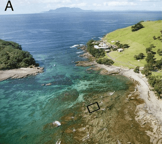
Chaos – not balance – and competition for resources are the natural order

This 2015 Amsterdam/New Zealand/Cornell shore-life study found:

“Species abundances in natural ecosystems may never settle at a stable equilibrium.

Species in one of the world’s oldest marine reserves showed chaotic fluctuations for more than 20 years. The species replaced each other in cyclic order, yet the exact timing and abundances of the species were unpredictable.

Our findings provide a field demonstration of nonequilibrium coexistence of competing species through a cyclic succession at the edge of chaos.

Our findings show that natural ecosystems can sustain continued changes in species abundances.”

chaos

http://www.pnas.org/content/112/20/6389.full “Species fluctuations sustained by a cyclic succession at the edge of chaos”


The University of Amsterdam also participated in a 2013 study Evolution of microbial markets where evolutionary biologists studied microbes. Their related findings included:

“Cooperative interactions between individuals of different species.

Strategies important for microbes to optimize their success in potential biological markets:

  • (i) avoid bad trading partners;
  • (ii) build local business ties;
  • (iii) diversify or specialize;
  • (iv) become indispensable;
  • (v) save for a rainy day; and
  • (vi) eliminate the competition.”

A 2015 study How a well-adapted immune system is organized (the *.pdf file is linked because the html has errors) had a related finding that applied to our body’s immune system. The researchers found that the primary reason why each of our immune systems is unique is due to the effect of:

“Competition between receptor clones..NOT a biologically implausible centralized mechanism distributing resources system-wide.

The repertoire of lymphocyte receptors in the adaptive immune system protects organisms from diverse pathogens. A well-adapted repertoire should be tuned to the pathogenic environment to reduce the cost of infections.

Competitive dynamics can allow the immune repertoire to self-organize into a state that confers high protection against infections.”

Chaos and competition for resources are facts of life observed within ourselves and in nature from ocean life down to the microbe level.

Why are we often presented – as a fact of life – that what’s natural is for all aspects of our lives to be in balance? Emotional, economic, social, intellectual – you name it, we’re told that the natural model is one of “stable equilibrium.”


Two hypotheses of Dr. Arthur Janov’s Primal Therapy are relevant:

Trying for closure, though, becomes an act-out – a temporary fulfillment of a substitute need. But the underlying need remains unsatisfied, and soon drives further act-outs. Balance is never achieved.

With this viewpoint, can you see how behavior like the following shows the internal state of the actor as they attempt to thwart the natural reality of the situation?

  • A person in authority who demands that people cease their competition for a resource and instead, accept what the authority figure determines is fair and balanced. An example is limiting supplies with price controls after a disaster.
  • A person who disrupts cooperative behavior that provides a solution for the cooperators’ needs/wants and instead, interposes themselves in a directed solution. An example is requiring licenses for cooperative childcare.
  • A person who insists that peoples’ responses to chaos to form an optimal adaptation cease, and instead, conform to some other responses. An example is prohibiting free movement after a disaster.

It reveals even more about the internal states of people that the above examples become codified. Children are taught that the natural and solely acceptable way to behave is in accordance with these unnatural solutions.


There are some signs that unnatural solutions in society can be reversed. For example, here is a 2013 article about a UK village that benefited from removing all of its traffic signals and reverting to the natural order of human cooperation and competition.

At the individual level, though, it’s up to each one of us to recognize and reverse our unnatural states. We and the people around us will be pleased when we and they are no longer adversely affected by our unconscious act-outs that are driven by our internal states. There’s enough natural chaos without adding more with act-outs.

Our internal systems will suffer damage, for example, when our unconscious act-out is to be busy, always doing something, and we can’t relax. Stress adversely affects our internal systems until we understand and reverse the driving unnatural states.

A possible link between stress responses and human cancers?

This 2015 UK rodent study found:

“An unexpected role for the GR [glucocorticoid receptor] in promoting accurate chromosome segregation during mitosis.

We also identify reduced GR expression in several common human cancers, thereby implicating GR as a novel tumor suppressor gene.”

One of the researchers said:

“Cancer is caused by cell division going wrong, but no one has previously looked at the role GR has to play in this process. It’s now clear that it is vital.”

From the study:

“Our findings now show that GR function regulates accurate mitotic progression, with clear implications for human health.

Add a previously unidentified perspective to GR action in cell division, affecting mitotic spindle function.

It may be that this action can be targeted by specific ligands, potentially opening up new therapeutic approaches to treat common cancers.”

The Translating PTSD research findings from animals to humans study also found reduced expression of glucocorticoid receptor genes, which appeared in some rodents after stress. Unfortunately, those researchers’ priorities weren’t to research the causes of this reduced expression.


One relevant hypothesis of Primal Therapy is that trauma in the earliest parts of human life epigenetically impairs the proper functioning of human development processes. A follow-on hypothesis is that the arrival of diseases in later life may be traceable back to the damage done during early-life development processes.

An example of this would be that a developing fetus adapts to being constantly stressed by an anxious and stressed mother. When the changes persist after birth, they may present as maladaptations of the infant to a non-stressful environment. These enduring changes may be among the causes of symptoms decades later such as over- and/or under-reactions to stress.

It seems possible that further research in these areas may find links among human stress responses and human cancers. The current study suggested that the glucocorticoid receptor may play a part in these links.

http://www.pnas.org/content/112/17/5479.full “Glucocorticoid receptor regulates accurate chromosome segregation and is associated with malignancy”

Translating PTSD research findings from animals to humans

This 2014 rodent study stressed the animals, measured their stress responses, then killed them and sampled genes in their amygdala, hippocampus, and blood. The researchers found that glucocorticoid receptor signaling genes were the primary pathway associated with “exposure-related individual differences in stress responses for the amygdala and blood. This pathway also placed first for the hippocampus in female rats (glucocorticoid receptor was second in male rats and prostate cancer signaling was first).

I’ll quote one press article’s coverage to show where the researchers wanted to go with the study’s findings:

“We found that most of the genes and pathways that are different in PTSD [post-traumatic stress disorder]-like animals compared to resilient animals are related to the glucocorticoid receptor, which suggests we might have identified a therapeutic target for treatment of PTSD.”

How about this lead sentence:

“There may some day be a blood test to determine whether someone suffers from Post-Traumatic Stress Disorder or is at risk for the psychiatric condition.”

Here’s another article’s paraphrase of a different researcher:

“Those are genes that become activated in the presence of stress. Like a key fitting into a lock, the hormone corticosterone, produced naturally by the body, connects to the receptor and has a calming effect.

In some rodents, and apparently in some people, the pathway appears to be defective, and this puts them at higher risk for PTSD.”

Also, from the study’s abstract:

“Corticosterone treatment 1 h[our] after PSS [predator-scent-stress]-exposure prevented anxiety and hyperarousal 7 d[ays] later in both sexes, confirming the GR [glucocorticoid receptor] involvement in the PSS behavioral response.”


Like other researchers continue to do, they stopped this study short of finding causes for the effects:

  1. What were the causes for genes in the glucocorticoid receptor signaling pathway being differentially expressed? “Exposure-related individual differences” isn’t a causal finding.
  2. If this pathway is “defective,” what exactly happened to make it that way?
  3. Did dampening the effects of stress with a shot of cortisol one hour after the stress treat the cause such that the rats were cured? Since the readers of the study and associated articles were led to infer that this treatment was a cure, why destroy the treated animals afterwards before the proofs of long-term efficacy were thoroughly documented and tested?

When studies like this are carried forward with humans, researchers should try to find the causes for these effects. It isn’t sufficient to pretend that there aren’t early-life causes for these effects. Such a pretense leads to the follow-on pretense that later-life consequences are mysteries such as “exposure-related individual differences” and not effects.

Researchers should act like the subjects are feeling human beings who can participate in treatments of both the causes and effects. They should remember that humans are not lab rats who need to be fixed.

http://www.pnas.org/content/111/37/13529.full “Expression profiling associates blood and brain glucocorticoid receptor signaling with trauma-related individual differences in both sexes”

Can a Romanian orphan give informed consent to be an experimental subject?

This 2015 study used Romanian orphans as lab rats for findings of which I failed to see the value. The world didn’t really need any further research to demonstrate that foster care would be better for a child than staying in an orphanage.

The researchers placed the orphans in five separate stressful situations, and measured their cortisol and DHEA-S levels, along with their electrocardiograph and impedance cardiograph activity. The findings were:

“Children who were removed from the Romanian institutions and placed with foster parents before the age of 24 months had stress system responses similar to those of children being raised by families in the community.

The children raised in institutions showed blunted responses in the sympathetic nervous system, associated with the flight or fight response, and in the HPA axis, which regulates cortisol.”

One unsupported assertion from the researchers was:

“We provide evidence for a causal link between the early caregiving environment and stress response system reactivity in humans with effects that differ markedly from those observed in rodent models.”

The researchers stated that rodent studies have converged to find:

“Early-life adversity results in hyperreactivity of the sympathetic nervous system (SNS) and hypothalamic–pituitary–adrenal (HPA) axis.”

It’s baloney that the same results from early life adversity in rodents haven’t also been present in humans. Even the lead researcher herself said in a news article:

“More significantly, McLaughlin said, their [the orphans] stress response systems might have been initially hyperactive at earlier points in development, then adapted to high levels of stress hormones.”

The difference was that the rodents were monitored 24/7 until researchers killed and dissected them. The children’s periods of adversity likely started while in the womb, and their lives had been monitored for research purposes sporadically after their births.

Everybody knows that just because adverse events and effects in these children’s lives weren’t recorded by researchers didn’t mean these effects weren’t present at some point.

Particularly irksome was another unsupported assertion from the lead reviewer:

“The children involved in the study are now about 16 years old, and researchers next plan to investigate whether puberty has an impact on their stress responses. It could have a positive effect, McLaughlin said, since puberty might represent another sensitive period when stress response systems are particularly tuned to environmental inputs. “It’s possible that the environment during that period could reverse the impacts of early adversity on the system,” she said.”

No, this is NOT possible. We may as well expect an apple to fall upward.

The impacts of early adversity persist with enduring physiological changes as shown in experimental studies. Studies have NOT provided evidence that the subjects’ environment can cause the effects of complete reversal of all these changes, no matter the stage of life of the subjects.

This point was addressed in The effects of early-life stress are permanent alterations in the child’s brain circuitry and function rodent study:

The current study manipulates the type and timing of a stressor and the specific task and age of testing to parallel early-life stress in humans reared in orphanages.

The results provide evidence of both early and persistent alterations in amygdala circuitry and function following early-life stress.

These effects are not reversed when the stressor is removed nor diminished with the development of prefrontal regulation regions.

That study had the same reviewer as the current study. The current study’s lead researcher knew or should have known of this and other relevant research. She knew or should have known of the irreversibility of critical periods, during which developments either occurred or were forever missed.

Did the lead researcher make assertions not supported by the study or relevant research – assertions made counter to her scientific knowledge – show her unease about treating the orphans as lab rats? Was there was some other agenda in play?

The larger problem was the study’s informed consent with this group of Romanian orphans. If you were in contact with a damaged person, and implicitly gave them hope that you would improve their life, then who are you as a feeling human being when you don’t personally carry through? Does the legal documentation matter?


Also, I’ve noticed problems with several studies that had this particular reviewer:

Add the current study to the list.

http://www.pnas.org/content/112/18/5637.full “Causal effects of the early caregiving environment on development of stress response systems in children”


This post has somehow become a target for spammers, and I’ve disabled comments. Readers can comment on other posts and indicate that they want their comment to apply here, and I’ll re-enable comments.