Immune system aging

This 2021 review by three coauthors of Take responsibility for your one precious life – Trained innate immunity cast a wide net:

“Non-specific innate and antigen-specific adaptive immunological memories are vital evolutionary adaptations that confer long-lasting protection against a wide range of pathogens. However, these mechanisms of memory generation and maintenance are compromised as organisms age.

This review discusses how immune function regulates and is regulated by epigenetics, metabolic processes, gut microbiota, and the central nervous system throughout life. We aimed to present a comprehensive view of the aging immune system and its consequences, especially in terms of immunological memory.

aging immune system

A comprehensive strategy is essential for human beings striving to lead long lives with healthy guts, functional brains, and free of severe infections.”

https://link.springer.com/article/10.1007/s12016-021-08905-x “Immune Memory in Aging: a Wide Perspective Covering Microbiota, Brain, Metabolism, and Epigenetics”


Attempts to cover a wide range of topics well are usually uneven. For example, older information in the DNA Methylation In Adaptive Immunity section was followed by a more recent Histone Modifications in Adaptive Immunity section.

This group specializes in tuberculosis vaccine trained immunity studies, and much of what they presented also applied to β-glucan trained immunity. A dozen previously curated papers were cited.

PXL_20211218_190653401

Glutathione primes β-glucan-trained immunity

Two 2021 papers on glutathione interactions with β-glucan, with the first studying human cells from healthy donors:

“(1→3)-β-D-Glucan stimulation induces epigenetic and transcriptomic changes in monocytes associated with increased glutathione (GSH) synthesis and metabolism. Intracellular glutathione levels were crucial in regulating several monocyte antifungal functions including resilience to oxidative stress, immunometabolism, nitric oxide production, phagocytosis, and cytokine production.

Our findings demonstrate an important role for GSH in immunity, and outline a better understanding of the acute response of monocytes to infections.”

https://www.frontiersin.org/articles/10.3389/fimmu.2021.694152/full “Glutathione Metabolism Is a Regulator of the Acute Inflammatory Response of Monocytes to (1→3)-β-D-Glucan”


A second study investigated the subject with a dozen rodent experiments:

“We demonstrated that antioxidation by GSH supported an environment essential for β-glucan-induced metabolic and epigenetic changes in monocytes. We found that GSH induced glycolysis and glutaminolysis in β-glucan-trained immunity in a mTOR-dependent manner.

These results uncovered the GSH/mTOR/c-Myc signaling axis as the central effector of metabolic reprogramming in trained immunity. We revealed that the delicate GSH/ROS redox balance determines discrete, long lasting metabolic modifications that are causal to β-glucan-trained immunity.

Our results suggest that H3K27me3 demethylation is a necessary event. We identified H3K27me3 demethylation as a novel histone modification mark that was impaired by GSH deficiency in β-glucan-trained bone marrow derived macrophages.

We identified EZH2 as a potential tool to boost trained immunity under GSH deficiency conditions, or to enhance trained immunity in clinical settings where excessive inflammatory responses could be beneficial.

ezh2 survival

Overall, these insights contribute to unraveling metabolic and epigenetic changes during trained immunity.”

https://www.sciencedirect.com/science/article/pii/S2213231721003669 “Glutathione synthesis primes monocytes metabolic and epigenetic pathway for β-glucan-trained immunity”


The second paper of Remembering encounters provides future benefits also explored this subject.

PXL_20211215_183316335

Offspring brain effects from maternal adversity

This 2021 rodent study investigated conception through weaning effects on offspring from stressing their mothers:

“We investigated consequences of two prenatal insults, prenatal alcohol exposure (PAE) and food-related stress, on DNA methylation profiles of the rat brain during early development. We analyzed patterns in prefrontal cortex, a key brain region involved in cognition, executive function, and behavior, of both males and females, and found sex-dependent and sex-concordant influences of these insults.

The pair-fed (PF) group in the PAE model is a standard control for effects of alcohol in reducing food intake. However, compared to the PAE group that, albeit eating less, eats ad libitum, pair-feeding is a treatment in itself, with PF dams receiving a restricted ration, which results in both hunger and a disrupted feeding schedule. These stress-related effects could potentially parallel or model food scarcity or food insecurity in human populations.

We observed more DMRs (Differentially Methylated Regions) that showed decreased DNAm rather than increased DNAm in PF animals, suggesting that food-related stress may interfere with one-carbon metabolism and the pathways that deposit methylation on DNA. We also identified a sex-concordant DMR that showed decreased DNAm in PF animals in the glucocorticoid receptor Nr3c1, which plays a key role in stress responsivity and may reflect a reprogramming of the stress response.

This result is in line with previous studies that have shown that pair-feeding is a considerable stressor on dams, with lasting consequences on development, behavior, and physiology of their offspring. Altered DNAm of this key HPA axis gene may reflect broader alterations to stress response systems, which may in turn, influence programming of numerous physiological systems linked to the stress response, including immune function, metabolic processes, and circadian rhythms.

In PAE and PF animals compared to controls, we identified 26 biological pathways that were enriched in females, including those involved in cellular stress and metabolism, and 10 biological pathways enriched in males, which were mainly involved in metabolic processes. These findings suggest that PAE and restricted feeding, both of which act in many respects as prenatal stressors, may influence some common biological pathways, which may explain some of the occasional overlap between their resulting phenotypes.

genes-12-01773-g005

This study highlights the complex network of neurobiological pathways that respond to prenatal adversity/stressors and that modulate differential effects of early life insults on functional and health outcomes. Study of these exposures provides a unique opportunity to investigate sex-specific effects of prenatal adversity on epigenetic patterns, as possible biological mechanisms underlying sex-specific responses to prenatal insults are understudied and remain largely unknown.”

https://www.mdpi.com/2073-4425/12/11/1773/htm “Prenatal Adversity Alters the Epigenetic Profile of the Prefrontal Cortex: Sexually Dimorphic Effects of Prenatal Alcohol Exposure and Food-Related Stress”


PXL_20211215_182428532

The impact of transgenerational epigenetic inheritance and early life experiences

A 2021 interview with McGill University’s Moshe Szyf:

There is a rejection of transgenerational inheritance as it goes against progressive thinking because it ties us to previous generations. The theory faces rejection because it sounds deterministic.

But if you understand what epigenetics is, it’s not deterministic. There is stability, and there’s also room for dynamic change.

The only way things change in the body for the long term is via epigenetics. We don’t know everything yet, new discoveries are yet to happen, and then we will just say, ‘Wow, it’s so obvious!’

The immune system is tightly connected to the brain and is directly affected by early adversity. Even though we will not be able to learn what’s going on in the brain, as far as epigenetics in living people, we will gain a lot of information from how the immune system responds to early adversity, and how this is correlated with behavioral phenotype and with mental health.

This brings into question the whole field of neuroimmunology, of which there is a lot of data. But it seems that a lot of psychiatrists are totally oblivious to these data, which is astounding, because the glucocorticoid hormone – the major player in this mechanism due to its involvement in early life stress as well as control of behavior – also controls immune function.

Nobody can live long enough to oversee a human transgenerational study. In humans, correlations are usually in peripheral tissue, where changes are small. The jury’s not out yet, but if evolution used it for so many different organisms, some of which are very close to us in the evolutionary ladder, it’s impossible that humans don’t use it.

How are current findings in animal models relevant to humans? How do we develop human paradigms that will allow us to achieve a higher level of evidence than what we have now?

  • One way is the immune-inflammatory connection to other diseases. I think this is where the secret of epigenetic aging lies, as well as epigenetics of other diseases.
  • Every disease is connected to the immune system. The brain translates the behavioral environment to the immune system, and then the immune system sends chemical signals across the body to respond to these challenges.

We need to understand that epigenetic programs are a network. Move beyond candidate genes, understand the concept of a network, and really understand the challenge: Reset the epigenetic network.

Epigenetics is going to be rapidly translated to better predictors, better therapeutics, and more interesting therapeutics. Not necessarily the traditional drug modeled against a crystal structure of an enzyme, but a more networked approach. Ideas about early life stress are critical and have impacted the field of childcare by highlighting the importance of early childhood relationships.”

https://www.futuremedicine.com/doi/10.2217/epi-2021-0483 “The epigenetics of early life adversity and trauma inheritance: an interview with Moshe Szyf”


Week 87 of Changing to a youthful phenotype with sprouts

This week I dialed back eating microwaved 3-day-old broccoli / red cabbage / mustard sprouts from twice a day to once a day. For my reasoning, here are two papers on broccoli sprouts and thyroid function, with the first a 2018 human study:

“We analyzed biochemical measures of thyroid function and thyroid autoimmunity in a subset of participants in a broccoli sprout clinical trial. The present work is a retrospective analysis of a subset of serum samples collected during a clinical trial conducted from mid-October 2011 to early January 2012.

130 individuals received placebo beverage, and 137 received broccoli sprout beverage for 84 consecutive days (12 weeks). Blood samples from day 0 and day 84 were analyzed in a subset of 45 female participants (19 placebo, 26 broccoli sprout beverage) for serum thyroid-stimulating hormone (TSH), free thyroxine (fT4), thyroglobulin (TG), anti-TG, and anti-thyroid peroxidase (anti-TPO) antibodies.

The percentage of patients with subclinical hypothyroidism (elevated TSH with normal fT4) was not significantly different between the two groups either before or after treatment.

subclinical hypothyroidism

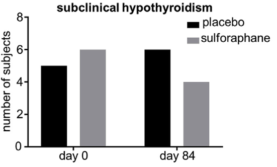
Daily ingestion of a broccoli sprout extract beverage over 84 days had no deleterious effect on thyroid function tests or measures of thyroid autoimmunity. It may be prudent to evaluate thyroidal safety of plant-based food supplements on a case-by-case basis.”

https://www.sciencedirect.com/science/article/abs/pii/S0278691519300547 “Broccoli sprout beverage is safe for thyroid hormonal and autoimmune status: Results of a 12-week randomized trial” (not freely available)


A 2020 review by three of these coauthors summarized further details:

“One difference between the thyroid and other tissues is that ROS are not primarily a byproduct of its physiology, but an indispensable part of it. Thyroid follicular cells actively produce H2O2 to facilitate a cascade of redox reactions that sequentially oxidize iodide, iodinate tyrosine residues within Tg, and couple iodinated tyrosine residues of Tg to each other to form T4 and T3 (triiodothyronine).

There exists a fail-safe mechanism in which specific combinations of four Keap1 cysteines can form a disulfide bond to sense H2O2. This sensing mechanism appears to be distinct from that triggered by other Nrf2 inducers, such as electrophiles.

Findings from Keap1KD mice suggest that chronic genetic activation of Nrf2 signaling may have negative consequences for the thyroid gland. However, analysis of data from a clinical trial has shown that consumption of a broccoli sprout beverage (yielding pharmacologically active amounts of the Nrf2-activating compound sulforaphane) is safe for thyroid hormonal and autoimmune status during a 12-week administration period.

Nevertheless, it appears prudent to monitor thyroid function and thyroid volume (at least by palpation) in patients treated with Nrf2-modulating compounds in clinical trials or clinical practice.”

https://www.mdpi.com/2076-3921/9/11/1082/htm “The Keap1/Nrf2 Signaling Pathway in the Thyroid—2020 Update”


My Day 70 lab results for inflammation markers were great:

IL-6 2020

A year later, IL-6 was below the test’s detection limit, and high-sensitivity C-reactive protein could hardly have been better at 0.24 mg/L.

But TSH (reference interval 0.45 – 4.50 μIU/mL) increased from 3.01 to 7.50. Here’s what Labcorp Technical Review L8186 said:

“The panel concluded that despite the fact that serum TSH concentrations higher than 2.5 μIU/mL but less than 4.5 μIU/mL may identify some individuals with the earliest stage of hypothyroidism, there is no evidence for associated adverse consequences. Additionally, consequences of subclinical hypothyroidism with serum TSH levels between 4.5 μIU/mL and 10 μIU/mL are minimal, and the panel recommends against routine treatment of patients with TSH levels in these ranges.”

I went in last weekend to retest. Although the provider verbally agreed to test TSH, free T3, and free T4, a different test was ordered.

TSH was still high at 5.85 μIU/mL. Other measurements (Total T4, T3 Uptake, and Free Thyroxine Index) aren’t suitable substitutes for free T3 and free T4. I’ll specify Labcorp test numbers next time.


My hypothesis is that preconditioning my endogenous ARE system twice daily worked alright elsewhere, but not for my thyroid. We’ll find out in 2022 whether halving the electrophilic activations of my Nrf2 signaling pathway has any effect on thyroid measurements.

I don’t take anything with, or an hour before or after these very reactive isothiocyanates. I continue to eat 3-day-old oat sprouts twice a day with other foods.

PXL_20211125_171335252

Uses of the neutrophils-to-lymphocytes ratio

This 2021 review subject was recent history and uses of the neutrophils-to-lymphocytes ratio parameter to indicate systemic inflammation and stress:

“NLR is widely used across almost all medical disciplines as a reliable and easily available marker of immune response to various infectious and non-infectious stimuli. NLR reflects dynamic relationships between innate (neutrophils) and adaptive (lymphocytes) cellular immune response during illness and various pathological states.

A normal range of NLR is between 1–2, and values higher than 3.0 and below 0.7 in adults are pathological. NLR between 2.3–3.0 may serve as an early warning of cancer, atherosclerosis, infection, inflammation, psychiatric disorders, and stress.

nlr

Neutrophils play a pivotal role in innate immune response including phagocytosis, and release of a variety of cytokines and molecule mediators. Lymphocytopenia is a hallmark of stress while inflammation is due to demargination, redistribution, and accelerated apoptosis.

Opposite changes in neutrophil and lymphocyte counts are a multifactorial dynamic process depending on regulation of various immunologic, neuroendocrine, humoral, and biologic processes such as margination / demargination, mobilization / redistribution, accelerated / delayed apoptosis, influence of stress hormones, and sympathetic / parasympathetic nervous system imbalance. NLR is the best expression of a tight functional relation between two fundamental immunocompetent leukocyte populations.”

https://doi.org/10.4149/bll_2021_078 “Neutrophil-to-lymphocyte ratio, past, present and future perspectives”


The fourth study of Uses of the lymphocytes-to-monocytes ratio commented on NLR, as did What is the health utility of white blood cell type count ratios?.

Sunday morning stroll

PXL_20211205_141539732

Eat broccoli sprouts to prevent radiation damage

This 2021 rodent study investigated effects of sulforaphane on skin damage from irradiation:

“Radiotherapy is currently the main treatment for various cancers. We observed the protective effect of sulforaphane (SFN) on radiation-induced skin injury (RISI), including oxidative stress and inflammatory response indexes, and Nrf2 expression with its downstream antioxidant genes:

  • SFN prevented DNA damage caused by radiation.
  • SFN prevented and treated radiation-induced skin inflammation.
  • SFN prevented radiation-induced oxidative stress in skin.
  • Activation of Nrf2 and expressions of its downstream genes in skin induced by SFN.

Nrf2 downstream antioxidant genes induced by SFN

Mice were randomly assigned to one of four groups (n = 8), including control group (CON), SFN group, irradiation (IR) group, and IR plus SFN (IR/SFN) group (* p < 0.05 vs. CON; & p < 0.05 vs. IR).

Our most innovative discovery was that SFN provided skin protection from IR. At present, there are a few drugs to treat RISI in clinical patients, but the effect is not very ideal, or some may cause certain side effects.

SFN extracted from natural broccoli has no toxicity and is easily accepted for usage in clinic. According to our findings, SFN will provide a new strategy for clinical treatment and prevention of RISI in the future.”

https://www.mdpi.com/2076-3921/10/11/1850/htm “Sulforaphane-Mediated Nrf2 Activation Prevents Radiation-Induced Skin Injury through Inhibiting the Oxidative-Stress-Activated DNA Damage and NLRP3 Inflammasome”


This study’s findings probably also apply to less-severe skin damage caused by sun exposure.

PXL_20211201_201315538

Is liver damage reversible?

This 2021 rodent study measured sequential liver changes caused by a high-fat diet:

“Using a longitudinal mouse study of diet-induced obesity in male mice, we investigated kinetics of hepatic DNA methylation and gene expression compared to those of obesity-induction to assess if they could be causal for development of insulin resistance. We aimed to find out if these changes were reversed by massive weight loss induced by vertical sleeve gastrectomy or metformin treatment.

We identified two CpG sites within exon 1 of Fgf21 that became gradually hypomethylated upon HFD feeding. DNA demethylation started between week two and four, to become significant at week five, and significantly correlated with hepatic Fgf21 gene expression.

These DNA methylation changes preceded development of insulin resistance, and were potentially causally involved in increased Fgf21 expression and plasma levels associated with insulin resistance. This points to a key regulatory function of gene body DNA methylation, which was eventually a compensatory response to counteract the developing insulin resistance.

HFD-induced decrease in Fgf21 DNA methylation could not be reversed by vertical sleeve gastrectomy or metformin treatment. As soon as weight loss slowed down or mice started to re-gain weight, differences in DNA methylation were no longer detected compared to sham-operated mice.

1-s2.0-S0955286321003272-ga1_lrg

As the altered DNA methylation pattern was acquired during adulthood in differentiated cells, our data emphasize that metabolic programming via DNA methylation is dynamic and not restricted to fetal development. This supports the concept that individuals can actively influence their DNA methylation patterns by lifestyle choices.

Our data indicate that DNA methylation alterations in key metabolic tissues can be acquired by an obesogenic diet, and not easily be reversed by interventions common in obese and diabetic subjects.”

https://www.sciencedirect.com/science/article/pii/S0955286321003272 “Dietary induction and reversal of obesity and insulin resistance is associated with changes in Fgf21 DNA methylation in liver of mice”


This study attempted two interventions that didn’t have desired effects. All about the betaine mentioned others that may reverse liver epigenetic changes.

PXL_20211125_170854068

Eat broccoli sprouts for your liver

This 2021 rodent study investigated sulforaphane pretreatment’s role in reducing liver injuries:

“As a double blood supply organ of the portal vein and artery, the liver is highly sensitive to ischemia, and is one of the common organs to suffer from hepatic ischemia-reperfusion injury (HI/RI). HI/RI leads to overproduction of reactive oxygen species (ROS).

  • Overdoses of ROS promote reaction of lipid peroxidation and generation of the extremely aggressive oxidants nitric oxide (NO) and malondialdehyde (MDA).
  • HI/RI decreased antioxidant enzyme activity of glutathione (GSH), catalase (CAT), and superoxide dismutase (SOD).
  • Sulforaphane (SFN) intervention significantly decreased levels of MDA and NO by increasing activity of GSH, CAT, and SOD.

Inflammation is the most serious secondary injury experienced in HI/RI.

  • Monocyte chemotactic protein 1 (MCP-1) is involved in an inflammatory reaction with regulation of monocytes, T lymphocytes, and NK cells. MCP-1 can also increase infiltration of inflammatory cells by activating NF-κB.
  • Tumor necrosis factor-α (TNF-α) is a promoter of the inflammatory response. Interleukin-6 (IL-6) is an inflammatory mediator in the acute reaction period.
  • SFN treatment significantly decreased HI/RI-induced expression of TNF-a, IL-6, and MCP-1.

sulforaphane and liver inflammation

In conclusion, SFN has a protective effect on HI/RI. The mechanism is associated with activating Nrf2/HO-1 signaling to suppress oxidative stress and inflammation.”

https://www.sciencedirect.com/science/article/abs/pii/S0966327421000794 “Sulforaphane alleviates hepatic ischemia–reperfusion injury through promoting the activation of Nrf-2/HO-1 signaling” (not freely available)


A human equivalent of this study’s 5 mg / kg sulforaphane dose was (.161 x 5 mg) x 70 kg ≈ 56 mg. For comparison, my estimated daily sulforaphane intake from microwaved sprouts is 52 mg.

PXL_20211031_110903996

Sprout bioaccessibility

Twin 2021 in vitro studies of cruciferous sprout bioaccessibility, with the first addressing hydroxycinnamic acids and flavonols:

“The present work studies effects of physicochemical and enzymatic characteristics of gastrointestinal digestion on two major groups of phenolic compounds – flavonols and cinnamoyl derivatives – on red radish, red cabbage, broccoli, and white mustard sprouts. Effects of gastrointestinal digestion on release and stability of phenolic compounds depends on different factors, such as physicochemical traits of the food matrix, pH, temperature, or enzymatic activity.

Although initial concentrations of phenolic acids in red radish were lower than in other sprouts, their bioaccessibility after digestion was higher, followed by red cabbage, white mustard, and broccoli. Most degradation of phenolic compounds corresponded to the flavonol fraction, which was almost erased during digestion (with the exception of digestion products of broccoli sprouts, which retained around 30% of the original flavonol concentration):

nutrients-13-04140-g003

Red radish sprouts exhibited the greatest bioaccessibility.

Gastric digestion prepares the food matrix for more efficient polyphenol extraction during intestinal digestion, in which the highest release and stability of these compounds takes place. Hydroxycinnamic acids reach higher concentrations than flavonols, making them tentatively more available to be absorbed at the intestinal level.”

https://www.mdpi.com/2072-6643/13/11/4140/htmIn Vitro Evidence on Bioaccessibility of Flavonols and Cinnamoyl Derivatives of Cruciferous Sprouts”


A cited predecessor used similar methods to study glucosinolate breakdown products like sulforaphane, iberin, and indole-3-carbinol:

“Significantly higher bioaccessibility of isothiocyanates (ITCs) and indoles from glucosinolates (GSLs) of red cabbage sprouts were observed. Bioaccessibility of GSLs from Brasicaceae sprouts is not exclusively associated with initial content of these compounds in plant material (almost negligible), but also with release of GSLs and ongoing breakdown reactions during gastric and intestinal phases of digestion, respectively:

ITCs

The intestinal phase was the most relevant for bioaccessibility of ITCs. Aliphatic GSLs provided higher bioaccessibility of their corresponding ITCs in comparison to indolic and aromatic GSLs.”

https://www.mdpi.com/1422-0067/22/20/11046/htm “Evidence on the Bioaccessibility of Glucosinolates and Breakdown Products of Cruciferous Sprouts by Simulated In Vitro Gastrointestinal Digestion”


Gastric and intestinal simulations were instructive. But rather than depending on digestion for ITCs, I “enzymatically convert to SF before oral intake” per A follow-on study to 3-day-old broccoli sprouts have the optimal yields.

Regarding phenolic compound digestion, my focus this year has been to give my gut microbiota what they want. I expect and get reciprocity from treating them well with whole oats, broccoli-red cabbage-mustard-oat sprouts, blackberries-blueberries-strawberries, quercetin from capers, etc. polyphenols. Not to mention inulin, artichoke hearts, and yeast cell wall β-glucan. Haven’t considered sprouting red radish seeds.

Per Red cabbage effects on gut microbiota, a related research group had an in vitro system that included gut microbiota. Maybe these researchers will get together in a future study?

PXL_20211118_114742112

Human agency vs. brain dysfunction

This 2021 human study used epigenetic clock technology to assess chronic inflammation as a driver of cognitive decline through its effects on brain structure:

“An epigenetic measure of C-reactive protein (DNAm CRP) was assembled for each participant. We found that higher inflammatory burden, indexed by DNAm CRP scores, associated with poor cognitive and neuroimaging brain health outcomes.

inflammation vs cognitive ability

DNAm CRP exhibited significantly larger associations with brain structural MRI metrics (including global grey and white matter atrophy, poorer white matter microstructure, and increased white matter hyperintensity burden) than serum CRP. Given that the 7 CpGs which make up DNAm CRP score reside in inflammation and vascular-related genes, these DNAm CRP-brain MRI associations may be capturing the impact of upstream inflammatory activity above and beyond that of serum CRP levels.

Our results indicate that some cognitive domains (processing speed) may be more mediated by brain structural consequences of chronic inflammation than others (verbal memory, visuospatial ability).

Our results add to the evidence base that DNAm-based predictors of inflammation may act as a quantifiable archive of longitudinal effects of these exposures – and other unaccounted for health and genetic profiles – that serum CRP levels fail to capture. By utilising an epigenetic inflammation measure, which integrates information from multiple immune-related CpG sites, we may provide a more reliable measure of chronic inflammation and thus a more comprehensive overview of consequences of chronic inflammation on brain structure and function.”

https://n.neurology.org/content/early/2021/11/17/WNL.0000000000012997.long “DNA Methylation and Protein Markers of Chronic Inflammation and Their Associations With Brain and Cognitive Aging”


These researchers essentially negated many of their findings by acknowledging:

“Although we endeavoured to remove participants with cognition-related pathology, these were screened via self-reported diagnoses, and we may be missing undiagnosed or subclinical incident neurodegenerative pathology.”

It wasn’t sufficient to claim in the Abstract section “Participants (N = 521) were cognitively normal, around 73 years of age” then include in the Discussion section a one-sentence limitation of relying on self-reports. Everyone defends themself against current and past realities and experiences.

Hard to imagine that objective measures such as the three comprising cognitive ability weren’t better screens. But then too many 73-year-old subjects may not have been “cognitively normal” and this study wouldn’t be adequately powered?

Can humans counteract inflammation? Non-communicable diseases? Smoking? Immune system degradation? Yes. No personal-agency actions were mentioned.

Also note this study’s social norming. The above-pictured 30-year-old female was busy at work, and subsequently hoisted a cat instead of a child in later years.

Take responsibility for your own one precious life.

PXL_20211118_114858264

Saving bees by regulating epigenetics

This 2021 study investigated an epigenetic treatment for bees forgetting about their hives:

“Over the last few decades, numbers of both wild and managed bee pollinators have been declining. Although reasons for this decline are under debate, it is highly likely that a combination of multiple stressors is to blame, in particular, deformed wing virus (DWV).

Histone deacetylase inhibitors (HDACi) are a class of compounds which prevent deacetylation of histones and therefore increase gene expression. The present study found that HDACi sodium butyrate (NaB) significantly increased survival and reversed the learning / memory impairment of DWV-infected bees. We demonstrated the mechanism of how epigenetic regulation can resume honeybees’ memory function.

bee survival rates

  • When bees were infected with DWV, 50% of bees died by the end of day 2 and only 10% survived to the end of day 5.
  • When NaB was added to the diet prior to DWV infection, survival rate of DWV-infected bees (N/D group) remained >90% after 5 days.
  • Under laboratory rearing conditions, around 30% of control bees died over a period of 5 days.
  • When NaB was included in uninfected bees’ diet, less than 15% of bees died.

These results indicate that feeding bees with NaB could significantly increase survival with or without DWV infection.”

https://www.cell.com/iscience/fulltext/S2589-0042(21)01024-5 “Real-time monitoring of deformed wing virus-infected bee foraging behavior following histone deacetylase inhibitor treatment”


Interesting that these researchers didn’t attempt to eliminate either the virus cause of bee behavior or parasitical mites that carried the virus. They mainly depended on bees’ endogenous systems providing beneficial responses when stimulated.

PXL_20211113_113321810

Save the bees with Brassicaceae seeds

Two 2021 papers, with the first studying bee infections:

“Fungus Nosema ceranae represents one of the primary bee infection threats worldwide, and antibiotic fumagillin is the only registered product for nosemosis disease control. Natural bioactive compounds deriving from glucosinolate–myrosinase (GSL–MYR) in Brassicaceae plants, mainly isothiocyanates (ITCs), are known for antimicrobial activity against numerous pathogens and health-protective effects in humans.

This work explored Brassica nigra [black mustard] and Eruca sativa [arugula] defatted seed meal (DSM) GSL-containing diets against natural Nosema infection in honeybees. Feeding was administered in May to mildly N. ceranae-infected colonies for four weeks at 250 g/week.

  • N. ceranae abundance showed a slight but significant decrease.
  • No significant effects on colony development and bee mortality were observed compared to controls.
  • MYR activity was detected both in bees fed DSMs and controls.
  • ITCs were found in gut tissues from bees treated with DSMs, corroborating presence of a MYR-like enzyme capable of hydrolyzing ingested GSLs.

Use of DSMs containing GSLs represents a promising alternative to fumagillin as it would overcome the problem of toxic bee product residues encountered with antibiotic treatment.”

https://www.mdpi.com/2218-273X/11/11/1657/htm “Glucosinolate Bioactivation by Apis mellifera Workers and Its Impact on Nosema ceranae Infection at the Colony Level”


A review was cited for “ITCs are GSL hydrolysis products known for their broad-spectrum biological activities against pests and soil/food-borne fungi, bacteria, and human microorganisms.”

“ITCs are efficient agents against a wide range of fungal strains. Many plant and human pathogens, as well as other fungi, were shown to be inhibited in vitro by these agents.

ITC-containing, chemically-characterized plant matrices used to test antifungal activity are summarized. The same activities were tested in pure ITCs; in fact, several well-designed studies did both approaches.

Although there is no one-size-fits-all rule to predict antifungal activity, in general, less polar compounds are usually more potent in solution, but lag behind more volatile compounds with small molecule size in vapor-phase applications. The main biochemical targets seem to be directly related to chemical reactivity, with antioxidant machinery a well-identified target.

Excellent studies on transcriptomics show general stress responses. Inhibiting production of fungal toxins is shown in many real-life applications as well.

Additional applications include:

  • Inhibition of fungal growth, pathogenesis, and/or toxin production in a variety of stored plants and grains;
  • Inhibition of disease on post-harvest fruits; as well as
  • Increasing shelf-life of different food products.

What is more, decrease in decay is frequently accompanied by the lack of measurable changes in various quality characteristics.

ITCs’ natural origin and biodegradability make them good candidates for a wide range of possible applications. Long-term studies show effects are delivered usually without apparent side-effects to plants.”

https://www.mdpi.com/2309-608X/7/7/539/htm “Effects of Glucosinolate-Derived Isothiocyanates on Fungi: A Comprehensive Review on Direct Effects, Mechanisms, Structure-Activity Relationship Data and Possible Agricultural Applications”


PXL_20211031_110852279

Reworking evolutionary theory

Dr. Michael Skinner coauthored a 2021 review arguing for inclusion of epigenetic transgenerational inheritance into evolutionary theory:

“Over the past 50 years, molecular technology has been used to investigate evolutionary biology. Many examples of finding no correlated genetic mutations or a low frequency of DNA sequence mutations suggest that additional mechanisms are also involved.

  • Identical twins have essentially the same genetics, but generally develop discordant disease as they age.
  • Only a low frequency (generally 1% or less) of individuals that have a specific disease have a correlated genetic mutation.
  • Dramatic increases in disease frequency in the population cannot be explained with genetics alone.

DNA methylation, histone modifications, changes to chromatin structure, expression of non-coding RNA, and RNA methylation can directly regulate gene expression independent of DNA sequence. These different epigenetic factors do not only act independently, but integrate with each other to provide a level of epigenetic complexity to accommodate the needs of cellular development and differentiation.

dvab012f1

Environmental epigenetics is the primary molecular mechanism in any organism that is used to promote physiological and phenotypic alterations. Actions of environmental factors early in development can permanently program the cellular molecular function, which then impacts later life disease or phenotypes.

dvab012f2

Integration of epigenetics and genetics contribute to a Unified Theory of Evolution that explains environmental impacts, phenotypic variation, genetic variation, and adaptation that natural selection acts on. The current review expands this proposed concept and provides a significant amount of supporting literature and experimental models to support the role of environmentally induced epigenetic transgenerational inheritance in evolution.”

https://www.ncbi.nlm.nih.gov/pmc/articles/PMC8557805/ “Role of environmentally induced epigenetic transgenerational inheritance in evolutionary biology: Unified Evolution Theory”


Organisms cited in this review’s references are similar to humans in ancestral influences and developmental influences during the first 1000 days of our lives. Humans are different in that even after all these influences, we can choose to influence our own change and individually evolve. We can also change our internal environments per Switch on your Nrf2 signaling pathway and An environmental signaling paradigm of aging.

PXL_20211031_111835802

Remembering encounters provides future benefits

Two 2021 papers on trained immunity, with the first a review:

“Effective memory immune responses rely on interaction between innate and adaptive immune cells. While activation of innate immunity provides the first line of defense against infections, it also primes the adaptive immune response.

Adaptive immunity can enhance antimicrobial machinery of innate cells, making them more effective at clearing pathogenic microorganisms. An additional layer of complexity adds to this network of interactions, with innate cells adopting a memory phenotype, which used to apply to only adaptive immunity. Furthermore, non-immune cells can develop some features of this memory-like phenotype.

fimmu-12-745332-g001

Cell subsets in which trained immunity has been described. Different stimuli including Bacillus Calmette Guerin (BCG), β-glucan, cytokines, cytomegalovirus (CMV), and bacterial components can induce a trained immunity phenotype. A common hallmark of trained immunity in these cases is H3K4me3 in promoters of genes encoding for different cytokines.

  • Mechanisms Underlying Establishment of Trained Immunity
  • Trained Immunity in Neutrophils
  • Trained Immunity in Monocytes and Macrophages: General Features
  • Metabolic Pathways Involved in Training of Monocytes and Macrophages
  • Hormonal Control of Trained Immunity Responses in Monocytes and Macrophages
  • Trained Immunity on Alveolar Macrophages and Involvement of Resident Cells
  • Trained Immunity in NK Cells
  • Trained Immunity in Innate Lymphoid Cells
  • Trained Immunity on Hematopoietic Stem Cells
  • Trained Immunity in Bronchial Epithelial Cells
  • Trained Immunity in Skin Stem Cells
  • Trained Immunity in the Gastrointestinal Tract
  • Immunity Training Against Protozoan-Mediated Pathologies
  • Trained Immunity in Non-Infectious Pathologies

Many gaps of knowledge remain in this field. For example, how long changes associated to trained immunity last, and if, in addition to epigenetic modulation, there are other post-translational modifications on proteins relevant for induction of trained immunity.”

https://www.frontiersin.org/articles/10.3389/fimmu.2021.745332/full “Molecular and Cellular Mechanisms Modulating Trained Immunity by Various Cell Types in Response to Pathogen Encounter”


This second paper was a human study cited for its glutathione findings as follows:

  • “Plasma concentration of IL-1β from BCG-vaccinated individuals are positively associated with serum glutathione concentrations.
  • Trained immunity up-regulates expression of genes involved in glutathione metabolism, suggesting an increase in glutathione synthesis and a higher glutathione recycling rate.
  • Single nucleotide polymorphisms in these genes are associated with changes in pro-inflammatory cytokine production after in vitro training by β-glucan and BCG.

Enzymes whose activities are dependent on glutathione could be used as novel targets to modulate trained immunity.”

IL-1β production

“We found a positive association between plasma glutathione concentration and ex vivo IL-1β production 90 days after BCG vaccination upon in vitro exposure to heterologous stimulus Staphylococcus aureus. Up-regulation of IL-1β production by BCG vaccination was also positively associated with circulating concentrations of other metabolites involved in glutathione metabolism, such as methionine, cysteine, glutamate, and glycine.

GSH metabolism was associated with trained immunity traits in 278 healthy individuals. Trained immunity mechanisms that are shaped by GSH metabolism remain to be further explored.”

https://www.mdpi.com/2073-4409/10/5/971/htm “Glutathione Metabolism Contributes to the Induction of Trained Immunity”


PXL_20211031_113233113