Why drugs aren’t ultimately therapeutic

This 2016 Oregon review’s concept was the inadequacy of drug-based therapies, explored with the specific subject of epilepsy:

“Currently used antiepileptic drugs:

  • [aren’t] effective in over 30% of patients
  • [don’t] affect the comorbidities of epilepsy
  • [don’t] prevent the development and progression of epilepsy (epileptogenesis).

Prevention of epilepsy and its progression [requires] novel conceptual advances.”

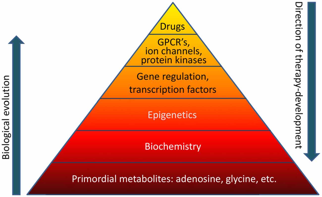
The overall concept that current drug-based therapies poorly address evolutionary biological realities was illustrated by a pyramid, with the comment that:

“If the basis of the pyramid depicted in Figure 1 is overlooked, it becomes obvious that a traditional pharmacological top-down treatment approach has limitations.”

Why drug ultimately aren't therapeutic


I would have liked the reviewer to further address the “therapeutic reconstruction of the epigenome” point he made in the Abstract:

“New findings based on biochemical manipulation of the DNA methylome suggest that:

  1. Epigenetic mechanisms play a functional role in epileptogenesis; and
  2. Therapeutic reconstruction of the epigenome is an effective antiepileptogenic therapy.”

As it was, the reviewer lapsed into the prevalent belief that the causes of and cures for human diseases will always be found on the molecular level – for example, the base of the above pyramid – and never in human experiences. This preconception leads to discounting human elements – notably absent in the above pyramid – that generate epigenetic changes.

A consequence of ignoring experiential causes of diseases is that the potential of experiential therapies to effect “therapeutic reconstruction of the epigenome” isn’t investigated.

http://journal.frontiersin.org/article/10.3389/fnmol.2016.00026/full “The Biochemistry and Epigenetics of Epilepsy: Focus on Adenosine and Glycine”

Using epigenetic outliers to diagnose cancer

This 2016 Chinese/UK human cancer cell study tested five algorithms and found:

“Most of the novel proposed algorithms lack the sensitivity to detect epigenetic field defects at genome-wide significance. In contrast, algorithms which recognise heterogeneous outlier DNA methylation patterns are able to identify many sites in pre-neoplastic lesions, which display progression in invasive cancer.

Many DNA methylation outliers are not technical artefacts, but define epigenetic field defects which are selected for during cancer progression.”

The usual method of epigenetic studies involves:

“Identify genomic sites where the mean level of DNAm [DNA methylation] differs as much as possible between the two phenotypes. As we have seen however, such an approach is seriously underpowered in cancer studies where tissue availability is a major obstacle.

In addition to allelic frequency, we also need to take the magnitude of the alteration into consideration. As shown here, infrequent but bigger changes in DNAm (thus defining outliers) are more likely to define cancer field defects, than more frequent yet smaller DNAm changes.”

A similar point was made in Genetic statistics don’t necessarily predict the effects of an individual’s genes:

“Epigenomic analyses are limited by averaging of population-wide dynamics and do not inform behavior of single cells.”

One of the five tested algorithms was made freely available by the researchers. The limitations on its use were discussed, and included:

“Studies conducted in a surrogate tissue such as blood are scenarios where DNAm outliers are probably not of direct biological relevance to cancer development.”

http://bmcbioinformatics.biomedcentral.com/articles/10.1186/s12859-016-1056-z “Stochastic epigenetic outliers can define field defects in cancer”

A human study of pain avoidance

This 2016 UK human study found:

“People differ in how they learn to avoid pain, with some individuals refraining from actions that resulted in painful outcomes, whereas others favor actions that helped prevent pain.

Learning in our task was best explained as driven by an outcome prediction error that reflects the difference between expected and actual outcomes. Consistent with the expression of such a teaching signal, blood-oxygen level-dependent (BOLD) responses to outcomes in the striatum were modulated by expectation.

Positive learners showed significant functional connectivity between the insula and striatal regions, whereas negative learners showed significant functional connectivity between the insula and amygdala regions.

The degree to which a participant tended to learn from success in avoiding than experiencing shocks was predicted by the structure of a participants’ striatum, specifically by higher gray matter density where the response to shocks was consistent with a prediction error signal.

Higher gray matter density in the putamen (and lower gray matter density in the caudate) predicted better learning from shocks and poorer learning from success in avoiding shocks.”

The researchers termed the subjects’ pain responses “learning” instead of conditioning. The difference between the two terms in the experimental contexts was that the subjects weren’t presented with 100%-certain choices to avoid pain.

The experiments were also rigged to force choices at similar rates among subjects because:

“Participants who learned more from painful outcomes developed a propensity to avoid gambling, whereas participants who learned more from success in preventing pain developed a propensity to gamble.”


Human responses to pain don’t arise out of nowhere. The subjects’ pain histories were clearly relevant, but weren’t investigated.

The closest the study came to considering the subjects’ histories was:

“Before the experiment, participants completed an 80-item questionnaire composed of several measures of different mood and anxiety traits. Age, sex and mood and anxiety traits did not differ between participants later classified as positive and negative learners.”

Emotional content was neither included nor solicited. Emotions were inferred:

“Participants biased in favor of passive avoidance learning (i.e., learning what gambles should be avoided), striatal response to painful outcomes was consistent with an aversive prediction error, as seen in fear conditioning.”

As a result, there weren’t causal explanations for the subjects’ differing pain responses. How, when, and why did the behavioral, functional, and structural differences develop?


I didn’t see the level of detail needed to characterize striatal regions into the Empathy, value, pain, control: Psychological functions of the human striatum segments. I’d guess that the findings of “higher gray matter density in the putamen (and lower gray matter density in the caudate)” applied to the posterior putamen and the anterior caudate nucleus.

Two of the coauthors were also coauthors of If a study didn’t measure feelings, then its findings may not pertain to genuine empathy which I rated < 0 Detracted from science. The technique of Why do we cut short our decision-making process? was referenced.

http://www.pnas.org/content/early/2016/04/06/1519829113.full “Striatal structure and function predict individual biases in learning to avoid pain”

What is epigenetic inheritance?

This 2016 review by Eric Nestler, a well-known and well-funded researcher, entitled Transgenerational Epigenetic Contributions to Stress Responses: Fact or Fiction? concluded:

“Further work is needed to understand whether and to what extent true epigenetic inheritance of stress vulnerability adds to the well-established and powerful influence of genetics and environmental exposures in determining an individual’s susceptibility versus resilience to stress throughout life.

There is growing evidence for at least some contribution of epigenetic regulation – perhaps achieved by miRNAs – in mediating part of the ability of parental behavioral experience to influence stress vulnerability in their offspring.”


The reviewer applied the terms involved to exclude behavioral inheritance mechanisms. The extent of what is “epigenetic inheritance” seemed to be lost in the process.

For example, his own 2011 research Paternal Transmission of Stressed-Induced Pathologies was cited for evidence that:

“Adult male mice subjected to chronic social defeat stress generate offspring that are more vulnerable to a range of stressful stimuli than the offspring of control mice.”

Yet that finding was dismissed in the review and in that study as behavioral:

“While epigenetic changes in sperm might be a small factor in transgenerational transmission of stress vulnerability, a large portion of the observed transmission may be behavioral.

The fact that most of the transgenerational transmission of stress vulnerability observed in our experiments was not seen with IVF argues against the preponderance of epigenetic mechanisms. Rather, our data would suggest that the bulk of the vulnerabilities are passed on to subsequent generations behaviorally.”


A few questions:

  1. If the experimental subjects had no more control over their behavioral stress-response effects than they had over their DNA methylation, histone modification, or microRNA stress-response effects, then why was such behavior not included in the “epigenetic mechanisms” term?
  2. How do behavioral inheritance mechanisms fall outside the “true epigenetic inheritance” term when behavioral stress-response effects are shown to be reliably transmitted generation after generation?
  3. Wouldn’t the cessation of behavioral inheritance mechanisms confirm their status by falsifiability? This was done with studies such as the 1995 Adoption reverses the long-term impairment in glucocorticoid feedback induced by prenatal stress.

A study of how genetic factors determined diet-induced epigenetic changes

This 2016 California rodent study found:

“HF [high fat] diet leads to persistent alterations of chromatin accessibility that are partially mediated by transcription factors and histone post-translational modifications. These chromatin alterations are furthermore strain specific, indicating a genetic component to the response.

These results suggest that persistent epigenetic modifications induced by HF diet have the potential to impact the long-term risk for metabolic diseases.”

The experimental procedure was that 7-8 week old subjects of two mice strains “were placed on three diet regimens:

  1. control diet for sixteen weeks,
  2. HF diet for sixteen weeks, or
  3. HF diet for an initial eight weeks followed by control diet for eight weeks (diet reversal).”

On diet regimen 3, one of the mouse strains wasn’t able to reverse the epigenetic changes caused by eight weeks of a high-fat diet. The symptoms included:

  • Elevated lipid accumulation and triglyceride levels
  • 15% of chromatin sites were more accessible, with the HNF4α transcription factor implicated
  • 6% of chromatin sites were less accessible due to H3K9 methylation
  • Persistently up-regulated genes were more likely to be in the vicinity of a persistently accessible site
  • A set of persistently up-regulated genes enriched for mitochondrial genes was present only with diet regimen 3 subjects.

A second mouse strain “known to display differences in metabolic dysfunction under HF diet” compared to the first strain didn’t experience the same symptoms on diet regimen 3:

  • Lipid accumulation and triglyceride levels weren’t elevated
  • The majority of diet-induced chromatin remodeling [was] reversible
  • Little overlap with the first strain in the set of genes that changed expression.

The study didn’t suggest any specific human applicability.

http://www.jbc.org/content/early/2016/03/22/jbc.M115.711028.long (pdf) “Persistent chromatin modifications induced by high fat diet”


Mechanisms of stress memories in plants

This 2016 Australian review’s subject was plant memory mechanisms:

“Plants are adept at rapidly acclimating to stressful conditions and are able to further fortify their defenses by retaining memories of stress to enable stronger or more rapid responses should an environmental perturbation recur.

The recovery process entails a balancing act between resetting and memory formation. During recovery, RNA metabolism, posttranscriptional gene silencing, and RNA-directed DNA methylation have the potential to play key roles in resetting the epigenome and transcriptome and in altering memory.”

Many of the principles applied to animals, and several animal studies were cited for illustration. Here’s one of the graphics:

F6.large


I disagreed with the Summary statement:

“Memory, in particular epigenetic memory, is likely a relatively rare event.”

The reviewers cited a 2015 Australian study Stress induced gene expression drives transient DNA methylation changes at adjacent repetitive elements which found the opposite conclusion with rice:

“Despite 21 days of starvation, resupplying phosphate for just 1 day reversed expression of 40% of induced genes, further increasing to 80% after 3 days and corresponding with a reestablished internal root phosphate concentration. Interestingly though, 80 genes remained differentially regulated even after 31 days of resupply.”

The cited study’s researchers attributed their epigenetic memory finding to several factors, including their study design:

“The majority of DNA methylation analyses performed in plants to date have focused on Arabidopsis, despite being relatively depleted of TEs [transposable elements] (15–20% of the genome) and being poorly methylated compared to other plant genomes.

To date, only a limited number of studies have comprehensively investigated the involvement of DNA methylation in response to adverse environmental conditions. Several studies have reported that changes in the environment can affect the methylation status of some regions of the genome, using low resolution and non-quantitative techniques. These studies have lacked the resolution to provide the specific context and genomic location of the changes in DNA methylation, thus offering limited insights into the potential role of stress-induced changes in DNA methylation.”

So, the current review judging “memory, in particular epigenetic memory” to be “a relatively rare event” probably had more to do with study designs rather than what actually occurs in nature. See one of the coauthor’s response below.

http://advances.sciencemag.org/content/2/2/e1501340.full “Reconsidering plant memory: Intersections between stress recovery, RNA turnover, and epigenetics”

The epigenetic influence of obese parents at conception

This 2016 German rodent study found:

“Using in vitro fertilization to ensure exclusive inheritance via the gametes, we show that a parental high-fat diet renders offspring more susceptible to developing obesity and diabetes in a sex- and parent of origin–specific mode.”

It would have benefited the researchers to have made the full study freely available. As it is, an interested reader has to ferret out information such as the basic study design (provided in a graphic) which had in the caption that both the parental and offspring diets were normal until:

  • “Between 9 [young adult] and 15 weeks of age.
  • This experiment was replicated at least three times for each F1 cohort, using sperm and oocytes from independent donors.”

Compare that information with the description provided by the study’s most thorough news coverage:

“Researchers raised genetically similar mice for six weeks [beginning at 9 weeks of age] on one of three diets: standard mouse chow [13.5% fat], a low-fat diet [11% fat], or a high-fat [60% fat], high-calorie diet. The latter became obese and developed severe glucose intolerance (a precursor to type 2 diabetes), while the other mice stayed slim.

Harvesting the eggs and sperm from mice in each of the diet groups, the researchers then used in vitro fertilization to make specific, controlled crosses. All of the embryos were transferred to healthy, skinny [actually, the foster mothers were on the standard mouse chow diet] foster mothers. To see if the diet of their biological parents affected their metabolism, all of the pups [actually, young adults at age 9 weeks] were challenged with a high-fat, high-calorie diet [beginning at 9 weeks of age].

Unsurprisingly, the female pups with two obese parents had a high degree of insulin resistance and gained at least 20 percent more weight than the offspring of parents on standard or low-fat diets. Female pups with only one obese parent, either the mother or the father, also gained more weight than the control groups—but only between 8 and 14 percent. The result suggests that the metabolic influence of each parent may be additive.

But in a puzzling finding, the male pups didn’t have the same pattern. The male pups of obese parents did tend to be a bit heavier than those from the control groups, but the difference wasn’t statistically significant, the authors report. They did, however, also have a high degree of insulin resistance.

Examining the glucose intolerance more closely, the researchers noted that offspring (both male and female) tended to have more severe glucose intolerance if their mothers were obese. This backs up epidemiological data in humans that suggests a stronger maternal influence over type 2 diabetes development.”


The study didn’t determine causal biological mechanisms for the observed epigenetic effects. No measurements of DNA methylation, histone modifications, or microRNAs were taken.

I look forward to further research into epigenetic contributions at conception to adulthood symptoms.

http://www.nature.com/ng/journal/vaop/ncurrent/full/ng.3527.html “Epigenetic germline inheritance of diet-induced obesity and insulin resistance” Thanks to the lead author Peter Huypens for providing a copy of the full study

Gene therapy by DNA methylation using CRISPR-Cas9

This 2016 Croatian human cell study was a proof-of-concept to induce specific DNA methylation of two genes:

“In this work we have created and characterized a novel CRISPR-Cas9-based epigenome editing tool, the dCas9-DNMT3A, which enabled targeted and specific CpG methylation at the promoter of two loci, the BACH2 and the IL6ST.

We have demonstrated the ability of the dCas9-DNMT3A construct to silence gene expression.

The BACH2 and IL6ST loci were previously associated with IgG glycosylation and inflammatory as well as autoimmune diseases.”

A few limitations:

“CpG methylation achieved using the active dCas9-DNMT3A construct was not stable in cultured cells. We found a ‘window’ of high methylation activity between days 5 and 15.

The relatively higher number of sgRNA [short complementary single guide RNA] targets in the BACH2 promoter compared to the IL6ST promoter (8 versus 4, respectively) might account for the higher statistical significance of gene silencing with inactive construct in the case of BACH2.”

http://nar.oxfordjournals.org/content/early/2016/03/10/nar.gkw159.full “Repurposing the CRISPR-Cas9 system for targeted DNA methylation”

Epigenetic regulation of natural killer cells

This 2016 German review focused on how epigenetic processes affected the natural killer cell part of the immune system:

“Natural killer (NK) cells recognize and eliminate tumor- and virus-infected cells, parasites as well as certain types of bacteria. NK cell activity is related to a complex interaction of activating and inhibiting receptors on the NK cell surface.

During the development of HPCs [hemopoietic progenitor cells] to mature NK cells, the DNA demethylation of KIR [killer cell immunoglobin-like receptors] genes leads to KIR expression. But DNA methylation does not just determine which KIR gene is expressed, it also determines which allele expresses the KIR gene. KIR genes are also regulated by microRNA.

KIR genes exhibit highly similar histone acetylation signatures, which are typically found in expressed genes. This fact puts the KIR genes into a state of readiness for transcription which is depending on the DNA methylation as critical epigenetic modification in the regulation of KIR gene expression.

Epigenetic modifications have been reported to be involved in the expression of NKG2D, which is one the most important activating NK cell receptor.”

The reviewers included a section on NK cell activity and external stimuli. They summarized:

“The significance of the described findings is limited by study designs. Although human NK cells were frequently used, in most cases treatment took place in ex vivo experiments.”

The reviewers also provided a good three-paragraph explanation of general epigenetic mechanisms.

http://www.mdpi.com/1422-0067/17/3/326/htm “Natural Killer Cells—An Epigenetic Perspective of Development and Regulation”

Epigenetic effects of diet, and reversing DNA methylation

This 2015 French review focused on:

“The role of maternal health and nutrition in the initiation and progression of metabolic and other disorders.

The effects of various in utero exposures and maternal nutritional status may have different effects on the epigenome. However, critical windows of exposure that seem to exist during development need to be better defined.

The epigenome can be considered as an interface between the genome and the environment that is central to the generation of phenotypes and their stability throughout the life course.”

The reviewer used the term “transgenerational” to refer to effects that were more appropriately termed parental or intergenerational. Per the definition in A review of epigenetic transgenerational inheritance of reproductive disease, for the term to apply there needed to be evidence in at least the next 2 male and/or 3 female generations of:

“Altered epigenetic information between generations in the absence of continued environmental exposure.”

The review had separate sections for animal and human studies.

http://www.ncbi.nlm.nih.gov/pmc/articles/PMC4663595/ “Impact of Maternal Diet on the Epigenome during In Utero Life and the Developmental Programming of Diseases in Childhood and Adulthood”


I arrived at the above review as a result of it citing the 2014 Harvard Reversing DNA Methylation: Mechanisms, Genomics, and Biological Functions. I’ll quote a few items from that review’s informative “Role of DNA demethylation in neural development” section:

“Distinct parts of mammalian brains, including frontal cortex, hippocampus, and cerebellum, all exhibit age-dependent acquisition of 5hmC [an oxidized derivative of 5mC [methylation of the fifth position of cytosine]].

In fact, the genome of mature neurons in adult central nervous system contains the highest level of 5hmC of any mammalian cell-type (~40% as abundant as 5mC in Purkinje neurons in cerebellum). These observations indicate that 5mC oxidation and potentially DNA demethylation may be functionally important for neuronal differentiation and maturation processes.

A comprehensive base-resolution analyses of 5mC and 5hmC in mammalian frontal cortex in both fetal and adult stages indicate that non-CpG methylation (mCH) and CpG hydroxymethylation (hCG) drastically build up in cortical neurons after birth, coinciding with the peak of synaptogenesis and synaptic pruning in the cortex. This study demonstrated that mCH could become a dominant form of cytosine modifications in adult brains, accounting for 53% in adult human cortical neuronal genome.

In mature neurons, intragenic mCH is preferentially enriched at inactive non-neuronal lineage-specific genes, indicating a role in negative regulation of the associated transcripts. By contrast, genic hCG is positively correlated with gene expression levels.”

What’s the underlying question for every brain study to answer?

Is the underlying question for every brain study to answer:

  • How do our brains internally represent the external world?

Is it:

  • How did we learn what we know?
  • How do we forget or disregard what we’ve learned?
  • What keeps us from acquiring and learning newer or better information?

How about:

  • What affects how we pay attention to our environments?
  • How do our various biochemical states affect our perceptions, learning, experiences, and behavior?
  • How do these factors in turn affect our biology?

Or maybe:

  • Why do we do what we do?
  • How is our behavior affected by our experiences?
  • How did we become attracted and motivated toward what we like?
  • How do we develop expectations?
  • Why do we avoid certain situations?

Not to lose sight of:

  • How do the contexts affect all of the above?
  • What happens over time to affect all of the above?

This 2015 UCLA paper reviewed the above questions from the perspective of Pavlovian conditioning:

“The common definition of Pavlovian conditioning, that via repeated pairings of a neutral stimulus with a stimulus that elicits a reflex the neutral stimulus acquires the ability to elicit that the reflex, is neither accurate nor reflective of the richness of Pavlovian conditioning. Rather, Pavlovian conditioning is the way we learn about dependent relationships between stimuli.

Pavlovian conditioning is one of the few areas in biology in which there is direct experimental evidence of biological fitness.”


The most important question unanswered by the review was:

  • How can its information be used to help humans?

How can Pavlovian conditioning answer: What can a human do about the thoughts, feelings, behavior, epigenetic effects – the person – the phenotype – that they’ve been shaped into?

One example of the unanswered question: the review pointed out in a section about fear extinction that this process doesn’t involve unlearning. Fear extinction instead inhibits the symptoms of fear response. The fear memory is still intact, awaiting some other context to be reactivated and expressed.

How can this information be used to help humans?

  • Is inhibiting the symptoms and leaving the fear memory in place costless with humans?
  • Or does this practice have both potential and realized adverse effects?
  • Where’s the human research on methods that may directly address a painful emotional memory?

One relevant hypothesis of Dr. Arthur Janov’s Primal Therapy is that a person continues to be their conditioned self until they address the sources of their pain. A corollary is that efforts to relieve symptoms seldom address causes.

How could it be otherwise? A problem isn’t cured by ameliorating its effects.

http://cshperspectives.cshlp.org/content/8/1/a021717.full “The Origins and Organization of Vertebrate Pavlovian Conditioning”

Use it or lose it: the interplay of new brain cells, age, and activity

This 2015 German review was of aging and activity in the context of adult neurogenesis:

“Adult neurogenesis might be of profound functional significance because it occurs at a strategic bottleneck location in the hippocampus.


Age-dependent changes essentially reflect a unidirectional development in that everything builds on what has occurred before. In this sense, aging can also be seen as continued or lifelong development. This idea has limitations but is instructive with regard to adult neurogenesis, because adult neurogenesis is neuronal development under the conditions of the adult brain.

The age-related alterations of adult neurogenesis themselves have quantitative and qualitative components. So far, most research has focused on the quantitative aspects. But there can be little doubt that qualitative changes do not simply follow quantitative changes (e.g., in cell or synapse numbers), but emerge on a systems level and above when an organism ages. With respect to adult neurogenesis, only one multilevel experiment including morphology and behavior has been conducted, and, even in that study, only three time points were investigated.

In old age, adult neurogenesis occurs at only a small fraction of the level in early adulthood. The decline does not seem to be ‘regulated’ but rather the by-product of many age-related changes of other sorts.


From a behavioral level down to a synaptic level, activity increases adult neurogenesis. This regulation does not seem to occur in an all-or-nothing fashion but rather influences different stages of neuronal development differently. Both cell proliferation and survival are influenced by or even depend on activity.

The effects of exercise and environmental enrichment are additive, which indicates that increasing the potential for neurogenesis is sufficient to increase the actual use of the recruitable cells in the case of cognitive stimulation. Physical activity would not by itself provide specific hippocampus-relevant stimuli that induce net neurogenesis but be associated with a greater chance to encounter specific relevant stimuli.


Adult hippocampal neurogenesis might contribute to a structural or neural reserve that if appropriately trained early in life might provide a compensatory buffer of brain plasticity in the face of increasing neurodegeneration or nonpathological age-related functional losses. There is still only limited information on the activity-dependent parameters that help to prevent the age-dependent decrease in adult neurogenesis and maintain cellular plasticity.

The big question is what the functional contribution of so few new neurons over so long periods can be. Any comprehensive concept has to bring together the acute functional contributions of newly generated, highly plastic neurons and the more-or-less lasting changes they introduce to the network.”

I’ve quoted quite a lot, but there are more details that await your reading. A few items from the study referenced in the first paragraph above:

“The hippocampus represents a bottleneck in processing..adult hippocampal neurogenesis occurs at exactly the narrowest spot.

We have derived the theory that the function of adult hippocampal neurogenesis is to enable the brain to accommodate continued bouts of novelty..a mechanism for preparing the hippocampus for processing greater levels of complexity.”


The role of the hippocampus in emotion was ignored as it so often is. The way to address many of the gaps mentioned by the author may be to Advance science by including emotion in research.

For example, from the author’s The mystery of humans’ evolved capability for adults to grow new brain cells:

“Adult neurogenesis is already effective early in life, actually very well before true adulthood, and is at very high levels when sexual maturity has been reached. Behavioral advantages associated with adult neurogenesis must be relevant during the reproductive period.”

When human studies are designed to research how “behavioral advantages associated with adult neurogenesis must be relevant” what purpose does it serve to exclude emotional content?

http://cshperspectives.cshlp.org/content/7/11/a018929.full “Activity Dependency and Aging in the Regulation of Adult Neurogenesis”

Which communities deserve your membership?

This 2015 California/Oxford review described the interplay between an individual and their group membership from an evolutionary biology viewpoint:

“Many central questions in evolutionary biology rely on understanding how individual-level and group-level selective processes interact to shape phenotypic variation and specialisation. Individuals can aggregate into groups, and the composition of these groups, populations, or communities (herein group phenotypic composition or GPC) can affect group-level dynamics and self-organisation.

Research across a range of disparate topics will benefit from simultaneously developing an understanding of how GPC affects individual fitness [genetic fitness, not physical fitness] and exerts selection on individual phenotypes, and assessing how individual phenotypes respond to GPC.

GPC can be a function of the phenotypes of its members or an emergent property that is not attributable to any single individual, such as the mating system. GPC is also an emergent property of genotypes and their patterns of expression.

GPC can affect individual fitness by influencing the overall performance of the group on collective tasks, affecting all the members of any given group equally, or by affecting the relative performance of different phenotypes within groups. For instance, a group with more aggressive individuals can be more successful at foraging, but aggressive individuals can have a higher fitness than non-aggressive individuals because they can monopolise a larger share of the total resources.

Individuals can respond to the effect of GPC by altering the phenotypic composition of the group (for example by controlling access to the group) and/or by changing their own phenotype.”

See my Individual evolution page for more on the topic of human individuals “changing their own phenotype.”


The review provided specific examples to illustrate each point of the overall framework. The authors seldom mentioned human examples, although many of the discussion items applied. Two of their points that weren’t necessarily applicable to human groups were:

  • Benefits from reducing competition
  • Altruism wasn’t viewed as an individual trait.

The authors didn’t use human-specific examples in their framework. For example, they mentioned division of labor, which benefits both animals and humans. There was no mention of applying capital to efforts, which is thought to be specific to humans, although reuse of tools by crows and chimpanzees may be animal examples.

I’d guess that the authors didn’t refer to humans often because that may have added the human trait of unforced individual choice. Unlike other species, we have the capability to direct much of our own lives, and choose the communities to which we belong.


A few questions about our group membership decisions:

  • Do we choose group memberships based on how the group recognizes and facilitates the unique individual each of us is?
  • How do we benefit as an individual when we become default members of communities by not making choices?
  • What individual benefits may we receive by opting out of default groups?

http://www.sciencedirect.com/science/article/pii/S0169534715001846 “From Individuals to Groups and Back: The Evolutionary Implications of Group Phenotypic Composition”

The effects of imposing helplessness

This 2016 New York rodent study found:

“By using unbiased and whole-brain imaging techniques, we uncover a number of cortical and subcortical brain structures that have lower activity in the animals showing helplessness than in those showing resilience following the LH [learned helplessness] procedure. We also identified the LC [locus coeruleus] as the sole subcortical area that had enhanced activity in helpless animals compared with resilient ones.

Some of the brain areas identified in this study – such as areas in the mPFC [medial prefrontal cortex], hippocampus, and amygdala – have been previously implicated in clinical depression or depression-like behavior in animal models. We also identified novel brain regions previously not associated with helplessness. For example, the OT [olfactory tubercle], an area involved in odor processing as well as high cognitive functions including reward processing, and the Edinger–Westphal nucleus containing centrally projecting neurons implicated in stress adaptation.

The brains of helpless animals are locked in a highly stereotypic pathological state.”

Concerning the study’s young adult male subjects:

“To achieve a subsequent detection of neuronal activity related to distinct behavioral responses, we used the c-fosGFP transgenic mice expressing c-FosGFP under the control of a c-fos promoter. The expression of the c-fosGFP transgene has been previously validated to faithfully represent endogenous c-fos expression.

Similar to wild-type mice, approximately 22% (32 of 144) of the c-fosGFP mice showed helplessness.”

The final sentence of the Introduction section:

“Our study..supports the view that defining neuronal circuits underlying stress-induced depression-like behavior in animal models can help identify new targets for the treatment of depression.”


Helplessness is both a learned behavior and a cumulative set of experiences during every human’s early life. Therapeutic approaches to detrimental effects of helplessness can be different with humans than with rodents in that we can address causes.

The researchers categorized activity in brain circuits as causal in the Discussion section:

“Future studies aimed at manipulating these identified neural changes are required for determining whether they are causally related to the expression of helplessness or resilience.”

Studying whether or not activity in brain circuits induces helplessness in rodents may not inform us about causes of helplessness in humans. Our experiences are often the ultimate causes of helplessness effects. Many of our experiential “neural changes” are only effects, as demonstrated by this and other studies’ induced phenotypes such as “Learned Helplessness” and “Prenatally Restraint Stressed.”

Weren’t the researchers satisfied that the study confirmed what was known and made new findings? Why attempt to extend animal models that only treat effects to humans, as implied in the Introduction above and in the final sentence of the Discussion section:

“Future studies aimed at elucidating the specific roles of these regions in the pathophysiology of depression as well as serve as neural circuit-based targets for the development of novel therapeutics.”

http://journal.frontiersin.org/article/10.3389/fncir.2016.00003/full “Whole-Brain Mapping of Neuronal Activity in the Learned Helplessness Model of Depression” (Thanks to A Paper a Day Keeps the Scientist Okay)

Does shame keep you up at night?

This 2016 Netherlands human study found:

“Restless REM [rapid eye movement] sleep reflects a process that interferes with the overnight resolution of distress. Its accumulation may promote the development of chronic hyperarousal.

We use the term “restless REM sleep” here to refer to REM sleep with a high number of phasic events, including arousals and eye movements.

The present study focused on shame, because it may interfere the most with healthy psychological functioning and was shown to be predictive of developing depression and PTSD symptoms, including hyperarousal. By obstructing effective coping mechanisms, shame often hinders therapeutic progress, to the point that it may even lead to a negative therapeutic outcome.

A dedicated assessment of the subjective duration of distress after a shameful experience was complemented by assessments on nocturnal mentation, insomnia severity, hyperarousal, and major life events, as well as an Internet-implemented structured interview on health.”

From the Limitations section:

  1. “Restless REM sleep was not directly quantified but approximated by means of a validated questionnaire rating of thought-like nocturnal mentation.
  2. Non-REM sleep has also been implicated in the resolution of emotional distress.
  3. A third limitation regards the observational nature of the present study..a more definite conclusion will require studies using experimental manipulation of emotions and sleep.
  4. Whereas there was good reason to focus first on distress induced by shame in our innovative approach to the role of sleep in self-conscious emotions rather than the basic emotions usually studied, our findings should not be interpreted as supporting a unique role for shame or self-conscious emotions. Future studies could address whether the duration of distress elicited by other self-conscious and basic emotions has a similar two-factor structure.”

I applaud the inclusion of emotion in research. I’m not convinced that studying shame will lead to etiologic advances in science, though.

How does shame arise in our lives? Is it a biologic human need on the same level as nourishment, protection, and socialization?

Shame is a symptom along with “nocturnal mentation, insomnia severity, hyperarousal.” If a person’s thoughts, feelings, behavior, and sleep are adversely affected by shame, a resolution should be achieved by addressing the underlying causes, not by tamping down the symptoms.

http://www.pnas.org/content/113/9/2538.full “Slow dissolving of emotional distress contributes to hyperarousal”