People will forgive you for being wrong, but they will never forgive you for being right – especially if events prove you right while proving them wrong. Thomas Sowell
This 2018 Loma Linda review subject was gestational hypoxia:
“Of all the stresses to which the fetus and newborn infant are subjected, perhaps the most important and clinically relevant is that of hypoxia. This review explores the impact of gestational hypoxia on maternal health and fetal development, and epigenetic mechanisms of developmental plasticity with emphasis on the uteroplacental circulation, heart development, cerebral circulation, pulmonary development, and the hypothalamic-pituitary-adrenal axis and adipose tissue.
An understanding of the specific hypoxia-induced environmental and epigenetic adaptations linked to specific organ systems will enhance the development of target-specific inhibition of DNA methylation, histone modifications, and noncoding RNAs that underlie hypoxia-induced phenotypicprogramming of disease vulnerability later in life.
A potential stumbling block to these efforts, however, relates to timing of the intervention. The greatest potential effect would be accomplished at the critical period in development for which the genomic plasticity is at its peak, thus ameliorating the influence of hypoxia or other stressors.
With future developments, it may even become possible to intervene before conception, before the genetic determinants of the risk of developing programmed disease are established.”
Table 3 “Antenatal hypoxia and developmental plasticity” column titles were Species | Offspring Phenotypes of Disorders and Diseases | Reference Nos.
This review was really an ebook, with 94 pages and 1,172 citations in the pdf file. As I did with Faith-tainted epigenetics, I read it with caution toward recognizing 1) the influence of the sponsor’s biases, 2) any directed narrative that ignored evidence contradicting the narrative, and 3) any storytelling.
One review topic that was misconstrued was transgenerational epigenetic inheritance of hypoxic effects. The “transgenerational” term was used inappropriately by several of the citations, and no cited study provided evidence for gestational hypoxic effects through the F3 great-grandchild generation.
“One substance that fetuses are frequently exposed to is caffeine, which is a non-selective adenosine receptor antagonist. We discovered that in utero alteration in adenosine action leads to adverse effects on embryonic and adult murine hearts. We find that cardiac A1ARs [a type of adenosine receptor] protect the embryo from in utero hypoxic stress, a condition that causes an increase in adenosine levels.
After birth in mice, we observed that in utero caffeine exposure leads to abnormal cardiac function and morphology in adults, including an impaired response to β-adrenergic stimulation. Recently, we observed that in utero caffeine exposure induces transgenerational effects on cardiac morphology, function, and gene expression.”
The timing of in utero caffeine treatment leads to differences in adult cardiac function, gene expression, and phenotype. Exposure to caffeine from E6.5–9.5 leads the F1 generation to develop dilated cardiomyopathy with decrease % FS and increased Myh7 expression. In utero caffeine exposure from E10.5–13.5 leads to a hypertrophic cardiomyopathy in the F2 generation along with increased % FS and decreased Myh7 expression
Why was this review and its studies omitted? It was on target for both gestational hypoxia and transgenerational epigenetic inheritance of hypoxic effects!
It was alright to review smoking, cocaine, methamphetamine, etc., but the most prevalent drug addiction – caffeine – couldn’t be a review topic?
The Loma Linda review covered a lot, but I had a quick trigger due to the sponsor’s bias. I started to lose “faith” in the reviewers after reading the citation for the review’s last sentence that didn’t support the statement.
My “faith” disappeared after not understanding why a few topics were misconstrued and omitted. Why do researchers and sponsors ignore, misrepresent, and not continue experiments through the F3 generation to produce evidence for and against transgenerational epigenetic inheritance? Where was the will to follow evidence trails regardless of socially acceptable beverage norms?
The review acquired the taint of storytelling with the reviewers’ assertion:
“..timing of the intervention. The greatest potential effect would be accomplished at the critical period in development for which the genomic plasticity is at its peak, thus ameliorating the influence of hypoxia or other stressors.”
Contradictory evidence was in the omitted caffeine study’s graphic above which described two gestational critical periods where an “intervention” had opposite effects, all of which were harmful to the current fetus’ development and/or to following generations. Widening the PubMed link’s search parameters to “caffeine hypoxia” and “caffeine pregnancy” returned links to human early life studies that used caffeine in interventions, ignoring possible adverse effects on future generations.
This is my final curation of any paper sponsored by this institution.
“The proposed epigenetic clock theory of ageing views biologicalageing as an unintended consequence of both developmental programmes and maintenance programmes, the molecular footprints of which give rise to DNAm [DNA methylation] age estimators.
It is best to interpret epigenetic age estimates as a higher-order property of a large number of CpGs much in the same way that the temperature of a gas is a higher-order property that reflects the average kinetic energy of the underlying molecules. This interpretation does not imply that DNAm age simply measures entropy across the entire genome.
To date, the most effective in vitro intervention against epigenetic ageing is achieved through expression of Yamanaka factors, which convert somatic cells into pluripotent stem cells, thereby completely resetting the epigenetic clock. In vivo, haematopoietic stem cell therapy resets the epigenetic age of blood of the recipient to that of the donor.
Future epidemiological studies should consider other sources of DNA (for example, buccal cells), because more powerful estimates of organismal age can be obtained by evaluating multiple tissues. Other types of epigenetic modifications such as adenine methylation or histone modifications may lend themselves for developing epigenetic age estimators.”
This 2015 UCLA human study used the epigenetic clock methodology to find:
“All brain regions have similar DNAm ages in subjects younger than 80, but brain region becomes an increasingly significant determinant of age acceleration in older subjects. The cerebellum has a lower epigenetic age than other brain regions in older subjects.
To study age acceleration effects in non-brain tissues as well, we profiled a total of 30 tissues of a 112 year old woman. The cerebellum exhibited the lowest (negative) age acceleration effect compared to the remaining 29 other regions. In contrast, bone, bone marrow, and blood exhibit relatively older DNAm ages.”
Limitations included:
“While the epigenetic age of blood has been shown to relate to biological age, the same cannot yet be said about brain tissue.
Cellular heterogeneity may confound these results since the cerebellum involves distinct cell types.
This cross-sectional analysis does not lend itself for dissecting cause and effect relationships.”
The study didn’t determine why the cerebellum was relatively younger. Some hypotheses were:
“Our findings suggest that cerebellar DNA is epigenetically more stable and requires less ‘maintenance work.’
The cerebellum has a lower metabolic rate than cortex.
It has far fewer mitochondrial DNA (mtDNA) deletions than cortex especially in older subjects, and it accumulates less oxidative damage to both mtDNA and nuclear DNA than does cortex.”
“The role of maternal health and nutrition in the initiation and progression of metabolic and other disorders.
The effects of various in utero exposures and maternal nutritional status may have different effects on the epigenome. However, critical windows of exposure that seem to exist during development need to be better defined.
The epigenome can be considered as an interface between the genome and the environment that is central to the generation of phenotypes and their stability throughout the life course.”
“Distinct parts of mammalian brains, including frontal cortex, hippocampus, and cerebellum, all exhibit age-dependent acquisition of 5hmC [an oxidized derivative of 5mC [methylation of the fifth position of cytosine]].
In fact, the genome of mature neurons in adult central nervous system contains the highest level of 5hmC of any mammalian cell-type (~40% as abundant as 5mC in Purkinje neurons in cerebellum). These observations indicate that 5mC oxidation and potentially DNA demethylation may be functionally important for neuronal differentiation and maturation processes.
A comprehensive base-resolution analyses of 5mC and 5hmC in mammalian frontal cortex in both fetal and adult stages indicate that non-CpG methylation (mCH) and CpG hydroxymethylation (hCG) drastically build up in cortical neurons after birth, coinciding with the peak of synaptogenesis and synaptic pruning in the cortex. This study demonstrated that mCH could become a dominant form of cytosine modifications in adult brains, accounting for 53% in adult human cortical neuronal genome.
In mature neurons, intragenic mCH is preferentially enriched at inactive non-neuronal lineage-specific genes, indicating a role in negative regulation of the associated transcripts. By contrast, genic hCG is positively correlated with gene expression levels.”
This 2015 US/Canadian rodent study investigated the effects of natural variation in maternal care:
“The effects of early life rearing experience via natural variation in maternal licking and grooming during the first week of life on behavior, physiology, gene expression, and epigenetic regulation of Oxtr [oxytocin receptor gene] across blood and brain tissues (mononucleocytes, hippocampus, striatum, and hypothalamus).
Rats reared by high licking-grooming (HL) and low licking-grooming (LL) rat dams exhibited differences across study outcomes:
LL offspring were more active in behavioral arenas,
Exhibited lower body mass in adulthood, and
Showed reduced corticosterone responsivity to a stressor.
Oxtr DNA methylation was significantly lower at multiple CpGs in the blood of LL versus HL males, but no differences were found in the brain. Across groups, Oxtr transcript levels in the hypothalamus were associated with reduced corticosterone secretion in response to stress, congruent with the role of oxytocin signaling in this region.
Methylation of specific CpGs at a high or low level was consistent across tissues, especially within the brain. However, individual variation in DNA methylation relative to these global patterns was not consistent across tissues.
These results suggest that:
Blood Oxtr DNA methylation may reflect early experience of maternal care, and
Oxtr methylation across tissues is highly concordant for specific CpGs, but
Inferences across tissues are not supported for individual variation in Oxtr methylation.
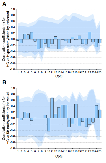
Individual DNA methylation values were not correlated across brain tissues, despite tissue concordance at the group level.
For each CpG, we computed the Pearson correlation coefficient r between methylation values for matched samples in pairs of brain regions (bars). Dark and light shaded regions represent 95% and 99% thresholds, respectively, of distributions of possible correlation coefficients determined from 10,000 permutations of the measured values among the individuals. These distributions represent the null hypothesis that an individual DNA methylation value in one brain region does not help to predict the value in another region in the same animal.
(A) Correlations based on pyrosequencing data for matched samples passing validation in both hippocampus (HC) and hypothalamus (Hypo). Correlations for individuals at each CpG were either weak (.2 < r < .3) or absent (r < .2), and none were significant, even prior to correction for multiple comparisons.
(B) Correlations for matched samples passing validation in both hippocampus and striatum (Str). Two correlations (CpG 1 and 11) were individually significant prior to but not following correction, and this result could be expected by chance.
Correlations between hippocampus and blood (described in the text) yielded similar results, and no particular CpG yielded consistently high correlation across multiple tissues.”
The study focused on whether or not an individual’s experience-dependent oxytocin receptor gene DNA methylation in one of the four studied tissues could be used to infer a significant effect in the three other tissues. The main finding was NO, it couldn’t!
The researchers’ other findings may have been strengthened had they also examined causes for the observed effects. The “natural variation in maternal licking and grooming” developed from somewhere, didn’t it?
The subjects’ mothers were presumably available for the same tests as the subjects, but nothing was done with them. Investigating at least one earlier generation may have enabled etiologic associations of “the effects of early life rearing experience” and “individual variation in DNA methylation.”
“Emotion categories [fear, anger, disgust, sadness, and happiness] are not contained within any one region or system, but are represented as configurations across multiple brain networks.
For example, among other systems, information diagnostic of emotion category was found in both large, multi-functional cortical networks and in the thalamus, a small region composed of functionally dedicated sub-nuclei.
The dataset consists of activation foci from 397 fMRI and PET [positron emission tomography] studies of emotion published between 1990 and 2011.”
From the fascinating Limitations section:
“Our analyses reflect the composition of the studies available in the literature, and are subject to testing and reporting biases on the part of authors. This is particularly true for the amygdala (e.g., the activation intensity for negative emotions may be over-represented in the amygdala given the theoretical focus on fear and related negative states). Other interesting distinctions were encoded in the thalamus and cerebellum, which have not received the theoretical attention that the amygdala has and are likely to be bias-free.
Some regions—particularly the brainstem—are likely to be much more important for understanding and diagnosing emotion than is apparent in our findings, because neuroimaging methods are only now beginning to focus on the brainstem with sufficient spatial resolution and artifact-suppression techniques.
We should not be too quick to dismiss findings in ‘sensory processing’ areas, etc., as methodological artifacts. Emotional responses may be inherently linked to changes in sensory and motor cortical processes that contribute to the emotional response.
The results we present here provide a co-activation based view of emotion representation. Much of the information processing in the brain that creates co-activation may not relate to direct neural connectivity at all, but rather to diffuse modulatory actions (e.g., dopamine and neuropeptide release, much of which is extrasynaptic and results in volume transmission). Thus, the present results do not imply direct neural connectivity, and may be related to diffuse neuromodulatory actions as well as direct neural communication.”
Why did the researchers use only 397 fMRI and PET studies? Why weren’t there tens or hundreds of times more candidate studies from which to select?
The relative paucity of candidate emotion studies demonstrated the prevalence of other researchers’ biases for cortical brain areas. The lead researcher of the current study was a coauthor of the 2016 Empathy, value, pain, control: Psychological functions of the human striatum, whose researchers mentioned that even their analyses of 5,809 human imaging studies was hampered by other imaging-studies researchers’ cortical biases.
Functional MRI signals depend on the changes in blood flow that follow changes in brain activity. Study designers intentionally limit their findings when they scan brain areas and circuits that are possibly activated by human emotions, yet exclude emotional content that may activate these areas and circuits.
Here are a few examples of limited designs that led to limited findings when there was the potential for so much more:
It’s well past time to change these practices now in the current year.
This study provided many methodological tests that should be helpful for research that includes emotion. It showed that there aren’t impenetrable barriers – other than popular memes, beliefs, and ingrained dogmas – to including emotional content in studies.
Including emotional content may often be appropriate and informative, with the resultant findings advancing science. Here are a few recent studies that did so:
This 2016 Italian human review considered the lower brain’s contributions to an individual’s behavior and temperament:
“In evidencing associations between personality factors and neurobiological measures, it seems evident that the cerebellum has not been up to now thought as having a key role in personality.
Cerebellar volumes correlate positively with novelty seeking scores and negatively with harm avoidance scores. Subjects who search for new situations as a novelty seeker does (and a harm avoiding does not do) show a different engagement of their cerebellar circuitries in order to rapidly adapt to changing environments.
Cerebellar abilities in planning, controlling, and putting into action the behavior are associated to normal or abnormal personality constructs. In this framework, it is worth reporting that increased cerebellar volumes are even associated with high scores in alexithymia, construct of personality characterized by impairment in cognitive, emotional, and affective processing.”
The full paper wasn’t freely available, but a list of the 173 references was. 17 references were of alexithymia, also mentioned in the title.
“Alexithymia scores were linked directly with cerebellar areas and inversely with limbic and para-limbic system, proposing a possible functional modality for the cerebellar involvement in emotional processing.
The increased volumes in Crus 1 of subjects with high alexithymic traits may be related to an altered embodiment process leading to not-cognitively interpreted emotions.”
“Alexithymia scores” referred to one of the methods used to characterize alexithymia symptoms, self-reported answers to questionnaires such as this one. Sample questions from the questionnaire used by the referenced study are:
“I am often confused about what emotion I am feeling
It is difficult for me to reveal my innermost feelings, even to close friends”
The questionnaire mainly engages a person’s cerebrum. The person may recall emotions, and form ideas as framed by each question. Then they’ll describe these ideas in terms of a scaled answer.
Cerebral answers may provide historical contexts for feelings. However, the person’s cerebellum and other brain areas aren’t necessarily engaged by the diagnostic questionnaire.
Without this engagement, the person may not experience feelings when providing answers about feelings. The answers may be more along the lines of “This is what I think I should be feeling” or “This is what I think I should tell the researchers about what I think I should feel.”
Can a questionnaire accurately determine associations among engaged and unengaged brain areas?
What can be done regarding “impairment in cognitive, emotional, and affective processing?”
What’s the lower brain’s “involvement in emotional processing?”
How does the lower brain shape a person’s behavior and traits?
When and where in an individual’s lifespan does their cerebellum develop?