Does sulforaphane treat autism?

A 2021 human study investigated sulforaphane treatments of autistic 3-to-12-year-olds:

“Sulforaphane (SF) led to non-statistically significant changes in the total and all subscale scores of the primary outcome measure. Several effects of SF on biomarkers correlated to clinical improvements. SF was very well tolerated and safe and effective based on our secondary clinical measures.

13229_2021_447_Fig1

Clinical response to SF was associated with changes in mitochondrial function, and large intrasubject variability in this study was linked to underlying biological responses. The increase in ATP [adenosine triphosphate]-Linked Respiration associated with improvement in ABC [Aberrant Behavior Checklist] scores suggests that those individuals who showed improvements in behavior also had improved mitochondrial capacity to produce ATP.

Individuals who showed an improvement in ABC scores also showed a decrease in Proton Leak Respiration, suggesting that their mitochondria were better able to regulate oxidative stress. It is also possible that the increase in ATP production was related to improvement in the ability of mitochondria to handle oxidative stress.

SF had significant positive effects on oxidative stress, cytoprotective markers and cytokines, as well as mitochondrial function. These were promising findings that require further investigation of both clinical effects and mechanisms of action of SF.”

https://molecularautism.biomedcentral.com/articles/10.1186/s13229-021-00447-5 “Randomized controlled trial of sulforaphane and metabolite discovery in children with Autism Spectrum Disorder”


Differences between this clinical trial and its pilot study curated in Autism biomarkers and sulforaphane included:

“HO-1 [heme oxygenase 1] functions to couple activation of mitochondrial biogenesis to anti-inflammatory cytokine expression. It was initially increased in the pilot study, then paradoxically decreased in the main study, on continued treatment for longer periods with SF.

Increased HO-1 is consistent with decreases in proinflammatory cytokines we observed initially in IL-6, IL-1β and TNF-α. Decreased levels of cytokines continued after HO-1 returned to baseline with longer duration of treatment and suggest a decreased inflammatory state.

These cytokines are usually elevated in children with ASD, but were decreased on treatment with SF: IL-6 and TNF-α at 15 (but not 30) weeks.”

This study made a good effort with autistic children. Its insignificant effects of sulforaphane treatments pointed toward an understanding that human experiences when we are fetuses, infants, and young children can override many subsequent events, treatments, and life experiences.

All about the betaine

A trio of papers on betaine, the first being a 2021 series of thorough rodent experiments relating betaine and gut microbiota, and cause and effect:

“Compared with lean individuals, adipose tissues in obese individuals secrete high levels of pro-inflammatory cytokines such as TNF-α, IL-1β, and IL-6, inducing:

  • Systemic inflammation;
  • Insulin resistance;
  • Large amounts of carcinogenic factors; and
  • Increasing risk of certain types of cancer such as melanoma, colon cancer, and liver cancer.

Prebiotics obtained from fruits and vegetables can regulate host lipid metabolism and glucose homeostasis by reversing gut dysbiosis in obese individuals.

kgmi_a_1862612_f0005_oc

Results of this study show that dietary betaine alleviated gut microbiota imbalance in obese mice, and reduced development of obesity and obesity-related complications. Regulation of the miR-378a-YY1 regulatory axis by gut microbial acetate and butyrate was a critical mechanism for modulating:

  • White adipose tissue browning;
  • Classical brown adipose tissue activation; and
  • Lipid and glucose homeostasis

in obese mice after betaine supplementation.

These findings offer novel insights into underlying mechanisms by which gut microbiota affect host metabolism and host immune system, and demonstrate that the betaine-gut microbiota-derived signal axis is a potential therapeutic target in obesity and metabolic syndrome.”

https://www.tandfonline.com/doi/full/10.1080/19490976.2020.1862612 “Dietary betaine prevents obesity through gut microbiota-drived microRNA-378a family”


A second 2021 paper was a meta-analysis of effects on human cardiovascular biomarkers:

“Betaine supplementation had a significant effect on concentrations of:

  • Betaine;
  • Total cholesterol;
  • Low-density lipoprotein (LDL);
  • Homocysteine [negative effect]; and
  • Methionine.

Betaine supplementation did not affect serum concentrations of:

  • Triglycerides;
  • High-density lipoprotein (HDL);
  • Fasting blood glucose;
  • C-reactive protein;
  • Liver enzymes alanine aminotransferase (ALT), aspartate aminotransferase (AST), gamma-glutamyl transferase (GGT); and
  • Blood pressure.

Our meta-analysis supports the advantage of a lower dose of betaine supplementation (<4 g/d) on homocysteine concentrations without the lipid-augmenting effect observed with a higher dosage.”

https://www.tandfonline.com/doi/abs/10.1080/10408398.2021.1902938 “Effects of betaine supplementation on cardiovascular markers: A systematic review and Meta-analysis” (not freely available)


A third paper was a 2014 cereal analysis of betaine and its precursor choline that found a 224% increase in betaine from 62 to 139 μg/g and a 31% increase in choline from microwaving oats:

“Betaine and its precursor choline are important components of one-carbon metabolism, remethylating homocysteine into methionine and providing methyl groups for DNA methylation. Cereals are the main source of betaine in diet.

During cooking processes which did not involve removal of water (in this case oat porridge microwaved using instant oats) appeared to lead to creation of betaine. Explanations for this phenomenon could be that betaine is synthesised during the process, or that heating with water liberates betaine from cereal matrix, enhancing efficiency of extraction.”

https://www.sciencedirect.com/science/article/abs/pii/S0308814613012247 “Cereal foods are the major source of betaine in the Western diet – Analysis of betaine and free choline in cereal foods and updated assessments of betaine intake” (not freely available)


Another 2021 betaine (aka trimethyl glycine) study was curated in Ride the waves of gene expression with betaine for its role in preventing nerve disease. I take 1.5 grams of a betaine supplement every morning and evening when eating hulled Avena sativa 3-day-old oat sprouts.

I found the first two papers from their citing a 2016 human and rodent study Dietary Betaine Supplementation Increases Fgf21 Levels to Improve Glucose Homeostasis and Reduce Hepatic Lipid Accumulation in Mice, which was linked in a comment on this 2021 video:

Small intestine alkaline phosphatase

This 2021 rodent study used small intestine alkaline phosphatase (IAP)-overexpressed subjects on a high-fat, high-cholesterol diet to investigate effects:

“To examine direct effects of increased IAP expression on barrier function and development of metabolic diseases, we developed intestine-specific IAP transgenic mice (IAPTg) overexpressing human chimeric IAP. We evaluated effects of intestine-specific IAP overexpression on Western-type diet (WD)–induced atherosclerosis in Ldlr−/ mice.

Diets low in fiber deprive intestinal bacteria of essential nutrients. Luminal bacteria turn to alternate sources of energy, namely, the carbohydrate-rich mucosal layer. This enhances direct contact between gut bacteria and intestinal epithelial layer, and promotes inflammation and intestinal barrier dysfunction.

Increase in IAP improves intestinal barrier function by not only dephosphorylating LPS and limiting its translocation to systemic circulation, but also by improving mucosal layer. Furthermore, IAP overexpression results in attenuated WD-induced weight gain and significantly reduced absorption of dietary lipids, leading to attenuation of total plasma cholesterol and TG levels, as well as hepatic lipids. This improved metabolic profile results in significant reduction in WD-induced atherosclerosis in Ldlr−/−IAPTg mice.

overexpressed IAP

IAP overexpression results in attenuated WD-induced weight gain and significantly reduced absorption of dietary lipids, leading to attenuation of total plasma cholesterol and TG levels, as well as hepatic lipids. This improved metabolic profile results in significant reduction in WD-induced atherosclerosis in Ldlr−/−IAPTg mice.

Increases in IAP can significantly attenuate effects of WD feeding on intestinal barrier function. It is noteworthy that IAP is also shown to be involved in innate immunity, and its activity is positively correlated to intestinal levels of IgA in mice and fecal immunoglobulins in humans.

The list of nutrients and food components/supplements that increase IAP continues to grow (galactooligosaccharides, glucomannan, vitamin D3), providing a novel opportunity to develop simple strategies for modulation of diet/nutrition to target metabolic diseases, including diabetes, fatty liver disease, atherosclerosis, or heart disease.”

https://www.ahajournals.org/doi/10.1161/CIRCRESAHA.120.317144 “Over-Expression of Intestinal Alkaline Phosphatase Attenuates Atherosclerosis”


This study used ileal samples from the small intestine’s last section for its findings. It complemented Take FOS or inulin to increase your gut’s alkaline phosphate activity which used large intestine samples to demonstrate effects of  increased IAP activity.

Eat to make your gut microbiota happy, and expect reciprocity.

A biological age snapshot from a year ago

This May 3, 2020 two-hour video on Optimizing Biological Age was instructive:


Content was great! I recommend the longish Q & A, especially at 1:23 regarding inflammation.

It was a snapshot in that researchers on this conference call were interested in improving people’s health. Few recognized at the time a globally coordinated effort to herd humans into one tracking database using an injurious jab as a pretext.

optimizing biological age

Take FOS or inulin to increase your gut’s alkaline phosphatase activity

This 2020 rodent study ran a series of experiments relating gut health factors. I left some items out that Google Translate didn’t handle well.

“This study investigated effects of food factors on colonic microbiota, fermentation products, mucins, immunoglobulin A (IgA) and alkaline phosphatase (ALP) activity. Colonic ALP activity was positively correlated with colonic luminal variables such as fecal mucin level, ratio of Bifidobacterium spp., and level of n-butyrate, which are associated with a more favorable colonic environment. We propose that the increase of colonic ALP activity induced by fermentable non-digestible carbohydrates may be important for protection of gut epithelial homeostasis.

Since glucomannan was found to induce colon ALP activity, effects of other dietary fibers were also investigated. Ingestion of water-soluble dietary fibers pectin and inulin significantly increased colon tissue and fecal ALP activity of high-fat diet-fed rats.

Ingestion of chitosan, an insoluble dietary fiber, had no effect on colonic ALP activity. This indicates that colonic ALP activity may be induced by indigestible sugars such as fermentable water-soluble dietary fiber.

ALP activity of the large intestine tissue of rats fed a high-fat diet was significantly increased by ingestion of indigestible oligosaccharides fructooligosaccharide (FOS), galactooligosaccharide (GOS), raffinose (RAF) and lactulose (LAC). Mucin, n-butyric acid, and Bifidobacterium spp. significantly increased, and Clostridium coccoides was significantly reduced.

ALP activity

In the digestible isomaltooligosaccharide (IMOS)-added diet group, large intestine ALP activity, ALP gene expression, mucin, organic acid, and intestinal flora showed no effect.

In order to investigate nutritional conditions on inducing colon ALP activity by oligosaccharide intake, the difference in lipid quality ingested was examined under a high-fat diet.

  • When soybean oil and lard were used as dietary fats, the difference in quality of dietary lipids did not affect large intestine ALP activity and IAP-I expression in the FOS-free diet.
  • When FOS was added, ALP activity and IAP-I expression in the large intestine tissue were significantly increased under the condition of a high lard diet as compared with a high soybean oil diet.
  • Mucin content behaved similarly to colon tissue ALP activity and IAP-I expression.

mucin content

In this study, it was newly found that the effect of indigestible oligosaccharide intake on increasing colon ALP activity differs depending on nutritional conditions such as type of lipid. Furthermore, it was found that the increase in colon ALP activity under indigestible sugar intake has a positive correlation with factors involved in maintaining function of the intestinal environment, such as mucin.”

https://www.jstage.jst.go.jp/article/jsnfs/74/1/74_9/_article “Modulation of the Colonic Luminal Environment by Food Factors” (in Japanese)


I arrived at this study by it citing a 2011 study Vitamin K1 (Phylloquinone) or Vitamin K2 (Menaquinone-4) Induces Intestinal Alkaline Phosphatase Gene Expression. More on IAP in this 2020 video:

Dietary fibers and the aged microbiome

This 2021 rodent study investigated effects of four different types of dietary fiber on two different types of aged human microbiota:

Individual differences in gut microbiota may influence host metabolic responses to dietary fiber in humans. Dietary fibers are edible carbohydrates resistant to host digestive enzymes, and not broken down or absorbed in the small intestine.

We colonized genetically identical germ-free mice with two distinct human fecal communities and fed them isocaloric diets containing different types of fiber. We used fecal specimens from a cohort of previously analyzed samples obtained from adults in their mid-seventies.

We used 10% dietary fiber and 35% kcal derived from fat as comparable to the intake level of dietary fiber in US adults:

four diets

All mice had the same assorted fiber diet for two weeks. Mice were then switched to one of four diets described above: cellulose, inulin, pectin, and assorted fiber, and maintained in these diets for another 4 weeks.

There was a ~ 4-fold range in levels of cecal butyrate among the eight groups despite all animals consuming the same diet [before switching]. Butyrate is known to vary widely among humans and has been linked with beneficial health effects on the host:

SCFA individual differences

We chose inulin and pectin as the former is commonly used as a prebiotic, while the latter has been proven to support growth of a wide variety of gut microbes, and it is commonly used as a dietary supplement. We also chose these two dietary fibers due to their distinct structures, including differences in basic units, linkages, and degree of polymerization.

Assorted fiber diet had the same total amount of dietary fiber as treatment groups used in this study, but with more diversity [FOS and two resistant starches] in fermentable substrates, which we reasoned would support engraftment of taxa relevant to all dietary treatments. Inclusion of this group in the experimental phase also served as a control to inform whether this diet used during colonization drove major differences.

Diet and its interaction with gut community showed a significant effect on serum glucose levels. While pectin diet had an overall beneficial effect on metabolic phenotypes relative to non-fermentable cellulose for SubA-colonized mice, this diet was less favorable for SubB-colonized animals, which showed the strongest benefits on inulin fiber.

In inulin diet, mice inoculated with SubB showed decreased adiposity, decreased liver triglycerides (TG) and lower serum levels of fasting glucose relative to animals colonized with SubA. In contrast, pectin-fed mice colonized with SubB accumulated more fat mass relative to SubA-colonized counterparts, whereas serum glucose and liver TG were comparable between the two community groups.

Mice colonized with SubB showed significantly lower levels of adiposity than those colonized with SubA in the assorted fiber diet, whereas serum glucose and liver TG were comparable.

We found that these two transplanted communities elicited divergent metabolic epigenetic and transcriptional responses to the same dietary fiber. Furthermore, differences between mice colonized with these two communities varied depending on type of fiber consumed.

Populations contain a significant amount of genetic variation derived from their largely individual associated microbiomes. Dissecting effects of gut microbial vs. host genetic variation while controlling environmental exposure is practically impossible to achieve in human studies.

One-size-fits-all approaches to promote health are unlikely to elicit consistent effects across individuals. Identifying gut microbial biomarkers associated with beneficial responses to common interventions may help to stratify subjects into more effective personalized treatments.”

https://microbiomejournal.biomedcentral.com/articles/10.1186/s40168-021-01061-6 “Gut microbiome variation modulates the effects of dietary fiber on host metabolism”


1. This study nailed it! You are what you eat, and The future of your brain is in your gut right now.

2. Group differences in cecal butyrate in the second graphic were instructive. But what really needed to be analyzed was each individual subject’s responses within the eight groups, and each individual’s characteristics.

What did or didn’t matter to each individual could then be applied and analyzed to what did or didn’t matter to its group. Researchers need to flip from a top-down statistics-package approach, to a bottoms-up individual paradigm for evidence.

3. Haven’t mentioned Increasing soluble fiber intake with inulin recently. I eat the labeled 2.5 grams serving. More than that runs into a 10 g “Over this dose would induce mild gastrointestinal symptoms” threshold.

I eat a half-dozen cloves of garlic in daily AGE-less chicken vegetable soup. Garlic contains ≈ 16% inulin, contributing 4-5 g inulin.

4. My dietary fiber intake of current practices is well beyond this study’s 10%. Several times more than our human ancestors’ estimated 100 g/day if Switch on your Nrf2 signaling pathway measurements are correct?

Trying to make my gut microbiota happy, expecting that they’ll reciprocally respond. Dietary fat content is < 10 %.

An outstanding review of Vitamin K deficiency and disease

This 2019 review focused on one Vitamin K-deficiency biomarker. All parts I’ve quoted are outside the liver, so Vitamin K deficiency ≈ Vitamin K2 deficiency.

This is a hard read with many technical details, but sometimes that’s how researchers do it:

“Active MGP (matrix Gla protein), once released into extracellular space, acts as a local inhibitor of calcification. Widespread expression of MGP points to a role of MGP that by far exceeds its well-known function as local inhibitor of calcification.

Recent research confirmed this concept, usually by measuring plasma dp-ucMGP (desphospho-uncarboxylated MGP), a biomarker reflecting poor vitamin K status:

1160fig02

Vitamin K plays a pivotal role in maintaining bone health. Increasing evidence also implicates MGP in maintaining bone health.

In the Health, Aging and Body Composition study, 791 older community-dwelling adults underwent magnetic resonance imaging to measure bilateral knee structural features. The highest [25%] compared with the lowest fourth of the dp-ucMGP distribution had higher odds of having:

  • Meniscus damage;
  • Osteophytes;
  • Bone marrow lesions; and
  • Subarticular cysts.

Regarding Vitamin K supplementation:

  • Studies showed a dose-dependent decrease in circulating dp-ucMGP with an 86% decrease already observed after 4 weeks of substitution by 360 μg menaquinone-7 [in 50 hemodialysis patients];
  • In a randomized double-blind trial of 244 postmenopausal women followed up for 3 years, arterial stiffness as captured by aortic pulse wave velocity or stiffness index β, decreased in intervention compared with control group.

These results should be considered as hypothesis-generating in view of small sample size, and because there were no between-group differences in vitamin K–induced changes in elastic properties of the carotid artery.

Plasma dp-ucMGP levels ranging from 1.4 to 4.6 μg/L were optimal in terms of risk of mortality and macrovascular cardiovascular illness (4.6 μg/L threshold corresponding to the 65th percentile of dp-ucMGP distribution).

Vitamin K supplementation before irreversible organ damage sets in might find its application in prevention of a wide range of disabling diseases. Circulating dp-ucMGP levels might be measured over time to track risk of vascular complications.”

https://www.ahajournals.org/doi/10.1161/HYPERTENSIONAHA.119.12412 “Vitamin K–Dependent Matrix Gla Protein as Multifaceted Protector of Vascular and Tissue Integrity”


I usually don’t give 5+ stars to reviews. This one was different.

Yes, there could be factors other than this one Vitamin K deficiency biomarker involved in study findings. Sure, these coauthors cited their own studies. Its overall purpose, though, was to inform readers.

I’ll summarize this paper as providing evidence for a biomarker of Vitamin K2 deficiency being implicated in the development and progression of many diseases.

Part 2 of Vitamin K2 – What can it do?

Two papers on Vitamin K2, and an online database to continue Part 1:

“Precise quantitative assessments of vitamin K bioavailability in humans is challenging due to unquantified tissue conversion of PK [phylloquinone, Vitamin K1] to MK [menaquinone, Vitamin K2]-4, and contributions of gut microbiota. Absorption of long-chain MKs (MK-7, MK-8 and MK-9) from natto, cheese and egg yolk is close to 100%.

Long-chain MKs have a longer half-life. Long half-life may not necessarily indicate increased bioavailability, but instead non-preferential utilisation by tissues compared to PK and MK-4. A long half-life may also indicate that long-chain MKs may be of particular importance for extrahepatic tissues.

12 databases list vitamin K content of individual food items, which is required to more accurately determine vitamin K intake. The Dutch database is the most comprehensive, and includes PK and several types of MKs, ranging from MK-4 to MK-10.”

https://pubs.rsc.org/en/content/articlelanding/2020/FO/C9FO02321F “Quantifying dietary vitamin K and its link to cardiovascular health: a narrative review” (not freely available)


One online database mentioned is at https://www.rivm.nl/en/dutch-food-composition-database:

“The Dutch Food Composition Database (NEVO) contains data on the composition of foods eaten frequently by a large part of the Dutch population. These foods contribute significantly to the intake of energy and nutrients. Foods of importance for specific groups of the Dutch population are also included.

The NEVO online website contains data on 133 nutrients of 2152 food items. The most recent version of NEVO online dates from November 2019.”

I downloaded a copy in Excel format, selected all, and sorted by column EE “VITK2_mug” in descending order. There were 263 food items with Vitamin K2 measurements.

Vitamin K2 food content


A second paper detailed a 2021 double-blind, placebo-controlled, feasibility study:

“INTRICATE will assess the influence of combined vitamin K2 and vitamin D3 supplementation on micro-calcification in carotid artery disease. Considering recent advancements in medical imaging, ultrasound, PET/MRI, and computed tomography can be used for selection and stratification of patients with atherosclerosis.

Subjects will be randomized (1:1) to a vitamin K2 (400 µg MK-7/day) and vitamin D3 (80 µg [3200 IU]/day) dose or to placebo. Primary endpoint is change in Na[18F]F PET/MRI (baseline vs. after 3 months) in treatment group as compared to placebo arm. Secondary endpoints are changes in plaque composition and in blood-biomarkers.

Studies suggest positive effects of vitamin D on vitamin K-dependent metabolism. The MGP-gene promoter contains a vitamin D response element, capable of a two to threefold enhanced MGP expression after vitamin D binding.

Upregulation of MGP due to vitamin D needs vitamin K to ensure full activation of MGP for optimal functioning. This implies that the combination of both vitamin K and vitamin D could provide enhanced protection against progressive vascular calcification, cardiovascular disease, and mortality.”

https://www.mdpi.com/2072-6643/13/3/994/htm “Effects of Combined Vitamin K2 and Vitamin D3 Supplementation on Na[18F]F PET/MRI in Patients with Carotid Artery Disease: The INTRICATE Rationale and Trial Design”


The second study was somewhat of a tell in that after two decades, researchers are still testing Vitamin K2 dose efficacy. Researchers don’t consider it proper science to not use a statistics package to lump subjects into groups.

Someday researchers will thoroughly analyze each individual, and relate measurements to each individual’s causal and symptomatic characteristics. Then we’ll find out whether what did or didn’t matter to each individual, will or won’t matter to a group.

Until then they’ll focus on one dimension of health like Vitamin K2 foods per their sponsor’s directions. Nevermind that Vitamin K2-rich foods like cheeses are full of advanced glycation end products (AGEs) that humans can’t adequately metabolize, to our detriment.

Vitamin K2 – What can it do?

A trio of papers on Vitamin K2, the first being a 2021 review that emphasized dual effects:

“Osteoporosis (OP) is the most common bone disease that affects elderly men and women. It is a metabolic skeletal disorder caused by an imbalance between bone formation and resorption, leading to a loss of bone mass and quality, skeletal structure deterioration, and an increased risk of fractures.

Vascular calcification is defined as ectopic deposition of mineral matrix in vessel wall. It occurs prevalently in aging and primary chronic conditions (hypertension, diabetes mellitus, and chronic kidney disease), representing an important risk factor for cardiovascular morbidity and mortality.

Studies have provided support for a close link between bone and vascular health. Findings suggest that bone loss in OP may promote and increase the risk of cardiovascular events and vascular atherosclerosis.

Vitamin K2 is involved in a phenomenon in which a low calcium deposition in bone tends to be associated with a parallel increase of calcium deposition in vessel wall as a consequence of impaired calcium metabolism. Most production of Vitamin K2 in humans takes place in intestines. However, the amount derived from intestinal bacteria is poorly absorbed, and is not able to reach concentrations required to exert physiological functions.

Vitamin K2‘s ability to reduce loss of bone mineral density and fracture risk, as well as to improve bone quality, has been described by several clinical studies, which have confirmed that osteocalcin (OC) γ-carboxylation is the main mechanism of action through which this natural compound is able to improve bone health. Clinical evidence suggests an analogous protective role of Vitamin K2 at the vascular level, emphasizing a strict association between:

  • Vitamin serum level;
  • Matrix gla protein (MGP) γ-carboxylation levels;
  • Reduction of vascular smooth muscle cells osteogenic trans-differentiation; and
  • Possible risk of cardiovascular events.”

https://www.mdpi.com/2072-6643/13/4/1222/htm “The Dual Role of Vitamin K2 in ‘Bone-Vascular Crosstalk’: Opposite Effects on Bone Loss and Vascular Calcification”


A second 2021 review emphasized aging:

“Vitamin K can:

  • Carboxylate OC (a protein capable of transporting and fixing calcium in bone);
  • Activate MGP (an inhibitor of vascular calcification and cardiovascular events); and
  • Carboxylate Gas6 protein (involved in brain physiology and a cognitive decline and neurodegenerative disease inhibitor).

By improving insulin sensitivity, Vitamin K lowers diabetes risk. It also exerts antiproliferative, proapoptotic, autophagic effects, and has been associated with a reduced risk of cancer.

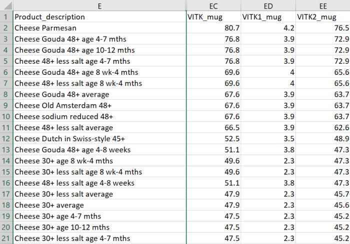
The most common [Vitamin K2] subtypes in humans are the short-chain MK[menaquinone]-4, which is the only MK produced by systemic conversion of phylloquinone [Vitamin K1] to menaquinone, and MK-7 through MK-10, which are synthesized by bacteria. The main sources of Vitamin K2 are fermented foods, cheeses, eggs, and meats.”

https://www.mdpi.com/2076-3921/10/4/566/htm “The Role of Vitamin K in Humans: Implication in Aging and Age-Associated Diseases”


The third paper – somehow not cited by these two reviews – was a 2006 human study that performed four experiments:

“The synthetic short-chain vitamin K1 is commonly used in food supplements, but recently the natural long-chain MK-7 has also become available as an over-the-counter supplement. The purpose of this paper was to compare in healthy volunteers absorption and efficacy of K1 and MK-7.

Serum vitamin K species were used as a marker for absorption and OC carboxylation as a marker for activity. Both K1 and MK-7 were absorbed well, with peak serum concentrations at 4 hours after intake.

A major difference was:

  • Very long half-life time of MK-7, resulting in much more stable serum levels; and
  • Accumulation of MK-7 to higher levels (7- to 8-fold) during prolonged intake.

MK-7 induced more complete carboxylation of OC.

Vitamin K2 vs K1

Accumulation and efficacy of K vitamins during long-term daily administration. Participants received in a crossover design either K1 (○) or MK-7 (•) or placebo; in the latter case only K1 (▴) could be detected.

  • (A) Circulating levels of vitamin K; baseline levels for K1 were subtracted; no MK-7 could be detected at baseline.
  • (B) Ratio between circulating carboxylated and undercarboxylated osteocalcin (cOC/ucOC); at baseline the ratio was 1.74 for MK-7, 1.8 for K1, and 1.7 for the placebo group.

MK-7 accumulated during the first 2 weeks until it reached a plateau level of about 10 nM (6 μg/L), whereas K1 remained slightly above placebo values during the entire study period. Efficacy of both K vitamins for OC carboxylation was monitored using the ratio between circulating cOC and ucOC, and it turned out that within 3 days both vitamins had induced increased cOC.

But only by taking MK-7 did the effect continue to increase during the entire study period.

Taken together, these data demonstrate considerable differences between MK-7 and K1:

  • Higher and more stable serum levels are reached with MK-7; and
  • MK-7 has a higher efficacy in both hepatic and extrahepatic protein carboxylation.”

https://ashpublications.org/blood/article/109/8/3279/23729/Vitamin-K-containing-dietary-supplements “Vitamin K–containing dietary supplements: comparison of synthetic vitamin K1 and natto-derived menaquinone-7″


I’ve tried various things over the years to address hypertension. I stopped high blood pressure medications briefly to see if each intervention worked. They all haven’t, presumably because I didn’t address causes.

More recently, I broke my left big toe on furniture while walking around in the dark last month, and haven’t recovered. No pictures from walking on the beach at sunrise because it still isn’t possible. 😦

A link between these two health conditions could be Vitamin K2. I don’t eat fermented foods because of their high sodium, or dairy products, and haven’t supplemented Vitamin K2.

Next week I’ll start a 300 μg MK-7 daily dose. Current Vitamin D3 dose is 3800 IU, compared to the second paper of Part 2 of Vitamin K2 – What can it do? which is 400 μg MK-7 and 3200 Vitamin D3.

Are overdosed rodent studies relevant to humans?

I partially read more than a dozen studies this week of overdosed rodents producing p < .05 significant results. Net effect was to thwart the purpose of rodent studies – to help humans.

The latest came from search term “SIRT1” “DHEA” 2021 after I read a 2021 study Dehydroepiandrosterone protects against acetaminophen-induced liver damage in rats by upregulation of Bcl-2 and activation of sirt signalling that found:

“The study examined protective effect of exogenous administration of dehydroepiandrosterone (DHEA) against acetaminophen (APAP) -induced liver damage in rats, and tested underlying mechanisms. DHEA prevents APAP-induced liver damage by concomitant upregulation of Bcl-2 and SIRT1-dependent effect.”

The daily DHEA dose was 50 mg/kg, which is a (.162 x 50 mg) x 70 kg = 567 mg human equivalent. Eleven times the most frequent human dose of Take responsibility for your one precious life – DHEA. Anyone who took this study’s DHEA amount would hurt themself.

The one-time acetaminophen dose was 800 mg/kg which is a (.162 x 800 mg) x 70 kg = 9 grams human equivalent. Someone who took 18 pills of the most frequent 500 mg acetaminophen dose would be attempting suicide.

How does this help humans?

apap dhea overdoses


See posts like Problematic rodent sulforaphane studies and Human relevance of rodent sulforaphane studies for further evidence and observations.

Eat oats and regain cognitive normalcy

This 2020 rodent study investigated effects of different diets:

“The present study aimed to evaluate effects of β-glucan on the microbiota gut-brain axis and cognitive function in an obese mouse model induced by a high-fat and fiber-deficient diet (HFFD). After long-term supplementation for 15 weeks, β-glucan prevented HFFD-induced cognitive impairment, assessed behaviorally by object location, novel object recognition, and nesting building tests:

  • Long-term β-glucan supplementation suppressed microglia activation and inflammation in hippocampus of HFFD-fed mice;
  • β-glucan attenuated deleterious engulfment of synapses by activation of microglia seen in HFFD mice;
  • β-glucan significantly prevented upregulation of TNF-α, IL-1β, and IL-6 mRNA expression in hippocampus; and
  • A broad-spectrum antibiotic intervention abrogated β-glucan-induced improvement in cognitive function, highlighting the essential role of gut microbiota to mediate cognitive function and behavior.

We found that short-term β-glucan supplementation did not change cognitive behavior in HFFD fed mice. HFFD feeding for 7 days dramatically changed gut microbial profile, with β-glucan-fed mice clustered apart from HFFD-fed mice sample, suggesting:

  • Quick changes in gut microbiota are induced by short-term β-glucan consumption and
  • Possible causality of gut microbiota profile on cognition.

7% β-glucan 7% nondigestible fiber

β-glucan supplementation increased place discrimination ratio in object location test compared with HFFD mice; however, there was no significant difference in total exploration time with objects during test phases between the two groups. Higher place discrimination index in β-glucan supplementation group was not due to better general performance, but increased recognition memory.

Results provide consistent evidence linking increased β-glucan intake to improved:

  • Gut microbiota profile;
  • Intestinal barrier function;
  • Reduced endotoxemia; and
  • Enhanced cognitive function via more optimized synaptic and signaling pathways in critical brain areas.

It is speculative that β-glucan improvement of gut microbiota composition, but not necessarily diversity per se, may be most critical for improved cognition. Enhanced consumption of β-glucan-rich foods is an easily implementable nutritional strategy to attenuate diet-induced cognitive decline.”

https://microbiomejournal.biomedcentral.com/articles/10.1186/s40168-020-00920-y “β-glucan attenuates cognitive impairment via the gut-brain axis in diet-induced obese mice”


This study did well by elaborating It’s the fiber, not the fat and Eat oats to prevent diabetes related findings. How many humans eat themselves into essentially the same situation as this HFFD group with no gut-microbiota-friendly dietary fiber?

Experiments were with β-glucan 1,3/1,4 found in oats. β-glucan 1,3/1,6 has separate effects, especially on innate immunity.

It’s a coin toss on whether observed cognitive improvement was due to 7% β-glucan soluble fiber, 7% indigestible fiber, or both since they were part of the same HFBG diet. I eat both fibers, beginning with Avena nuda oats for breakfast.

Astaxanthin bioavailability

By request, research on astaxanthin bioavailability. I used a “astaxanthin” “bioavailability” “quinone reductase” 2021 search term, and read citing papers.

“The bioaccessibility, bioavailability, and antioxidative activities of three astaxanthin geometric isomers were investigated using an in vitro digestion model.

  • 13Z-Astaxanthin showed higher bioaccessibility than 9Z- and all-E-astaxanthins during in vitro digestion, and
  • 9Z-astaxanthin exhibited higher transport efficiency than all-E- and 13Z-astaxanthins.

These might explain why 13Z- and 9Z-astaxanthins are found at higher concentrations in human plasma than all-E-astaxanthin.

9Z- and 13Z- astaxanthins exhibited a higher protective effect than all-E-astaxanthin against oxidative stress.”

https://pubs.acs.org/doi/10.1021/acs.jafc.7b04254 “Bioaccessibility, Cellular Uptake, and Transport of Astaxanthin Isomers and their Antioxidative Effects in Human Intestinal Epithelial Caco-2 Cells” (2017, not freely available)


jf1c00087_0005

“Astaxanthin with a high proportion of Z-isomer (especially rich in 9Z- and 13Z-isomers) was prepared from (all-E)-astaxanthin by thermal treatment and solid–liquid separation. Z-isomer-rich astaxanthin diet resulted in higher levels of astaxanthin in blood and many tissues (in particular, skin, lung, prostate, and eye) compared to all-E-isomer-rich diet.

Z-isomer-rich diet enhanced the level of 13Z-isomer in blood and tissues rather than that of 9Z-isomer. (13Z)-astaxanthin would have higher bioavailability and tissue accumulation than other isomers.”

https://pubs.acs.org/doi/10.1021/acs.jafc.1c00087Z-Isomers of Astaxanthin Exhibit Greater Bioavailability and Tissue Accumulation Efficiency than the All-E-Isomer” (2021, not freely available)


“Astaxanthin is highly susceptible to light, oxygen, and heat stress degradation. In addition, poor water solubility and bioavailability limit its efficacy in vivo. Investigating novel astaxanthin delivery systems is necessary in order to solve these drawbacks.”

https://www.mdpi.com/1420-3049/24/14/2640/htm “The Neuroprotective Effects of Astaxanthin: Therapeutic Targets and Clinical Perspective” (2019)


“Astaxanthin Z-isomers potentially have greater bioavailability and biological activity than (all-E)-astaxanthin. However, stability of Z-isomers is lower than all-E-isomer, which is a serious problem affecting its practical use.

In this study, we investigated impacts of different suspension media (oils and fats) and additives on astaxanthin isomer stability.

  • Z-isomers of astaxanthin isomerized to all-E-isomer during storage.
  • When soybean and sunflower oils were used as the suspension medium, astaxanthin isomers were hardly degraded. However the total Z-isomer ratio decreased from ~80% to ~50% during 6-week storage at 30 °C.
  • (9Z)-astaxanthin showed higher stability than 13Z- and 15Z-isomers.”

https://www.sciencedirect.com/science/article/abs/pii/S0308814621003770 “Evaluation and improvement of storage stability of astaxanthin isomers in oils and fats” (2021, not freely available)


I looked for but didn’t find a graph similar to this one that comparatively plotted astaxanthin:

OMCL2019-2716870.006

I also didn’t find recent human studies.

It seems that a special delivery system is required for taking astaxanthin as a supplement. It would require investigating manufacturers’ claims about isomer content and stability.

Eating colorful seafood is another way to get astaxanthin. Don’t know about eating raw or dried algae.

Are rodent models of human neurodegenerative diseases realistic?

This 2020 stem cell review argued against rodent models of human neurodegenerative diseases:

“Neuronal loss is not caused solely by intrinsic degenerative processes but rather via impaired interactions with surrounding glia and other brain cells. Dysfunctional astrocytes do not provide sufficient nutrients and antioxidants to neurons, while dysfunctional microglia cannot efficiently clear pathogens and cell debris from extracellular space, resulting in chronic inflammatory processes in the brain.

Human glia, especially astrocytes, differ significantly in morphology and function from their mouse counterparts. Recent advances in stem cell technology make it possible to reprogram human patients’ somatic cells to induced pluripotent stem cells (iPSC) and differentiate them further into patient‐specific glia and neurons, thus providing a source of human brain cells.

stem3309-fig-0002-m

Astrocytes do not efficiently utilize energy resources and cannot provide adequate metabolic support to neurons. A coculture of healthy human neurons with diseased astrocytes impaired neuronal calcium responses to glutamate and γ‐aminobutyric acid (GABA) as compared to coculture with healthy human astrocytes.

Treatment with sulforaphane:

  • Normalized basal level glycolysis;
  • Decreased basal level Aβ42 secretion; as well as
  • Ameliorated inflammatory response to pro‐inflammatory cytokines TNF-α and IL1-β in PSEN1 mutant iPSC astrocytes.

It is essential to make sure that what we see in the dish is the real patient‐specific phenotype. Transplantation of human brain organoids containing microglia into mice could provide a novel tool for drug screening in vivo.”

https://stemcellsjournals.onlinelibrary.wiley.com/doi/full/10.1002/stem.3309 “Metabolic and immune dysfunction of glia in neurodegenerative disorders: Focus on iPSC models”


This review’s thesis seems plausible. However, one problem with in vitro stem cell studies is that they often don’t have a control group.

Giving children allergies with pets

This 2021 human study investigated development and persistence of allergies:

“Allergic rhinitis (AR) is a common IgE-mediated disorder involving troublesome symptoms of nasal congestion, nasal itch, sneezing, and associated eye symptoms. Like many chronic health conditions, AR stems from complex gene–environment interactions.

130 subjects with AR were recruited. Control population included 154 healthy children who underwent a regular physical examination in the same ear, nose and throat clinic as AR patients. Individuals with history of asthma or atopic dermatitis were excluded.

AR analysis

Plenty of contradictory associations exist as whether furred pet exposure (cats and dogs) may be a risk or a protective factor for AR development. Discrepancies are likely due to the ubiquitous nature of pet allergens, while pet owners are more concerned about sanitation and many other hygiene-related reasons.

Interaction of early-life pet exposure with methylation level of ADAM33 increased the risk for AR onset 1.423 times more in children. This study provides evidence that:

  • Early-life pet exposure and low methylation level of ADAM33 increase AR risk in children; and
  • The interaction between pet exposure and methylation level of ADAM33 may play an important role in development of AR.”

https://aacijournal.biomedcentral.com/articles/10.1186/s13223-021-00526-5 “Interaction between early-life pet exposure and methylation pattern of ADAM33 on allergic rhinitis among children aged 3–6 years in China”


There’s nothing children can do about who their parents were. Exposing them to pet allergens, though, may be another example of early-life experiences causing lifelong effects.