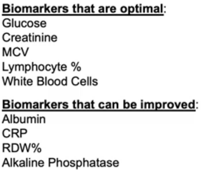
Take FOS or inulin to increase your gut’s alkaline phosphatase activity

This 2020 rodent study ran a series of experiments relating gut health factors. I left some items out that Google Translate didn’t handle well.

“This study investigated effects of food factors on colonic microbiota, fermentation products, mucins, immunoglobulin A (IgA) and alkaline phosphatase (ALP) activity. Colonic ALP activity was positively correlated with colonic luminal variables such as fecal mucin level, ratio of Bifidobacterium spp., and level of n-butyrate, which are associated with a more favorable colonic environment. We propose that the increase of colonic ALP activity induced by fermentable non-digestible carbohydrates may be important for protection of gut epithelial homeostasis.

Since glucomannan was found to induce colon ALP activity, effects of other dietary fibers were also investigated. Ingestion of water-soluble dietary fibers pectin and inulin significantly increased colon tissue and fecal ALP activity of high-fat diet-fed rats.

Ingestion of chitosan, an insoluble dietary fiber, had no effect on colonic ALP activity. This indicates that colonic ALP activity may be induced by indigestible sugars such as fermentable water-soluble dietary fiber.

ALP activity of the large intestine tissue of rats fed a high-fat diet was significantly increased by ingestion of indigestible oligosaccharides fructooligosaccharide (FOS), galactooligosaccharide (GOS), raffinose (RAF) and lactulose (LAC). Mucin, n-butyric acid, and Bifidobacterium spp. significantly increased, and Clostridium coccoides was significantly reduced.

ALP activity

In the digestible isomaltooligosaccharide (IMOS)-added diet group, large intestine ALP activity, ALP gene expression, mucin, organic acid, and intestinal flora showed no effect.

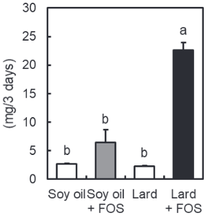
In order to investigate nutritional conditions on inducing colon ALP activity by oligosaccharide intake, the difference in lipid quality ingested was examined under a high-fat diet.

  • When soybean oil and lard were used as dietary fats, the difference in quality of dietary lipids did not affect large intestine ALP activity and IAP-I expression in the FOS-free diet.
  • When FOS was added, ALP activity and IAP-I expression in the large intestine tissue were significantly increased under the condition of a high lard diet as compared with a high soybean oil diet.
  • Mucin content behaved similarly to colon tissue ALP activity and IAP-I expression.
mucin content

In this study, it was newly found that the effect of indigestible oligosaccharide intake on increasing colon ALP activity differs depending on nutritional conditions such as type of lipid. Furthermore, it was found that the increase in colon ALP activity under indigestible sugar intake has a positive correlation with factors involved in maintaining function of the intestinal environment, such as mucin.”

https://www.jstage.jst.go.jp/article/jsnfs/74/1/74_9/_article “Modulation of the Colonic Luminal Environment by Food Factors” (in Japanese)


I arrived at this study by it citing a 2011 study Vitamin K1 (Phylloquinone) or Vitamin K2 (Menaquinone-4) Induces Intestinal Alkaline Phosphatase Gene Expression. More on IAP in this 2020 video:
Part 2 of Vitamin K2 – What can it do?

Two papers on Vitamin K2, and an online database to continue Part 1:

“Precise quantitative assessments of vitamin K bioavailability in humans is challenging due to unquantified tissue conversion of PK [phylloquinone, Vitamin K1] to MK [menaquinone, Vitamin K2]-4, and contributions of gut microbiota. Absorption of long-chain MKs (MK-7, MK-8 and MK-9) from natto, cheese and egg yolk is close to 100%.

Long-chain MKs have a longer half-life. Long half-life may not necessarily indicate increased bioavailability, but instead non-preferential utilisation by tissues compared to PK and MK-4. A long half-life may also indicate that long-chain MKs may be of particular importance for extrahepatic tissues.

12 databases list vitamin K content of individual food items, which is required to more accurately determine vitamin K intake. The Dutch database is the most comprehensive, and includes PK and several types of MKs, ranging from MK-4 to MK-10.”

https://pubs.rsc.org/en/content/articlelanding/2020/FO/C9FO02321F “Quantifying dietary vitamin K and its link to cardiovascular health: a narrative review” (not freely available)


One online database mentioned is at https://www.rivm.nl/en/dutch-food-composition-database:

“The Dutch Food Composition Database (NEVO) contains data on the composition of foods eaten frequently by a large part of the Dutch population. These foods contribute significantly to the intake of energy and nutrients. Foods of importance for specific groups of the Dutch population are also included.

The NEVO online website contains data on 133 nutrients of 2152 food items. The most recent version of NEVO online dates from November 2019.”

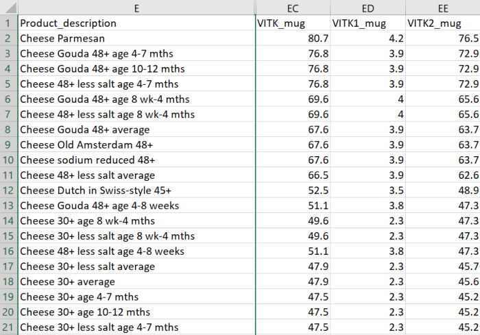
I downloaded a copy in Excel format, selected all, and sorted by column EE “VITK2_mug” in descending order. There were 263 food items with Vitamin K2 measurements.

Vitamin K2 food content


A second paper detailed a 2021 double-blind, placebo-controlled, feasibility study:

“INTRICATE will assess the influence of combined vitamin K2 and vitamin D3 supplementation on micro-calcification in carotid artery disease. Considering recent advancements in medical imaging, ultrasound, PET/MRI, and computed tomography can be used for selection and stratification of patients with atherosclerosis.

Subjects will be randomized (1:1) to a vitamin K2 (400 µg MK-7/day) and vitamin D3 (80 µg [3200 IU]/day) dose or to placebo. Primary endpoint is change in Na[18F]F PET/MRI (baseline vs. after 3 months) in treatment group as compared to placebo arm. Secondary endpoints are changes in plaque composition and in blood-biomarkers.

Studies suggest positive effects of vitamin D on vitamin K-dependent metabolism. The MGP-gene promoter contains a vitamin D response element, capable of a two to threefold enhanced MGP expression after vitamin D binding.

Upregulation of MGP due to vitamin D needs vitamin K to ensure full activation of MGP for optimal functioning. This implies that the combination of both vitamin K and vitamin D could provide enhanced protection against progressive vascular calcification, cardiovascular disease, and mortality.”

https://www.mdpi.com/2072-6643/13/3/994/htm “Effects of Combined Vitamin K2 and Vitamin D3 Supplementation on Na[18F]F PET/MRI in Patients with Carotid Artery Disease: The INTRICATE Rationale and Trial Design”


The second study was somewhat of a tell in that after two decades, researchers are still testing Vitamin K2 dose efficacy. Researchers don’t consider it proper science to not use a statistics package to lump subjects into groups.

Someday researchers will thoroughly analyze each individual, and relate measurements to each individual’s causal and symptomatic characteristics. Then we’ll find out whether what did or didn’t matter to each individual, will or won’t matter to a group.

Until then they’ll focus on one dimension of health like Vitamin K2 foods per their sponsor’s directions. Nevermind that Vitamin K2-rich foods like cheeses are full of advanced glycation end products (AGEs) that humans can’t adequately metabolize, to our detriment.

Vitamin K2 – What can it do?

A trio of papers on Vitamin K2, the first being a 2021 review that emphasized dual effects:

“Osteoporosis (OP) is the most common bone disease that affects elderly men and women. It is a metabolic skeletal disorder caused by an imbalance between bone formation and resorption, leading to a loss of bone mass and quality, skeletal structure deterioration, and an increased risk of fractures.

Vascular calcification is defined as ectopic deposition of mineral matrix in vessel wall. It occurs prevalently in aging and primary chronic conditions (hypertension, diabetes mellitus, and chronic kidney disease), representing an important risk factor for cardiovascular morbidity and mortality.

Studies have provided support for a close link between bone and vascular health. Findings suggest that bone loss in OP may promote and increase the risk of cardiovascular events and vascular atherosclerosis.

Vitamin K2 is involved in a phenomenon in which a low calcium deposition in bone tends to be associated with a parallel increase of calcium deposition in vessel wall as a consequence of impaired calcium metabolism. Most production of Vitamin K2 in humans takes place in intestines. However, the amount derived from intestinal bacteria is poorly absorbed, and is not able to reach concentrations required to exert physiological functions.

Vitamin K2‘s ability to reduce loss of bone mineral density and fracture risk, as well as to improve bone quality, has been described by several clinical studies, which have confirmed that osteocalcin (OC) γ-carboxylation is the main mechanism of action through which this natural compound is able to improve bone health. Clinical evidence suggests an analogous protective role of Vitamin K2 at the vascular level, emphasizing a strict association between:

  • Vitamin serum level;
  • Matrix gla protein (MGP) γ-carboxylation levels;
  • Reduction of vascular smooth muscle cells osteogenic trans-differentiation; and
  • Possible risk of cardiovascular events.”

https://www.mdpi.com/2072-6643/13/4/1222/htm “The Dual Role of Vitamin K2 in ‘Bone-Vascular Crosstalk’: Opposite Effects on Bone Loss and Vascular Calcification”


A second 2021 review emphasized aging:

“Vitamin K can:

  • Carboxylate OC (a protein capable of transporting and fixing calcium in bone);
  • Activate MGP (an inhibitor of vascular calcification and cardiovascular events); and
  • Carboxylate Gas6 protein (involved in brain physiology and a cognitive decline and neurodegenerative disease inhibitor).

By improving insulin sensitivity, Vitamin K lowers diabetes risk. It also exerts antiproliferative, proapoptotic, autophagic effects, and has been associated with a reduced risk of cancer.

The most common [Vitamin K2] subtypes in humans are the short-chain MK[menaquinone]-4, which is the only MK produced by systemic conversion of phylloquinone [Vitamin K1] to menaquinone, and MK-7 through MK-10, which are synthesized by bacteria. The main sources of Vitamin K2 are fermented foods, cheeses, eggs, and meats.”

https://www.mdpi.com/2076-3921/10/4/566/htm “The Role of Vitamin K in Humans: Implication in Aging and Age-Associated Diseases”


The third paper – somehow not cited by these two reviews – was a 2006 human study that performed four experiments:

“The synthetic short-chain vitamin K1 is commonly used in food supplements, but recently the natural long-chain MK-7 has also become available as an over-the-counter supplement. The purpose of this paper was to compare in healthy volunteers absorption and efficacy of K1 and MK-7.

Serum vitamin K species were used as a marker for absorption and OC carboxylation as a marker for activity. Both K1 and MK-7 were absorbed well, with peak serum concentrations at 4 hours after intake.

A major difference was:

  • Very long half-life time of MK-7, resulting in much more stable serum levels; and
  • Accumulation of MK-7 to higher levels (7- to 8-fold) during prolonged intake.

MK-7 induced more complete carboxylation of OC.

Vitamin K2 vs K1

Accumulation and efficacy of K vitamins during long-term daily administration. Participants received in a crossover design either K1 (○) or MK-7 (•) or placebo; in the latter case only K1 (▴) could be detected.

  • (A) Circulating levels of vitamin K; baseline levels for K1 were subtracted; no MK-7 could be detected at baseline.
  • (B) Ratio between circulating carboxylated and undercarboxylated osteocalcin (cOC/ucOC); at baseline the ratio was 1.74 for MK-7, 1.8 for K1, and 1.7 for the placebo group.

MK-7 accumulated during the first 2 weeks until it reached a plateau level of about 10 nM (6 μg/L), whereas K1 remained slightly above placebo values during the entire study period. Efficacy of both K vitamins for OC carboxylation was monitored using the ratio between circulating cOC and ucOC, and it turned out that within 3 days both vitamins had induced increased cOC.

But only by taking MK-7 did the effect continue to increase during the entire study period.

Taken together, these data demonstrate considerable differences between MK-7 and K1:

  • Higher and more stable serum levels are reached with MK-7; and
  • MK-7 has a higher efficacy in both hepatic and extrahepatic protein carboxylation.”

https://ashpublications.org/blood/article/109/8/3279/23729/Vitamin-K-containing-dietary-supplements “Vitamin K–containing dietary supplements: comparison of synthetic vitamin K1 and natto-derived menaquinone-7″


I’ve tried various things over the years to address hypertension. I stopped high blood pressure medications briefly to see if each intervention worked. They all haven’t, presumably because I didn’t address causes.

More recently, I broke my left big toe on furniture while walking around in the dark last month, and haven’t recovered. No pictures from walking on the beach at sunrise because it still isn’t possible. 😦

A link between these two health conditions could be Vitamin K2. I don’t eat fermented foods because of their high sodium, or dairy products, and haven’t supplemented Vitamin K2.

Next week I’ll start a 300 μg MK-7 daily dose. Current Vitamin D3 dose is 3800 IU, compared to the second paper of Part 2 of Vitamin K2 – What can it do? which is 400 μg MK-7 and 3200 Vitamin D3.

Eat broccoli sprouts instead of antibiotics

This 2020 cell study investigated antibiotic effects of broccoli sprout compounds:

“In this work, we asked whether isothiocyanates (ITCs) could act synergistically with each other to increase antibacterial effect. A set of aliphatic ITCs, such as iberin, iberverin, alyssin, erucin, sulforaphene, erysolin, and cheirolin was tested in combination with sulforaphane against E. coli.

All tested ITCs exhibit strong antimicrobial effect individually. Synergistic action observed for iberin, iberverin, and alyssin led to minimal inhibitory concentration necessary for antibacterial effect four- to eight-fold lower than for individual ITCs.

Effectiveness of antimicrobial effect is correlated with both type of ITC used and bacterial growth conditions. The combination of several fold lower concentration of ITCs gives a similar effect as much higher amounts of individual ITCs.

Antimicrobial action of sulforaphane analogs was impaired by specific amino acids. Antibacterial effect of ITC treatment is related to stringent response induction, which is triggered by amino acid starvation.

The use of ITCs as antibacterial agents can be advantageous, as there are very few examples of bacterial resistance to these compounds.”

https://www.frontiersin.org/articles/10.3389/fmicb.2020.591802/full “Induction of the Stringent Response Underlies the Antimicrobial Action of Aliphatic Isothiocyanates”


One of this study’s references was the 2016 Relationship between Chemical Structure and Antimicrobial Activities of Isothiocyanates from Cruciferous Vegetables against Oral Pathogens which found that broccoli and red cabbage compound indole-3-carbinol and mustard compound benzyl isothiocyanate were even more potent antibiotics than half of the aliphatic isothiocyanates in this study:

antibiotic isothiocyanates

Our ancestors evolved to deal with everyday bacteria, viruses, and other pathogens. Not sure about the current virus developed to herd humans into an agenda.

Train your immune system every day! disclosed that I was in Milan, Italy on the same February 22-23, 2020 weekend that ten towns were closed south of Milan. I’ve never experienced any symptoms.

  • One factor in immune response was that fifteen years previous, I’d taken daily steps with yeast cell wall β-glucan to guard against the phenotypical immune system collapse of old age.
  • Another factor was that I’d ridden the filthy Washington DC Metro twice a day to-and-from work for years, and had already been exposed to who knows what.

Treat your gut microbiota well. Give them what they want – including cruciferous sprouts – instead of prescription antibiotics, and expect reciprocity.

Ride the waves of gene expression with betaine

This 2021 cell study investigated a dietary supplement’s role in preventing nerve disease:

“A loss of epigenetic control has been implicated in development of neurodegenerative diseases. Previous studies have implicated aberrant DNA and histone methylation in multiple sclerosis (MS) disease pathogenesis.

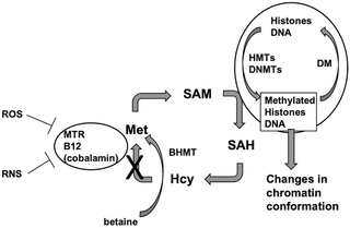
We have previously reported that methyl donor betaine is depleted in MS and is linked to changes in histone H3 trimethylation (H3K4me3) in neurons. We have also shown that betaine increases histone methyltransferase activity by activating chromatin bound betaine homocysteine S-methyltransferase (BHMT).

A hallmark of MS is the death of oligodendrocytes, the cells responsible for wrapping axons in myelin in the central nervous system and maintaining a healthy sheath. In demyelinating diseases like MS, oligodendrocyte progenitor cells (OPCs) fail to differentiate and make more myelin, resulting in sclerotic lesions.

Promoting differentiation of OPCs and generation of myelin is of great interest as a novel MS therapy. Waves of gene regulation (repression and activation) need to occur to promote myelination.

This BHMT-betaine methylation pathway ensures availability of S-adenosylmethionine (SAM) for a variety of DNA and histone methylation processes. OPC survival and differentiation are dependent upon DNA and histone methylation, and both processes require SAM.

journal.pone.0250486.g001

BHMT uses betaine to remethylate homocysteine to methionine. Betaine can be taken in through the diet or synthesized through the oxidation of choline in mitochondria.

We demonstrated that oligodendrocyte gene expression can be modulated by betaine supplementation through the BHMT-betaine methylation pathway. Our study suggests that dietary betaine supplementation may prove to be a therapeutic agent for MS and other demyelinating disorders.”

https://journals.plos.org/plosone/article?id=10.1371/journal.pone.0250486 “The BHMT-betaine methylation pathway epigenetically modulates oligodendrocyte maturation”


I started taking betaine 16 years ago. Didn’t know of these effects until reading this study.

Treating psychopathological symptoms will somehow resolve causes? had more on betaine (aka trimethyl glycine). Current dose is 1.5 grams twice daily.

Are rodent models of human neurodegenerative diseases realistic?

This 2020 stem cell review argued against rodent models of human neurodegenerative diseases:

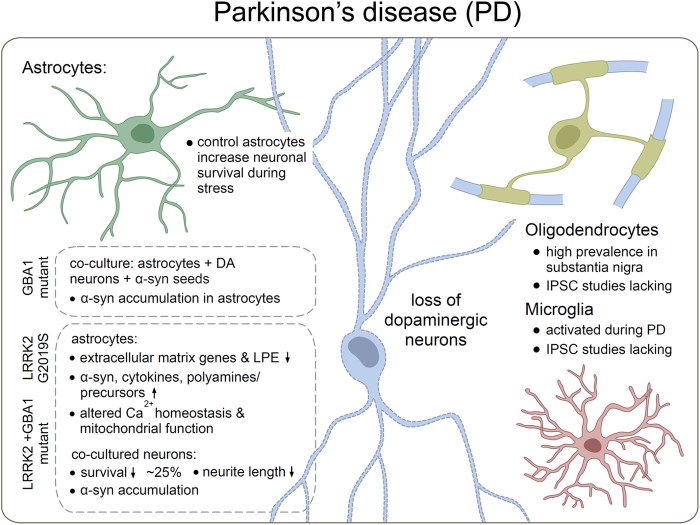
“Neuronal loss is not caused solely by intrinsic degenerative processes but rather via impaired interactions with surrounding glia and other brain cells. Dysfunctional astrocytes do not provide sufficient nutrients and antioxidants to neurons, while dysfunctional microglia cannot efficiently clear pathogens and cell debris from extracellular space, resulting in chronic inflammatory processes in the brain.

Human glia, especially astrocytes, differ significantly in morphology and function from their mouse counterparts. Recent advances in stem cell technology make it possible to reprogram human patients’ somatic cells to induced pluripotent stem cells (iPSC) and differentiate them further into patient‐specific glia and neurons, thus providing a source of human brain cells.

stem3309-fig-0002-m

Astrocytes do not efficiently utilize energy resources and cannot provide adequate metabolic support to neurons. A coculture of healthy human neurons with diseased astrocytes impaired neuronal calcium responses to glutamate and γ‐aminobutyric acid (GABA) as compared to coculture with healthy human astrocytes.

Treatment with sulforaphane:

  • Normalized basal level glycolysis;
  • Decreased basal level Aβ42 secretion; as well as
  • Ameliorated inflammatory response to pro‐inflammatory cytokines TNF-α and IL1-β in PSEN1 mutant iPSC astrocytes.

It is essential to make sure that what we see in the dish is the real patient‐specific phenotype. Transplantation of human brain organoids containing microglia into mice could provide a novel tool for drug screening in vivo.”

https://stemcellsjournals.onlinelibrary.wiley.com/doi/full/10.1002/stem.3309 “Metabolic and immune dysfunction of glia in neurodegenerative disorders: Focus on iPSC models”


This review’s thesis seems plausible. However, one problem with in vitro stem cell studies is that they often don’t have a control group.

Giving children allergies with pets

This 2021 human study investigated development and persistence of allergies:

“Allergic rhinitis (AR) is a common IgE-mediated disorder involving troublesome symptoms of nasal congestion, nasal itch, sneezing, and associated eye symptoms. Like many chronic health conditions, AR stems from complex gene–environment interactions.

130 subjects with AR were recruited. Control population included 154 healthy children who underwent a regular physical examination in the same ear, nose and throat clinic as AR patients. Individuals with history of asthma or atopic dermatitis were excluded.

AR analysis

Plenty of contradictory associations exist as whether furred pet exposure (cats and dogs) may be a risk or a protective factor for AR development. Discrepancies are likely due to the ubiquitous nature of pet allergens, while pet owners are more concerned about sanitation and many other hygiene-related reasons.

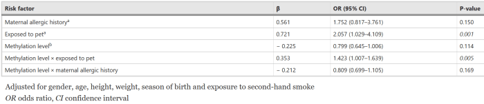
Interaction of early-life pet exposure with methylation level of ADAM33 increased the risk for AR onset 1.423 times more in children. This study provides evidence that:

  • Early-life pet exposure and low methylation level of ADAM33 increase AR risk in children; and
  • The interaction between pet exposure and methylation level of ADAM33 may play an important role in development of AR.”

https://aacijournal.biomedcentral.com/articles/10.1186/s13223-021-00526-5 “Interaction between early-life pet exposure and methylation pattern of ADAM33 on allergic rhinitis among children aged 3–6 years in China”


There’s nothing children can do about who their parents were. Exposing them to pet allergens, though, may be another example of early-life experiences causing lifelong effects.

Happy Mothers Day

This 2021 rodent study investigated effects on offspring of maternal high-fat diet (HFD) during gestation and lactation, and offspring HFD during young adulthood:

“We found that gestation was the most sensitive period to induce obesity in late life, and there was no difference between sexes in chance of obesity. Furthermore, we found that lactation and administration of a HFD post‐weaning increased incidence of lipid metabolism disorders and obesity in offspring.

gestational hfd effects on offspring

There are different windows of opportunity for programming epigenetically labile genes. Some studies support the alteration of epigenetic status during development as an important cause induced adult obesity.

Gestation is considered as the most sensitive period because high DNA synthesis and DNA methylation patterns are established for normal tissue development during the embryonic period. These two programming events are the times when the epigenetic state changes most widely in the life cycle.”

https://onlinelibrary.wiley.com/doi/10.1111/jcmm.16551 “Gestational high-fat diet impaired demethylation of Pparα and induced obesity of offspring”


Hey mothers! Do what you please. But don’t turn around and deny consequences of your behavior and choices on your descendants’ physiology and behavior, and possibly those of further descendants.

Gestation, birth, infancy, and early childhood are critical periods for humans. There’s no going back to correct errors and problems.

Does skin improvement cause overall effects?

This 2019 human skin study found:

“We demonstrated in aged mice that epidermal dysfunction largely accounted for age-associated elevations in circulating cytokine levels, and that improving epidermal function reduced circulating cytokine levels. We performed a pilot study to determine whether improving epidermal function reduces circulating proinflammatory cytokine levels in aged humans.

Both aged human and mouse skin display sustained abnormalities in epidermal permeability barrier homeostasis, stratum corneum (SC) hydration, and elevations in SC pH 4-6, each of which has been shown to independently provoke cutaneous inflammation. Disruption of the epidermal permeability barrier provoked an increase in:

  1. Cutaneous cytokine production; and
  2. Serum cytokine levels, independent of hepatic or T cell involvement.

We assessed whether improving epidermal function with an emollient, containing a mixture of lipids that mimics components of normal SC, lowered circulating levels of these same pro-inflammatory cytokines in aged humans.

skin treatment

After 30 days of twice-daily topical treatments, circulating levels of IL-1β and IL-6 decreased significantly in the treated aged cohort vs. untreated aged controls. Topical treatments reduced circulating levels of IL-1β and IL-6 to levels comparable to young controls. Though levels of TNF-α declined by over 40% in comparison to untreated aged humans, the difference did not attain statistical significance.

Results of this preliminary study suggest that a larger clinical trial should be performed to confirm whether improving epidermal function also can reduce circulating proinflammatory cytokine levels in aged humans, while also possibly attenuating downstream development of chronic inflammatory disorders.”

https://onlinelibrary.wiley.com/doi/abs/10.1111/jdv.15540 “Topical applications of an emollient reduce circulating pro‐inflammatory cytokine levels in chronically aged humans: a pilot clinical study” (not freely available)


I discussed enrolling in a trial whose objective would be to test this study’s findings. No big deal, just have to take IL-6 and TNF-α measurements in an upcoming annual physical. Then apply that trial’s skin treatment for 30 days per this study’s twice-daily protocol.

Day 70 results from Changing to a youthful phenotype with broccoli sprouts provided some of last year’s measurements. IL-6 was already at a negligible 1.0 pg / ml, one-fifth of both the above Baseline Young group’s 5.1 ± 0.9 and the Treated Aged Group’s 5.7 ± 0.9.

IL-6 2020

Probably won’t want my data, since their treatment wouldn’t be expected to lower an already very low inflammation marker.

Weight loss for the lazy

At the risk of becoming Dr. Paul Clayton’s echo chamber, another great blog post, Falling Down:

“When lab rats or mice are weighted down with lead pellets they lose substantial amounts of weight, almost exclusively adipose tissue. Unlike dieting, there is little if any loss of muscle mass, making lead an ideal weight loss strategy for the lazy.

A clinical trial generated the same result. Their paper concludes, ‘Increased weight loading reduces body weight and fat mass in obese subjects in a similar way as previously shown in obese rodents. These findings demonstrate that there is a loading-dependent homeostatic regulation of body weight, the gravitostat, also in humans.’

Polyphenol resveratrol protects against damaging effects of de-loading by acting as an exercise mimetic, and does so by activating AMP-K directly. Other nutrients which do the same thing include polyphenol quercetin, sapogenin dammaranes, and omega 3 fatty acid EPA.”


The doctor still doesn’t mention sulforaphane in this or any other blog post, although it activates the AMPK pathway on the way to its primary effect of Nrf2 activation. First time I’d seen the term covidiots.

“You can fool some people sometimes
But you can’t fool all the people all the time
And now you’ve seen the light
Stand up for your rights”

Part 2 of Broccoli sprouts activate the AMPK pathway

This 2021 review subject was metformin’s role in autophagy:

“Metformin had been used as the first choice for treating diabetes for almost a century. Autophagy is responsible for recycling and degrading cellular components, which significantly affects cell functions in physiology and pathology.

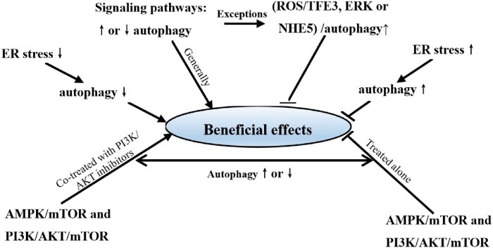
Effects of metformin on autophagy mainly depend on corresponding signaling pathways in specific organs or tissues. Metformin can induce autophagy in cells of many organs and tissues via affirmed signaling pathways, such as AMPK-related signaling pathways.

1-s2.0-S0753332221000718-gr5_lrg

Different signaling pathways (alone or in combination) mediated the process of metformin affecting autophagy in different organs or tissues. It is necessary to combine effects of metformin on autophagy with pharmacological effects on pathologies in different organs or tissues, which would provide indications for future metformin applications.”

https://www.sciencedirect.com/science/article/pii/S0753332221000718 “The effects of metformin on autophagy”


I characterized this review as Part 2 of Broccoli sprouts activate the AMPK pathway because that study’s experimental evidence showed sulforaphane activation of the AMPK pathway was a predecessor to sulforaphane’s main effects of Nrf2 pathway activation. This review didn’t even mention Nrf2 activation.

Do all of metformin’s cited effects apply to daily intake of broccoli sprouts? Probably not, but most people who take metformin every day aren’t healthy.

See Part 3 for updates.

Grow your 3-day-old sprouts in darkness

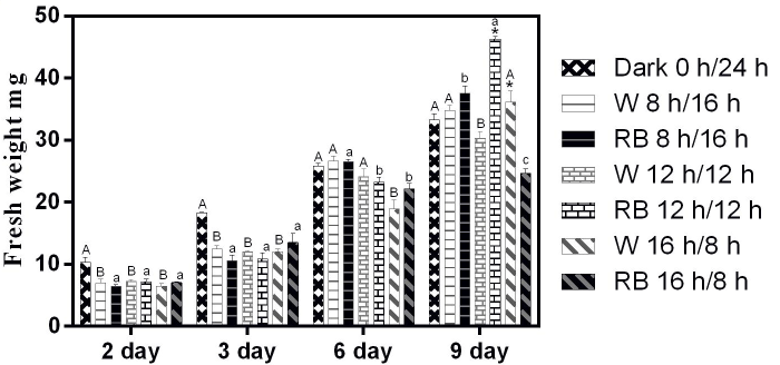
This 2021 study examined light frequency effects on Chinese kale sprouts’ development of glucosinolates:

“We investigated sprout growth and secondary metabolite glucosinolates (GSs) accumulation under white or combined red-and-blue (RB) light sources. Most GSs in sprouts are stored in seeds, which is gradually degraded to provide nutrients for other metabolic functions.

Phenotype of 3-day-old Chinese kale sprouts grown with different photoperiods condition under white or RB light:

capital A was grown in darkness

Sprouts grown under dark conditions showed only elongation of hypocotyls [shoots]. Sprouts grew with shorter hypocotyls and wider cotyledons [first leaves] irrespective of whether a white or combined RB light source was used.

Growth indicators (including plant height, cotyledon length, fresh weight, and dry weight) under different photoperiodic treatments were measured on days 2, 3, 6, and 9. Consistent with the phenotype presented, plant height and cotyledon length responded rhythmically to illumination time.”

https://www.frontiersin.org/articles/10.3389/fpls.2020.589746/full “Effect of Photoperiod on Chinese Kale (Brassica alboglabra) Sprouts Under White or Combined Red and Blue Light”


Circadian rhythms rule. Accept and adjust.

An overlooked gut microbiota product

This 2021 review subject was histone crotonylation:

“Histone crotonylation is a newly identified epigenetic modification that has a pronounced ability to regulate gene expression. It belongs to an expanding group of short chain lysine acylations that also includes the extensively studied mark histone acetylation.

Histone Kcr was first identified in 2011 where it was found to be mainly associated with active chromatin. Kcr occurs on the ε-amino group of the lysine side chain, where it neutralizes the positive charge of this residue. The loss in positive charge on histone Lys residues weakens DNA interaction, thus making chromatin less compact and accessible to DNA-binding factors.

Crotonate, like other short chain fatty acids (SCFAs), is mainly produced by gut microbiota during fermentation of partially and nondigestible carbohydrates. Circulating SCFAs (acetate, crotonate, butyrate, and propionate) can be taken up by tissues and converted into their cognate short-chain acyl-CoAs, the direct donors of histone Lys acylations.

fcell-09-624914-g001

Crotonyl-CoA is generated as a by-product of fatty acid and amino acid metabolism. Synthesis of crotonyl-CoA can occur in mitochondria or the cytoplasm. Evidence suggests that histone acylations are directly sensitive to changes in concentrations of their corresponding acyl-CoA metabolites, and therefore can act as indicators of cellular metabolic state.

Only a small number of Kcr sites in human histones have been identified so far. This is in part due to a lack of commercially available Kcr site-specific antibodies, which has meant much of the research in this field has focused on studying total histone crotonylation. This is likely to limit our understanding of the importance of histone Kcr, as functional impact of modification at specific sites cannot be readily assessed.”

https://www.frontiersin.org/articles/10.3389/fcell.2021.624914/full “The Regulation and Function of Histone Crotonylation”


At first I thought I had missed recent studies of gut microbiota producing crotonate. Searching again for “crotonate” “microbiota” 2020 2021, I didn’t find any that weren’t cited by this paper.

A lack of research could be due to factors mentioned above. It may also be that researchers just don’t look for evidence of the circulating SCFA crotonate.

Broccoli sprouts’ immune effects

Two 2021 papers, with the first’s subject being sulforaphane’s immune effects:

“Effects of sulforaphane (SFN) on immune response generate scientific interest because of its bioavailability, which is much higher than other phytochemicals, and its capacity to induce Nrf2 target genes. Clinical trials suggest that sulforaphane produces favorable results in cases where pharmaceutical products fail.

SFN exhibits the highest bioavailability among well-known antioxidant phytochemicals, such as quercetin (20-fold higher) and curcumin (80-fold higher). SFN confers a high potential to be used either as a nutraceutical to improve health status, or as pharmaceutical to treat disease states.

molecules-26-00752-g001

Sulforaphane exerts a pleiotropic effect on immunological response, and the final effect depends on cell type.

  • In lymphocyte T-cells, SFN induces ROS production, GSH depletion, and repression of inflammatory cytokines, resulting in suppression of immune and inflammatory responses.
  • In monocytes and macrophages, SFN stimulates immune response by inducing Nrf2, thus triggering antioxidant and anti-inflammatory responses.”

https://www.mdpi.com/1420-3049/26/3/752/htm “Potential of Sulforaphane as a Natural Immune System Enhancer: A Review”


A second study was Fertilization and Pre-Sowing Seed Soaking Affect Yield and Mineral Nutrients of Ten Microgreen Species:

“Ten tested microgreen species [amaranth, arugula, basil, broccoli, red cabbage, Daikon radish, kale, kohlrabi, mustard, and green pea] in this study varied in fresh and dry shoot weights, shoot height, and mineral nutrient concentrations.”

This study grew sprouts for 6 – 18 days before harvesting. Its study design didn’t require sampling along the way to discover informative compositional changes, as did 2020’s 3-day-old broccoli sprouts have the optimal yields and Broccoli sprout compounds include sinapic acid derivatives.

Their supplier was the same as I used for broccoli and red cabbage seeds. No endorsement is intended.

I’d rather use an unknown broccoli variety than this study’s broccoli cultivar, Waltham 29. It was found to be relatively glucoraphanin-deficient when measured in a 2004 study referenced in Tailoring measurements for broccoli sprouts, 32nd of 34 tested.

Received these today:

PXL_20210424_191628875

I’ve asked for clarification of the red cabbage seed variety I received. Not sure what “Agnostic” means in a “Red Cabbage Microgreen – Agnostic” context. 🙂

Mustard and red cabbage sprouting will follow Improving healthy compounds of broccoli sprouts efforts, minus that study’s laboratory setup and duration. I expect synergistic effects from handling both species’ sprouts with my protocol for microwaved 3-day-old broccoli sprouts.