Optimizing glucosinolate analysis

I’ll highlight microwave findings of this 2021 study:

“Glucosinolates (GSLs) are important precursor compounds with anticancer activities in Brassicaceae vegetables and are readily hydrolyzed by myrosinase. Given the diversity of these species, establishing an accurate and universal method to quantify intact GSLs in different plant tissues is necessary.

We compared and optimized three tissue disruption methods for sample preparation:

  • Recoveries of GSLs in a Chinese cabbage sample were significantly lower than 100% after microwave treatment for 60 s, due to insufficient inactivation of myrosinase.
  • After microwave treatment for 90 s, recoveries of 13 GSLs were in the range of 73–124%, indicating that this condition could inactivate myrosinase completely.
  • The increase in GSL recoveries with microwave treatment for 120 s might be due to increased extractability of GSLs.
  • A limitation of this method was that different tissues could not be processed under the same microwave conditions.

molecules-27-00231-g001

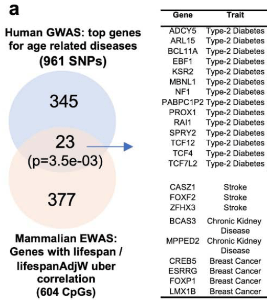
SIN, Sinigrin; NAP, Gluconapin; GBN, Glucobrassicanapin; PRO, Progoitrin; ERU, Glucoerucin; RAE, Glucoraphenin; RAA, Glucoraphanin; ALY, Glucoalyssin; GBC, Glucobrassicin; 4ME, 4-Methoxyglucobrassicin; NEO, Neoglucobrassicin; TRO, Glucotropaeolin; NAS, Gluconasturtiin.

Five GSLs without available standards were estimated using calibration curves of structurally similar compounds. Specifically, the pair glucoberteroin (GOB) and glucoerucin (ERU), glucoiberin (GIB) and glucoraphanin (RAA), gluconapoleiferin (GNL) and PRO, and 4OH and 4ME are homologs that differ in structure by one -CH2 group, with glucoraphasatin (GRH) containing an alkenyl group in its molecular structure, which is two hydrogen atoms less than ERU.

The verified method of intact GSLs by UHPLC-MS/MS established in this study was more accurate and time-saving than the commonly used ISO method for desulfo-GSLs.”

https://www.mdpi.com/1420-3049/27/1/231/htm “Determination of 18 Intact Glucosinolates in Brassicaceae Vegetables by UHPLC-MS/MS: Comparing Tissue Disruption Methods for Sample Preparation”


This study was in line with other studies that increased GLS amounts by microwaving. For example, most of the above graphic’s Chinese cabbage GLS measurements at 90 seconds were greater than raw samples, which kept going:

“The increase in GSL recoveries with microwave treatment for 120 s might be due to increased extractability of GSLs.”

Unlike this study, my goal is to optimize glucosinolate hydrolysis products such as sulforaphane. I increase myrosinase enzyme activity rather than decrease it, and want to have less GLS amounts than what I started with after processing.

I facilitate myrosinase activity by:

  • Adjusting immersion water to pH 5; and
  • Stopping at 60°C (140°F) to avoid a myrosinase deactivation cliff between 60°C and 65°C.

This study used a 900W microwave to process 30-gram broccoli floret samples at Figure S1 different times (20, 40, 60, 90, 120, 180 seconds). I use a 1000W microwave to process a 65-gram broccoli / red cabbage / mustard sprouts mix in 100 ml water for 40 seconds.

Kickstarting endogenous regenerative pathways

A 2022 amphibian study by the Electroceuticals team investigated limb regeneration:

“Organisms such as Xenopus laevis – whose limited regenerative capacities in adulthood mirror those of humans – are important models with which to test interventions that can restore form and function. We demonstrate long-term (18 months) regrowth, marked tissue repatterning, and functional restoration of an amputated X. laevis hindlimb following a 24-hour exposure to a multidrug, pro-regenerative treatment delivered by a wearable bioreactor.

  • Regenerated multidrug treatment (MDT) hindlimbs were longer than the no added factors (BD) and no device (ND) groups by 2.5 mpa, as indicated by growth beyond resection site (red dashed line).
  • At 4 mpa, vascularized structures developed at the distal extension of MDT (yellow arrow), but not BD or ND regenerates.
  • At 9 mpa, digit-like projections appeared (blue arrow), contrasting hypomorphic spikes of BD and ND regenerates (pink arrows).

limb regeneration

We suggest that the overall strategy of providing wound cells with an aqueous, amniotic-like environment, which is uniquely given through our bioreactor, that contains pro-regenerative signals is likely to be an effective method for kickstarting biomedically relevant growth and patterning cascades that are too complex to directly implement. One additional direction that may present new opportunities for enhanced regeneration is to assess immune function in relation to tissue remodeling.”

https://www.science.org/doi/10.1126/sciadv.abj2164 “Acute multidrug delivery via a wearable bioreactor facilitates long-term limb regeneration and functional recovery in adult Xenopus laevis”

Sulforaphane and hair loss

This 2021 human clinical trial evaluated sulforaphane’s cosmetic effects:

“We demonstrated that sulforaphane has the potential to become a highly effective functional hair cosmetic to relieve hair loss with androgen alopecia. Sulforaphane increases expression of the dihydrotestosterone (DHT)-degrading enzyme 3α-hydroxysteroid dehydrogenase (3α-HSD) in the liver, which accelerates DHT degradation, thereby inhibiting hair loss.

We performed a visual evaluation of parietal and frontal lines of 23 men and women from 18 to 54 years old before and after using the product, and then calculated total number of hairs. This clinical study showed that parietal lines and bangs visually improved, and the number of hairs increased by 6.71% from before using the test product to 18 weeks after using the test product.

hair growth

We tested expression levels of Ak1c21 and Dhrs9 isoforms of 3α-HSD in the in vitro cell culture experiment where Hepa1c1c7 cells were treated with sulforaphane or a mixture of biotin, dexpanthenol, and l-menthol. This study showed that sulforaphane alone achieved a hair loss-relieving effect in our experimental cell culture conditions.

Our finding that sulforaphane induces Akr1c2 in a dose-dependent manner is consistent with previous studies. Sulforaphane treatment induced expression of Dhrs9, which has several sites in the promoter region that bind to Nrf2, which is induced by sulforaphane.

It is highly likely that sulforaphane might enhance degradation of DHT, not only via the induction of degrading enzymes 3α-HSD, but also by functional activation of these enzymes. Further studies remain to test this possibility.”

https://www.mdpi.com/2079-9284/8/3/63/htm “Sulforaphane, L-Menthol, and Dexpanthenol as a Novel Active Cosmetic Ingredient Composition for Relieving Hair Loss Symptoms”

Gut signals

I’ll highlight signaling pathway aspects of this 2022 review:

“The gut bacterial community plays an important role in regulation of multiple aspects of metabolic disorders. This regulation depends, among other things, on production of a wide variety of metabolites by microbiota and on their interactions with receptors on host cells that can activate or inhibit signalling pathways, and either be beneficial and detrimental to the host’s health.

Colonocytes and endocrine cells express a variety of receptors able to sense and transmit signals from the microbial environment:

gutjnl-2021-326789-F4.large

  • TLRs cover a wide range of both external stimuli (PAMPs) and internal signals derived from tissue damage. Their activation induces antigen-presenting cell activation, thereby bridging innate and adaptive immune responses, and stimulates signalling cascades as an attempt to fend off microbial invaders or repair damaged tissue.
  • The endocannabinoid signalling system appears to play a key role in regulating energy, glucose, and lipid metabolism but also in immunity, inflammation, and more recently in microbiota-host interactions.
  • Although the primary function of bile acids (BAs) is to regulate digestion and absorption of cholesterol, triglycerides, and fat-soluble vitamins, it has been recently recognised that BAs also serve an endocrine function as they act as signalling molecules. BAs have been shown to modulate epithelial cell proliferation, gene expression, lipid, glucose, and energy metabolism by activating several receptors. Because of their signalling capacities and the fact that BAs are chemically transformed by gut microbiota, BAs can be considered as microbiota-derived signalling metabolites.
  • Numerous AhR ligands exist including environmental triggers, nutrition-derived signals, various phytochemicals, and bacterial metabolites such as tryptophan.

Most signalling metabolites can be produced by large numbers of different gut bacteria, and hence have limited specificity.”

https://gut.bmj.com/content/early/2022/01/31/gutjnl-2021-326789.long “Gut microbiome and health: mechanistic insights”

Predicting atherosclerosis

Starting this blog’s eighth year with a 2022 epigenetic clock study that assessed young people’s common blood tests fifteen and twenty years later:

GrimAge acceleration (GAA), an epigenetic marker that represents physiologic aging, is associated with atherosclerotic cardiovascular disease. We used multivariable regression models to examine associations of Y15 and Y20 GAA estimates with plasma lipid levels measured at prior examination years (Y0, Y5, and Y10) and concurrently: triglycerides (TG), low-density lipoprotein cholesterol (LDL-C), and high-density lipoprotein cholesterol (HDL-C) levels:

  • Each 1-SD higher cumulative TG level was associated with an average 0.73 ± 0.12 years older GAA;
  • Each 1-SD higher cumulative HDL-C level was associated with an average 0.57 ± 0.17 years younger GAA;
  • Associations between TG and GAA were stronger among female and Black participants; and
  • Associations between HDL-C and GAA were stronger among female and White participants.

lipid-profiles-and-GrimAge-acceleration

We observed that elevated TG and low HDL-C levels in young adulthood are associated with accelerated midlife epigenetic aging, and epigenetic aging mediates some of the well-described associations between elevated TG levels in early life and subclinical atherosclerosis in middle age. These findings suggest that maintaining optimal lipid levels in early adulthood may help to slow epigenetic aging, which reflects delays in the onset of age-related diseases like atherosclerosis.”

https://clinicalepigeneticsjournal.biomedcentral.com/articles/10.1186/s13148-021-01222-2 “Plasma lipid profiles in early adulthood are associated with epigenetic aging in the Coronary Artery Risk Development in Young Adults (CARDIA) Study”


Which is better for resolving a health situation?

  • Hope for luck / providence before subclinical symptoms become clinical problems?
  • Do nothing constructive, and depend on interventions after problems occur?
  • Take responsibility for your own one precious life?

PXL_20220131_201832917

An epigenetic regulator of vascular aging

This 2022 rodent and human cell study investigated the smooth muscle cell mineralocorticoid receptor:

“Vascular stiffness increases with age and independently predicts cardiovascular disease risk. Epigenetic changes, including histone modifications, accumulate with age, but the global pattern has not been elucidated nor are the regulators known.

Rising mineralocorticoid receptor (MR) in aging vascular smooth muscle cells downregulates EZH2 to globally shift to a more open chromatin thereby allowing MR to be recruited to promoters to transcriptionally upregulate target genes involved in vascular stiffness. This mechanism provides multiple potential targets to prevent vascular stiffness in aging humans.

cvac007f8

We demonstrate for the first time that:

  1. MR expression increases with age in primary, low passage, human aortic smooth muscle cell (SMC) and correlates with age in whole aortic tissue from aging humans;
  2. The global proteomic profile of histone modifications in mouse vessels changes profoundly with aging with a significant overall decrease in H3K27 methylation;
  3. Expression of H3K27 methyltransferase EZH2 decreases with age in mouse vessels and in human SMCs in a MR-dependent manner and negatively correlates with MR expression in whole human aortic tissue;
  4. The aging-induced decline in EZH2 associates with reduced H3K27 methylation and increased H3K27 acetylation in vitro and in vivo;
  5. These epigenetic changes in aging human SMC and mouse vessels correspond with increased expression of the vascular stiffness genes, CTGF and integrin-α5, previously identified vascular MR target genes;
  6. Induction of an aging phenotype in human SMC associates with increased MR enrichment and H3K27 acetylation at these stiffness gene promoters; and
  7. Inhibition of MR in aged mice and aged human aortic SMCs reverses the entire process; increasing EZH2 and H3K27 methylation, increasing locus-specific EZH2 enrichment and decreasing H3K27 acetylation at stiffness gene promoters, decreasing vascular expression of CTGF and integrin-α5, and decreasing the stiffness and adhesiveness of aged human SMC in vitro and mouse aortic stiffness and fibrosis in vivo.”

https://academic.oup.com/cardiovascres/advance-article-abstract/doi/10.1093/cvr/cvac007/6502304 “Smooth muscle mineralocorticoid receptor as an epigenetic regulator of vascular ageing” (not freely available) Thanks to Dr. Seung Kyum Kim for providing a copy.

Intergenerational epigenetic inheritance of trained immunity, Part 2

A 2022 McGill University rodent study couldn’t replicate Part 1 findings:

“We find that using similar mouse models of trained immunity induced by:

  1. Live vaccination (BCG);
  2. PAMPs (β-glucan); or
  3. Infection (C. albicans),

protection against:

  1. Viral (influenza virus);
  2. Bacterial (Mycobacterium tuberculosis (Mtb)); or
  3. Fungal (C. albicans)

infections was the same between offspring of trained and non-trained parents.

BCG-vaccination-in-the-offspring-of-vaccinated-parents-does-not-enhance-trained-immunity_W640

BCG vaccination in the offspring of vaccinated parents does not enhance trained immunity in macrophages.

a) Mice were vaccinated with BCG-iv (1 × 10⁶ CFU) for one month and mated with vaccinated or naive counterparts. 6–8 week-old F1.1 and F1.3 offspring were then vaccinated or not with BCG-iv (1 × 10⁶ CFU).

b), c) At 1 month post BCG vaccination, protective capacities of BMDM from BCG-iv vaccinated and nonvaccinated F1.1 (b), or F1.3 (c) offspring from naïve or BCG-iv vaccinated parents were assessed against M. tuberculosis (H37Rv, MOI 1) infection. * p < 0.05.”

https://www.nature.com/articles/s41590-021-01102-0 “Lack of evidence for intergenerational inheritance of immune resistance to infections” (not freely available)


Part 1 coauthors replied:

“We are very encouraged that this topic is gaining increased interest. The reason for the discrepancy between findings in the two studies is unclear. It likely involves local differences in mouse substrains, housing, diet, microbiome, infection models, or other factors.

These findings underscore the effect of environment on intergenerational inheritance of infection resistance. What these environmental factors are and how these factors are integrated with regards to intergenerational inheritance remains largely elusive at this time.

One intriguing possibility that needs to be tested in future studies is whether such effects may be more robust in outbred wild mice, in which subtle environmental changes may have less strong impact.”

https://www.nature.com/articles/s41590-021-01103-z “Reply to: ‘Lack of evidence for intergenerational inheritance of immune resistance to infections'”


PXL_20211031_112819792

Lifespan Uber Correlation

This 2022 study developed new epigenetic clocks:

“Maximum lifespan is deemed to be a stable trait in species. The rate of biological function decline (i.e., aging) would be expected to correlate inversely with maximum species lifespan. Although aging and maximum lifespan are intimately intertwined, they nevertheless appear in some investigations to be distinct processes.

Some cytosines conserved across mammals exhibit age-related methylation changes so consistent that they were used to successfully develop cross-species age predictors. In a similar vein, methylation levels of some conserved cytosines correlate highly with species lifespan, leading to the development of highly accurate lifespan predictors. Surprisingly, little to no commonality is found between these two sets of cytosines.

We correlated the intra-species age correlation with maximum lifespan across mammalian species. We refer to this correlation of correlations as Lifespan Uber Correlation (LUC).

We overlapped genes from the LUC signature with genes found in human genome-wide association studies (GWAS) of various pathologies and conditions. With all due caution, we report that some genes from the LUC signature were those highlighted by GWAS to be associated with type II diabetes, stroke, chronic kidney disease, and breast cancer.

Human aging genes vs mammalian LUC

We used the subset of CpGs found to be significant in our LUC to build age estimators (epigenetic clocks). We demonstrated that these clocks are able to capture effects of interventions that are known to alter age as well as lifespan, such as caloric restriction, growth hormone receptor knockout, and high-fat diet.

We found that Bcl11b heterozygous knockout mice exhibited an increased epigenetic age in the striatum. BCL11B is a zinc finger protein with a wide range of functions, including development of the brain, immune system, and cardiac system.

This gene is also implicated in several human diseases including, but not limited to, Huntington disease, Alzheimer’s diseases, HIV, and T-cell malignancies. BCL11B plays an important role in adult neurogenesis, but is less studied in the context of lifespan disparities in mammals.

Bcl11b knockout affected both DNA methylation and mRNA expression of LUC genes. Our current study does not inform us about the potential role of Bcl11b in aging processes during adulthood since observed patterns could be attributed to developmental defects.

We are characterizing other genetic and non-genetic interventions that perturb the LUC clocks. These we will feature in a separate report that will uncover biological processes regulated by LUC cytosines and their associated genes.”

https://www.biorxiv.org/content/10.1101/2022.01.16.476530v1 “Divergent age-related methylation patterns in long and short-lived mammals”


PXL_20220106_201346155

Gut microbiota’s positive epigenetic effects

Three papers with the first a 2021 review:

“Gut microbiota along with their metabolites are involved in health and disease through multiple epigenetic mechanisms including:

  • Affecting transporter activities, e.g. DNA methyltransferases (DNMTs), histone methyltransferases (HMTs), histone acetyltransferases (HATs), and histone deacetylases (HDACs);
  • Providing methyl donors to participate in DNA methylation and histone modifications; and
  • miRNAs that can lead to gene transcriptional modifications.

ijms-22-06933-g003

These mechanisms can participate in a variety of biological processes such as:

  • Maturation of intestinal epithelial cells (IECs);
  • Maintenance of intestinal homeostasis;
  • Inflammatory response;
  • Development of metabolic disorders; and
  • Prevention of colon cancer.”

https://www.mdpi.com/1422-0067/22/13/6933/htm “Dissecting the Interplay Mechanism between Epigenetics and Gut Microbiota: Health Maintenance and Disease Prevention”


A second 2022 review added subjects such as crotonate (aka unsaturated butyrate):

“Studies are carving out potential roles for additional histone modifications, such as crotonylation and ethylation, in facilitating crosstalk between microbiota and host. Lysine crotonylation is a relatively less studied histone modification that is often enriched at active promoters and enhancers in mammalian cells.

While addition or removal of crotonyl motifs can be catalyzed by specialized histone crotonyltransferases and decrotonylases, HATs and HDACs have also been reported to exhibit histone crotonyl-modifying activity. Microbiota stimulate multiple types of histone modifications and regulate activity of histone-modifying enzymes to calibrate local and extra-intestinal chromatin landscapes.”

https://www.tandfonline.com/doi/full/10.1080/19490976.2021.2022407 “Epigenetic regulation by gut microbiota”


A third 2021 review added subjects such as broccoli sprout compounds’ epigenetic effects:

“Glucosinolates are converted into isothiocyanates (ITCs) by bacteria that regulate host epigenetics. Levels of ITCs produced following broccoli consumption are highly dependent on the functional capacity of individual microbiomes, as much interindividual variability exists in gut microbiota composition and function in humans.

Sulforaphane inhibits HDAC activity both in vitro and in vivo, and protects against tumor development. Microbial-mediated production of ITCs represents a strong diet-microbe interaction that has a direct impact on host epigenome and health.”

https://www.sciencedirect.com/science/article/pii/S0955286321000516 “The interplay between diet, gut microbes, and host epigenetics in health and disease”


Clearing the channel

PXL_20220118_203446833

The aryl hydrocarbon signaling pathway

I’ll emphasize this densely packed 2021 review’s broccoli sprout compounds / gut microbiota / health interactions:

“The aryl hydrocarbon receptor (AhR) senses cues from environmental toxicants and physiologically relevant dietary/microbiota-derived ligands. AhR signaling mediates bidirectional host-microbiome interactions in a wide range of cellular functions in a ligand-, cell type-, species-, and context-specific manner.

Brassicaceae family plants are rich sources of glucobrassicin, the glucosinolate precursor of indole-3-carbinol (I3C). Glucobrassicin can be enzymatically hydrolyzed and converted into I3C by myrosinase, which is present in intact plant cells and gut microbiota.

I3C activates AhR but exhibits low binding affinity. However, in acidic conditions found in the stomach, I3C undergoes acid condensation reaction to generate a variety of more potent AhR ligands, such as 3,3′-diindolylmethane (DIM).

AhR activation by natural AhR ligands (e.g., I3C) has been shown to prevent pathogenic gut microbial dysbiosis by altering gut microbiome composition in mice with colitis. Depletion of AhR ligands in the diet decreased α diversity of gut microbiota, while I3C supplementation restored microbiota composition.

I3C treatment is effective for treating IBD patients, partly by upregulating IL-22. Targeting AhR could modulate the amplitude and duration of IL-22 signaling to treat IBD patients.

Administration of I3C or DIM significantly reduced the number of tumors in the cecum and small intestine. Supplementation of I3C reduces the number of colorectal tumors in WT, but not in AhR null mice.

nihms-1759454-f0003

Gut microbiota and diet are major sources of AhR ligands that influence the whole body, including gut, liver, brain, and the immune system. Many human diseases are associated with decreased circulating levels of AhR ligands, partly due to dysbiosis.

The ability of AhR signaling to regulate self-renewal and differentiation of intestinal stem cells intrinsically or extrinsically has recently been brought into the spotlight.”

https://www.ncbi.nlm.nih.gov/labs/pmc/articles/PMC8667662/ “Diet–Host–Microbiota Interactions Shape Aryl Hydrocarbon Receptor Ligand Production to Modulate Intestinal Homeostasis”


Young hawk

PXL_20220109_195611152

Every baby needs a sugar mama

This 2021 in vitro study examined butyrate producers:

“Butyrate produced by gut microbiota has multiple beneficial effects on host health. Oligosaccharides derived from host diets, and glycans originating from host mucus, are major sources of its production.

Butyrate is the major energy source for epithelial cells in the distal colon, induces differentiation of colonic regulatory T cells, and functions as an inhibitor of host histone deacetylase. These activities are essential for documented beneficial properties of butyrate, including anti-inflammation, gut immune homeostasis, inhibition of proliferation, and induction of apoptosis of colorectal cancer cells.

FOS-type oligosaccharides (kestose, nystose, fructooligosaccharide) were metabolized by only 6 of 14 butyrate-producing strains tested:

Growth of butyrate producers

Faecalibacterium prausnitzii, which is the most abundant butyrate producer in the healthy human gut, metabolized only FOS-type oligosaccharides among tested oligosaccharides. Anaerostipes spp. exhibited a similar pattern, except that A. caccae metabolized kestose but not nystose.

Glycoside hydrolase (GH)32 enzymes exhibiting FOS degradation activities were conserved in all six strains metabolizing FOS, and in three of the eight strains that did not metabolize FOS. This suggests that GH32 enzymes in those three strains are not actively used in metabolism.

The present study highlighted that even if functional genes are present in microbes, they are sometimes unable to metabolize substrates. This should be carefully considered in metagenomic studies to understand metabolic potential of gut microbiota.”

https://www.tandfonline.com/doi/full/10.1080/19490976.2020.1869503 “Characterization of fructooligosaccharide metabolism and fructooligosaccharide-degrading enzymes in human commensal butyrate producers”


These researchers had some work to do to show that selected strains’ characteristics were representative of their species. This post’s title was excerpted from Citation 37.

PXL_20220112_201642006

Nrf2 and circadian rhythm

This 2021 rodent study investigated aging’s effects:

“We investigated aging consequences on temporal patterns of antioxidant defenses, molecular clock machinery, and blood pressure.

We observed circadian rhythms of catalase (CAT) and glutathione peroxidase (GPx) mRNA expression, as well as ultradian rhythms of Nrf2 mRNA levels, in the hearts of young adult rats. We also found circadian oscillations of CAT and GPx enzymatic activities, reduced glutathione (GSH), and BMAL1 protein.

Aging abolished rhythms of CAT and GPx enzymatic activities, phase-shifted rhythm acrophases of GSH and BMAL1 protein levels, and turned circadian the ultradian oscillation of Nrf2 expression.

aging changes Nrf2 oscillation

Moreover, aging phase-shifted the circadian pattern of systolic blood pressure. In conclusion, aging modifies temporal organization of antioxidant defenses and blood pressure, probably as a consequence of disruption in the circadian rhythm of the clock’s transcriptional regulator, BMAL1, in heart.”

https://doi.org/10.1007/s10522-021-09938-7 “Aging disrupts the temporal organization of antioxidant defenses in the heart of male rats and phase shifts circadian rhythms of systolic blood pressure” (not freely available)


A human equivalent to this study’s 3-month-old young adult group is around 19 years. The older group’s 22-month age is roughly equivalent to a 68-year-old human.

Couldn’t say whether Nrf2 oscillations flattening out with age is specific to heart tissue, or is a more general trend. I’m pretty sure that humans have to make good things happen while aging, because bad things are pre-programmed.

I came across this study from a citation trail of a comment to Eat broccoli sprouts for your workouts. I didn’t curate the mentioned study because one of its coauthors tainted it by designing and supervising Problematic rodent sulforaphane studies.

How would you answer the comment’s question?


Repairs needed: The story of 2021

PXL_20220104_194204601

Sulforaphane vs. too much oxygen

This 2021 rodent study investigated perinatal effects of hyperoxia and sulforaphane:

“We demonstrated that early-life oxidant-induced acute lung injury had significant consequences later in life on NRF2-dependent respiratory syncytial virus (RSV) susceptibility in mice. We also determined that increased antioxidant conditions in utero potentially contribute to a decreased risk of postnatal airway disease as we found that prenatal antioxidant sulforaphane (SFN) protected developing lungs from bronchopulmonary dysplasia (BPD)-like oxidative pathogenesis in mice.

Unexpectedly, our results indicated that prenatal SFN-mediated postnatal protection against BPD-like phenotypes are not NRF2-dependent. Prenatal SFN markedly improved hyperoxia-caused severe BPD-like lung injury parameters in Nrf2−/− pups while we observed relatively marginal protection by in utero SFN in hyperoxia-resistant Nrf2+/+ pups.

SFN is a strong NRF2 and ARE gene inducer for cytoprotection by NRF2 stabilization. However, SFN also acts through other mechanisms, including NF-κB inhibition, MAPK activation, and histone deacetylase inhibition for anti-inflammation, chemoprevention, apoptosis, and autophagy.

Our study provided new insights into infant oxidant lung injury severity influence on persistence of pulmonary morbidity and therapeutic intervention for NRF2 agonists. Our results also provided justification for further studies on feto–placental barrier crossing of SFN metabolites and SFN-triggered molecular and epigenetic aspects of maternal cues for barrier and fetal lung signaling.”

https://www.mdpi.com/2076-3921/10/12/1874/htm “Murine Neonatal Oxidant Lung Injury: NRF2-Dependent Predisposition to Adulthood Respiratory Viral Infection and Protection by Maternal Antioxidant”


This study’s oral human-equivalent dose for treatment dams was 9 mg sulforaphane (1.67 mg x .081 x 70 kg) every other day during the last half of pregnancy. A small dose per How much sulforaphane is suitable for healthy people?

“The daily SFN dose found to achieve beneficial outcomes in most of the available clinical trials is around 20-40 mg.”

PXL_20211226_120528134

Your lungs and Nrf2 activity

Two 2021 papers of Nrf2 activation effects on lung diseases, with the first a McGill University review:

“Oxidative stress and subsequent activation of Nrf2 have been demonstrated in many human respiratory diseases. The purpose of this review is to summarize involvement of Nrf2 and its inducers in acute respiratory distress syndrome, chronic obstructive pulmonary disease (COPD), asthma, and lung fibrosis in both human and experimental models.

fphys-12-727806-g004

These inducers have proven particularly effective at reducing severity of oxidative stress-driven lung injury in various animal models. In humans, these compounds offer promise as potential therapeutic strategies for management of respiratory pathologies associated with oxidative stress, but there is thus far little evidence of efficacy through human trials.

Perhaps, by analogy with biologics, patients with demonstrated deficient antioxidant responses to their disease should be selected for study in future clinical trials.”

https://www.frontiersin.org/articles/10.3389/fphys.2021.727806/full “Role of Nrf2 in Disease: Novel Molecular Mechanisms and Therapeutic Approaches – Pulmonary Disease/Asthma”


A second paper was a human/rodent study of COPD:

“We investigated Nrf2 expression and epigenetic regulation, and mechanisms by which the Nrf2 signaling pathway in ferroptosis is related to COPD. These findings elucidated pathways of ferroptosis in bronchial epithelial cells in COPD, and revealed Nrf2 as a potential target for COPD treatment.

COPD_A_340113_t0001

DNA hypermethylation at specific CpG sites of the Nrf2 promoter in primary epithelial cells and in clinical lung tissues is correlated with decreased Nrf2 expression, which is related to COPD occurrence and development.”

https://www.ncbi.nlm.nih.gov/pmc/articles/PMC8684379/ “Hypermethylation of the Nrf2 Promoter Induces Ferroptosis by Inhibiting the Nrf2-GPX4 Axis in COPD”


Similar to this second paper’s CpG findings, Eat broccoli sprouts for your heart found:

“Sulforaphane (SFN) reduced Ang II‐induced CpG hypermethylation and promoted Ac‐H3 [histone H3 acetylation] accumulation in the Nrf2 promoter region, accompanied by inhibition of global DNMT [DNA methyltransferase] and HDAC [histone deacetylase] activity, and a decreased protein expression of key DNMT and HDAC enzymes. Overall, DNA methylation and histone deacetylation are considered to inhibit gene transcription with a synergistic effect.

Nrf2 can also be regulated independently of Keap1. Evidence indicates that SFN may indirectly activate Nrf2 by affecting activity of several upstream kinases.”

However, this second paper didn’t measure DNMT and HDAC inhibition, although their therapeutic effects in reducing oxidative injury and inflammation may have been present.

PXL_20211225_193439146

Gut microbiota vs. disease risks

This 2021 review subject was risk relationships between diseases from the perspective of gut microbiota:

“There is a significant inverse relationship between the onset of Alzheimer’s disease/Parkinson’s disease (AD/PD) and cancer, but the mechanism is still unclear. Considering that intestinal flora can connect them, we briefly introduced the relationship among AD/PD, cancer, and intestinal flora, studied metabolites or components of the intestinal flora, and the role of intestinal barriers and intestinal hormones in AD/PD and cancer.

According to existing evidence:

  • Bifidobacterium and Lactobacillus positively affect AD/PD and cancer;
  • Ruminococcaceae, Prevotellaceae, and Prevotella significantly improve on AD/PD but harm cancer; and
  • Blautia has universal anticancer ability, but it may aggravate AD pathology.

1-s2.0-S0753332221011276-gr1_lrg

This may partially explain the antagonistic relationship between neurodegenerative diseases and cancer. When some individuals suffer from one disease, their intestinal flora change to obtain a stronger resistance to the other disease than healthy individuals, which is consistent with statistical data.”

https://www.sciencedirect.com/science/article/pii/S0753332221011276 “Composition of intestinal flora affects the risk relationship between Alzheimer’s disease/Parkinson’s disease and cancer”


PXL_20211224_180111266