The epigenetic clock theory of aging

My 400th curation is a 2018 US/UK paper by coauthors of Using an epigenetic clock to distinguish cellular aging from senescence. They reviewed the current state of epigenetic clock research, and proposed a new theory of aging:

“The proposed epigenetic clock theory of ageing views biological ageing as an unintended consequence of both developmental programmes and maintenance programmes, the molecular footprints of which give rise to DNAm [DNA methylation] age estimators.

It is best to interpret epigenetic age estimates as a higher-order property of a large number of CpGs much in the same way that the temperature of a gas is a higher-order property that reflects the average kinetic energy of the underlying molecules. This interpretation does not imply that DNAm age simply measures entropy across the entire genome.

To date, the most effective in vitro intervention against epigenetic ageing is achieved through expression of Yamanaka factors, which convert somatic cells into pluripotent stem cells, thereby completely resetting the epigenetic clock. In vivo, haematopoietic stem cell therapy resets the epigenetic age of blood of the recipient to that of the donor.

Future epidemiological studies should consider other sources of DNA (for example, buccal cells), because more powerful estimates of organismal age can be obtained by evaluating multiple tissues. Other types of epigenetic modifications such as adenine methylation or histone modifications may lend themselves for developing epigenetic age estimators.”


https://www.nature.com/articles/s41576-018-0004-3 “DNA methylation-based biomarkers and the epigenetic clock theory of ageing” (not freely available)


I curated four other papers cited in this review:

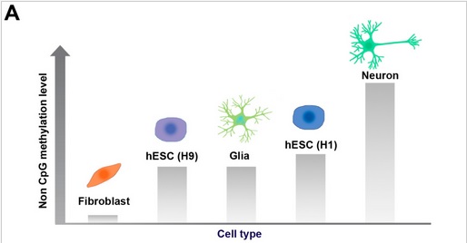
Do you want your quality of life to be under or over this curve?

What are you doing to reverse epigenetic processes and realize what you want?

  • Do you have ideas and/or behaviors that interfere with taking constructive actions to change your phenotype?
  • If you aren’t doing anything, are you honest with yourself about feelings of helplessness?
  • Do your beliefs in fate, or in technology, or in divine interventions justify inactions?

Genomic imprinting and growth

This 2018 UK paper reviewed genomic imprinting:

“Since their discovery nearly 30 years ago, imprinted genes have been a paradigm for exploring the epigenetic control of gene expression. Moreover, their roles in early life growth and placentation are undisputed.

However, it is becoming increasingly clear that imprinted gene function has a wider role in maternal physiology during reproduction – both by modulating fetal and placental endocrine products that signal to alter maternal energy homeostasis, and by altering maternal energetic set points, thus producing downstream actions on nutrient provisioning.”

“Imprinted genes in the conceptus produce products that alter maternal resource allocation by:

  1. altering the transport capacity of the placenta;
  2. increasing fetal demand for resources by their action on the intrinsic growth rate; and
  3. signalling to the mother by the production of fetal/placental hormones that modify maternal metabolism.”

Other studies/reviews I’ve curated that covered genomic imprinting are:

http://jeb.biologists.org/content/jexbio/221/Suppl_1/jeb164517.full.pdf “Genomic imprinting, growth and maternal-fetal interactions”


This post has somehow become a target for spammers, and I’ve disabled comments. Readers can comment on other posts and indicate that they want their comment to apply here, and I’ll re-enable comments.

Maternal obesity causes fetal liver damage

This 2018 US baboon study was on fetal effects from maternal obesity before and during pregnancy:

“Approximately 64% of women of childbearing age in the USA [are] overweight or obese. The baboon is a well-characterized animal model sharing many physiological, metabolic, and genetic characteristics with humans allowing direct translation of findings to human pregnancy.

Our study shows that fetal exposure to the MO [maternal obesity] intrauterine environment results in dysregulation of fetal hepatic genes central to metabolism.

These findings were further supported by identification of miRNAs that were inversely expressed with key genes in these pathways..suggest important early molecular mechanisms by which MO programs fetal hepatic lipid metabolism.

Future studies are required in MO post-natal offspring to determine the extent to which the fetal phenotype persists, and the degree to which this increases offspring risk of cardiometabolic disorders in later life.”


The study provided many measurements that may be relevant to humans. Other consequential measurements were missing that may have made the study’s findings even more applicable to humans:

  • No placental measurements other than weight. The organ through which the fetus received its nutrients, signaled its needs, modulated its growth rate, and developed its organs, was only measured by weight?
  • No other epigenetic analyses such as DNA methylation and histone modifications.

Were these omitted due to limited resources?

http://onlinelibrary.wiley.com/doi/10.1113/JP275422/pdf “Primate fetal hepatic responses to maternal obesity: epigenetic signalling pathways and lipid accumulation”

Viruses target epigenetic processes

This 2018 Colorado review subject was general and specific ways viruses target epigenetic processes:

“We describe viral mechanisms and virus-host interactions by which DNA tumor viruses regulate host DNA methylation to evade antiviral immunity.

It is well known that most endogenous retroviruses and retrotransposons in the human genome are inactivated by DNA hypermethylation. In addition to endogenous retroviruses, the genomes of DNA viruses, such as human papillomavirus (HPV), herpes simplex virus 1 (HSV-1), adenovirus, and hepatitis B virus (HBV), are also frequently methylated and silenced in infected cells.

A recently described mechanism for viruses to epigenetically subvert host immunity is repression of immune-related gene expression by induction of DNA hypermethylation. Some host genes are not silenced simply through promoter hypermethylation or histone deacetylation alone, and therefore, viruses may have evolved mechanisms to ensure host gene downregulation through multiple epigenetic modifications.”

http://www.mdpi.com/1999-4915/10/2/82/htm “DNA Tumor Virus Regulation of Host DNA Methylation and Its Implications for Immune Evasion and Oncogenesis”


A second 2018 New York study focused on the Zika virus and DNA methylation:

“We studied the impact of ZIKV infection on the DNA methylation pattern across the entire genome in selected neural cell types. The virus unexpectedly alters the DNA methylome of neural progenitors, astrocytes, and differentiated neurons at genes that have been implicated in the pathogenesis of a number of brain disorders.

It remains open, however, whether the methylation changes come first or whether the viral infection dysregulates epigenetic regulatory genes prior to any epigenetic shift.”

http://msystems.asm.org/content/3/1/e00219-17 “Zika Virus Alters DNA Methylation of Neural Genes in an Organoid Model of the Developing Human Brain”

Non-CpG DNA methylation

This 2017 Korean review compared and contrasted CpG and non-CpG DNA methylation:

“Non-CpG methylation is restricted to specific cell types, such as pluripotent stem cells, oocytes, neurons, and glial cells. Accumulation of methylation at non-CpG sites and CpG sites in neurons seems to be involved in development and disease etiology.

Non-CpG methylation is established during postnatal development of the hippocampus and its levels increase over time. Similarly, non-CpG methylation is scarcely detected in human fetal frontal cortex, but is dramatically increased in later life. This increase in non-CpG methylation occurs simultaneously with synaptic development and increases in synaptic density.

In contrast, CpG methylation occurs during early development and does not increase over time.

Neurons have considerably higher levels of non-CpG methylation than glial cells. The human male ES [embryonic stem] cell line (H1) is more highly methylated than the female ES cell line (H9).

Among the different types of non-CpG methylation (CpA [adenosine], CpT [thymine], and CpC [another cytosine]), methylation is most common at CpA sites. For instance, in human iPS [induced pluripotent stem] cells, 5mCs are found in approximately 68.31%, 7.81%, 1.99%, and 1.05% of CpG, CpA, CpT, and CpC sites, respectively.”


The reviewers’ referenced statement:

“CpG methylation occurs during early development and does not increase over time.”

was presented outside of its context. The 2013 cited source’s statement was restricted to “selected loci” in the rodent hippocampus:

“Consistent with a recent study of the cortex, time-course analyses revealed that CpH [non-CpG] methylation at the selected loci was established during postnatal development of the hippocampus and was then present throughout life, whereas CpG methylation was established during early development.”

Epigenetic study methodologies improved in 2017 had more information on CpA methylation.

http://www.mdpi.com/2073-4425/8/6/148/htm “CpG and Non-CpG Methylation in Epigenetic Gene Regulation and Brain Function”

Make consequential measurements in epigenetic studies

The subject of this 2017 Spanish review was human placental epigenetic changes:

“39 papers assessing human placental epigenetic signatures in association with either

  • (i) psychosocial stress,
  • (ii) maternal psychopathology,
  • (iii) maternal smoking during pregnancy, and
  • (iv) exposure to environmental pollutants,

were identified.

Their findings revealed placental tissue as a unique source of epigenetic variability that does not correlate with epigenetic patterns observed in maternal or newborn blood.

Each study’s confounders were summarized by a column in Table 1. Some of the reviewers’ comments included:

“33 out of 39 papers reviewed (85%) reported significant associations between either placental DNA methylation or placental miRNA expression and exposure to any of the risk factors assessed. However, the methodological heterogeneity present throughout the studies reviewed does not allow meta-analytic exploration of reported findings.

Heterogeneity regarding the origin of biological tissues analyzed confounds the replicability and validity of reported findings and their potential synthesis.”


Sponsors and researchers really have to take their work seriously if the developmental origins of health and disease hypothesis can advance to a well-evidenced theory. Study designers should:

  1. Sample consequential dimensions. “There were no studies examining histone modifications.” Why were there no human studies in this important category of epigenetic changes in the placenta, the “barrier protecting the fetus?
  2. Correct methodological deficiencies in advance. Eliminate insufficiencies like “Once collected, processing and storage of placental samples also differed across studies and was not reported in all of them.”
  3. Stop using convenient but non-etiologic proxy assays such as global methylation. How can a study advance the DOHaD hypothesis if everyone knows ahead of time that its outcome will be yet another finding that epigenetic changes “are associated with” non-causal factors?
  4. Forget about non-biological measurements like educational attainment per Does a societal mandate cause DNA methylation?.

Every human alive today has observable lasting epigenetic effects caused by environmental factors during the earliest parts of our lives, and potentially even before we’re conceived. Isn’t this sufficient rationale to expect serious efforts by research sponsors and designers?

https://www.sciencedirect.com/science/article/pii/S0892036217301769 “The impact of prenatal insults on the human placental epigenome: A systematic review” (click the Download PDF link to read the paper)

Epigenetics research and evolution

This 2017 UK essay was a longish review of how epigenetics and other research has informed evolutionary theory:

“There are several processes by which directed evolutionary change occurs – targeted mutation, gene transposition, epigenetics, cultural change, niche construction and adaptation.

Evolution is an ongoing set of iterative interactions between organisms and the environment. Directionality is introduced by the agency of organisms themselves.”

A few takeaway items concerned:

“It is of course the functional phenotype that is ‘seen’ by natural selection. DNA sequences are not directly available for selection other than through their functional consequences.

The comparative failure of genome-wide association studies to reveal very much about the genetic origins of health and disease is one of the most important empirical findings arising from genome sequencing.

Environmental epigenetic impacts on biology and disease include:

  • Worldwide differences in regional disease frequencies
  • Low frequency of genetic component of disease as determined with genome wide association studies (GWAS)
  • Dramatic increases in disease frequencies over past decades
  • Identical twins with variable and discordant disease frequency
  • Environmental exposures associated with disease
  • Regional differences and rapid induction events in evolution

The above list was from the cited 2016 review “Developmental origins of epigenetic transgenerational inheritance” https://www.ncbi.nlm.nih.gov/pmc/articles/PMC4933018


Further points about behavior’s role in evolution:

“Differential mutation rates are not essential to enable organisms to guide their own evolution.

If organisms have agency and, within obvious limits, can choose their lifestyles, and if these lifestyles result in inheritable epigenetic changes, then it follows that organisms can at least partially make choices that can have long-term evolutionary impact.”

These discussions provided support for the central question of The PRice “equation” for individually evolving: Which equation describes your life?:

“Applying the “How does a phenotype influence its own change?” question to a person:

How can a person remedy their undesirable traits – many of which are from their ancestral phenotype – and acquire desirable traits?”

http://www.mdpi.com/2079-7737/6/4/47/htm “Was the Watchmaker Blind? Or Was She One-Eyed?”

Epigenetic study methodologies improved in 2017

Let’s start out 2018 paying more attention to advancements in science that provide sound empirical data and methodology. Let’s ignore and de-emphasize studies and reviews that aren’t much more than beliefs couched in models and memes, whatever their presumed authority.

Let sponsors direct researchers to focus on ultimate causes of diseases. Let’s put research of treatments affecting causes ahead of those that only address symptoms.

Here are two areas of epigenetic research that improved in 2017.


Improved methodologies enabled DNA methylation studies of adenine, one of the four bases of DNA, to advance, such as this 2017 Wisconsin/Minnesota study N6-methyladenine is an epigenetic marker of mammalian early life stress:

“6 mA is present in the mammalian brain, is altered within the Htr2a gene promoter by early life stress and biological sex, and increased 6 mA is associated with gene repression. These data suggest that methylation of adenosine within mammalian DNA may be used as an additional epigenetic biomarker for investigating the development of stress-induced neuropathology.”

Most DNA methylation research is performed on the cytosine and guanine bases.


Other examples of improved methodologies were discussed in this 2017 Japanese study Genome-wide identification of inter-individually variable DNA methylation sites improves the efficacy of epigenetic association studies:

“A strategy focusing on CpG sites with high DNA methylation level variability may attain an improved efficacy..estimated to be 3.7-fold higher than that of the most frequently used strategy.

With ~90% coverage of human CpGs, whole-genome bisulfite sequencing (WGBS) provides the highest coverage among the currently available DNAm [DNA methylation] profiling technologies. However, because of its high cost, it is presently infeasible to apply WGBS to large-scale EWASs [epigenome-wide association studies], which require DNAm profiling of hundreds or thousands of subjects. Therefore, microarrays and targeted bisulfite sequencing are currently practicable for large-scale EWASs and thus, effective strategies to select target regions are essentially needed to improve the efficacy of epigenetic association studies.

DNAm levels measured with microarrays are invariable for most CpG sites in the study populations. As invariable DNAm signatures cannot be associated with exposures, intermediate phenotypes, or diseases, current designs of probe sets are inefficient for blood-based EWASs.”

How to cure the ultimate causes of migraines?

Most of the spam I get on this blog comes in as ersatz comments on The hypothalamus couples with the brainstem to cause migraines. I don’t know what it is about the post that attracts internet bots.

The unwanted attention is too bad because the post represents a good personal illustration of “changes in the neural response to painful stimuli.” Last year I experienced three three-day migraines in one month as did the study’s subject. This led to me cycling through a half-dozen medications in an effort to address the migraine causes.

None of the medications proved to be effective at treating the causes. I found one that interrupted the progress of migraines – sumatriptan, a serotonin receptor agonist. I’ve used it when symptoms start, and the medication has kept me from having a full-blown migraine episode in the past year.

1. It may be argued that migraine headache tendencies are genetically inherited. Supporting personal evidence is that both my mother and younger sister have migraine problems. My father, older sister, and younger brother didn’t have migraine problems. Familial genetic inheritance usually isn’t the whole story of diseases, though.

2. Migraine headaches may be an example of diseases that are results of how humans have evolved. From Genetic imprinting, sleep, and parent-offspring conflict:

“Evolutionary theory predicts: that which evolves is not necessarily that which is healthy.

Why should pregnancy not be more efficient and more robust than other physiological systems, rather than less? Crucial checks, balances and feedback controls are lacking in the shared physiology of the maternal–fetal unit.

Both migraine causes and effects may be traced back to natural lacks of feedback loops. These lacks demonstrate that such physiological feedback wasn’t evolutionarily necessary in order for humans to survive and reproduce.

3. Examples of other processes occurring during prenatal development that also lack feedback loops, and their subsequent diseases, are:

A. Hypoxic conditions per Lack of oxygen’s epigenetic effects are causes of the fetus later developing:

  • “age-related macular degeneration
  • cancer progression
  • chronic kidney disease
  • cardiomyopathies
  • adipose tissue fibrosis
  • inflammation
  • detrimental effects which are linked to epigenetic changes.”

B. Stressing pregnant dams per Treating prenatal stress-related disorders with an oxytocin receptor agonist caused fetuses to develop a:

and abnormalities:

  • in social behavior,
  • in the HPA response to stress, and
  • in the expression of stress-related genes in the hippocampus and amygdala.”

1. What would be a treatment that could cure genetic causes for migraines?

I don’t know of any gene therapies.

2. What treatments could cure migraines caused by an evolved lack of feedback mechanisms?

We humans are who we have become, unless and until we can change original causes. Can we deal with “changes in the neural response to painful stimuli” without developing hopes for therapies or technologies per Differing approaches to a life wasted on beliefs?

3. What treatments could cure prenatal epigenetic causes for migraines?

The only effective solution I know of that’s been studied in humans is to prevent adverse conditions like hypoxia from taking place during pregnancy. The critical periods of our physical development are over once we’re adults, and we can’t unbake a cake.

Maybe science will offer other possibilities. Maybe researchers could do more than their funding sponsors expect?

Differing approaches to a life wasted on beliefs

Let’s start by observing that people structure their lives around beliefs. As time goes on, what actions would a person have taken to ward off non-confirming evidence?

One response may be that they would engage in ever-increasing efforts to develop new beliefs that justified how they spent their one precious life’s time so far.

Such was my take on beliefs embedded in https://www.ncbi.nlm.nih.gov/pmc/articles/PMC5684598/pdf/PSYCHIATRY2017-5491812.pdf “Epigenetic and Neural Circuitry Landscape of Psychotherapeutic Interventions”:

“Animal models have shown the benefits of continued environmental enrichment (EE) on psychopathological phenotypes, which carries exciting translational value.

This paper posits that psychotherapy serves as a positive environmental input (something akin to EE).”

The author conveyed his belief that wonderful interventions were going to happen in the future. However, when scrutinized, most human studies have demonstrated NULL effects of psychotherapeutic interventions on causes. Without sound evidence that treatments affect causes, his belief seemed driven by something else.

The author cited findings of research like A problematic study of oxytocin receptor gene methylation, childhood abuse, and psychiatric symptoms as supporting external interventions to tamp down symptoms of patients’ presenting problems. Did any of the 300+ cited references concern treatments where patients instead therapeutically addressed their problems’ root causes?


For an analogous religious example, a person’s belief caused him to spend years of his life trying to convince men to act so that they could get their own planet after death, and trying to convince women to latch onto men who had this belief. A new and apparently newsworthy belief developed from his underlying causes:

“The founder and CEO of neuroscience company Kernel wants “to expand the bounds of human intelligence.” He is planning to do this with neuroprosthetics; brain augmentations that can improve mental function and treat disorders. Put simply, Kernel hopes to place a chip in your brain.

He was raised as a Mormon in Utah and it was while carrying out two years of missionary work in Ecuador that he was struck by what he describes as an “overwhelming desire to improve the lives of others.”

He suffered from chronic depression from the ages of 24 to 34, and has seen his father and stepfather face huge mental health struggles.”

https://www.theguardian.com/small-business-network/2017/dec/14/humans-20-meet-the-entrepreneur-who-wants-to-put-a-chip-in-your-brain “Humans 2.0: meet the entrepreneur who wants to put a chip in your brain”

The article stated that he had given up Mormonism. There was nothing to suggest, though, that he had therapeutically addressed any underlying causes for his misdirected thoughts, feelings, and behavior.

So he developed other beliefs instead.


What can people do to keep their lives from being wasted on beliefs? As mentioned in What was not, is not, and will never be:

“The problem is that spending our time and efforts on these ideas, beliefs, and behaviors won’t ameliorate their motivating causes. Our efforts only push us further away from our truths, with real consequences: a wasted life.

The goal of the therapeutic approach advocated by Dr. Arthur Janov’s Primal Therapy is to remove the force of presenting problems’ motivating causes. Success in reaching this goal is realized when patients become better able to live their own lives.

Epigenetic effects of microRNA on fetal heart development

This 2017 Australian review’s subject was epigenetic impacts involving microRNA in adverse intrauterine environments, and how these affected fetal heart tissue development:

“We describe how an adverse intrauterine environment can influence the expression of miRNAs (a sub-set of non-coding RNAs) and how these changes may impact heart development. Potential consequences of altered miRNA expression in the fetal heart include; Hypoxia inducible factor (HIF) activation, dysregulation of angiogenesis, mitochondrial abnormalities and altered glucose and fatty acid transport/metabolism.

This feedback network between miRNAs and other epigenetic pathways forms an epigenetics–miRNA regulatory circuit that organizes the whole gene expression profile. The human heart encodes over 700 miRNAs.”


A 2016 review Lack of oxygen’s epigenetic effects also provided a details about hypoxia. Those reviewers importantly pointed out the natural lack of a feedback mechanism to the HIF-1α signaling source, and how this evolutionary lack contributed to diseases.

http://www.mdpi.com/1422-0067/18/12/2628/htm “Adverse Intrauterine Environment and Cardiac miRNA Expression”

Do you have your family’s detailed medical histories?

Imagine that you were a parent who puzzled over the mystery of your pre-teen daughter’s hyperactive behavior. Without detailed family medical histories, would anyone recognize this as a preprogammed phenotype?

Could anyone trace the daughter’s behavior back to her maternal great-grandmother being treated with glucocorticoids near the end of the second trimester of carrying her grandfather?

Such was a finding of a 2017 Canadian guinea pig study that was undertaken to better inform physicians of the transgenerationally inherited epigenetic effects of glucocorticoid treatments commonly prescribed during human pregnancies:

“This study presents the first evidence that prenatal treatment with sGC [synthetic glucocorticoid] results in transgenerational paternal transmission of hyperactivity and altered hypothalamic gene expression through three generations of young offspring. Female offspring appear to be more sensitive than male offspring to the programming effects of sGC, which suggests an interaction between sGC and sex hormones or sex-linked genes. Paternal transmission to F3 strongly implicates epigenetic mechanisms in the process of transmission, and small noncoding RNAs likely play a major role.”


Some details of the study included:

Veh[icle] was the control group initially treated with saline.

The study was informative and conclusive for the aspects studied. From the Methods section:

“Data from same-sex littermates were meaned to prevent litter bias. Sample sizes (N) correspond to independent litters, and not to the total number of offspring across all litters.

Power analyses based on previous studies determined N ≥ 8 sufficient to account for inter-litter variability and detect effects in the tests performed.”

https://www.nature.com/articles/s41598-017-11635-w “Prenatal Glucocorticoid Exposure Modifies Endocrine Function and Behaviour for 3 Generations Following Maternal and Paternal Transmission”

What is a father’s role in epigenetic inheritance?

The agenda of this 2017 Danish review was to establish a paternal role in intergenerational and transgenerational epigenetic inheritance of metabolic diseases:

“There are four windows of susceptibility which have major importance for epigenetic inheritance of acquired paternal epigenetic changes:

  1. paternal primordial germ cell (PGC) development,
  2. prospermatogonia stages,
  3. spermatogenesis, and
  4. during preimplantation.”

The review was a long read as the authors discussed animal studies. When it came to human studies near the paper’s end, though, the tone was of a “we know this is real, we just have to find it” variety. The authors acknowledged:

“To what extent the described DNA methylation changes influence the future health status of offspring by escaping remodeling in the preimplantation period as well as in future generations by escaping remodeling in PGC remodeling has yet to be determined.

These studies have not yet provided an in-depth understanding of the specific mechanisms behind epigenetic inheritance or exact effect size for the disease risk in offspring.

Pharmacological approaches have reached their limits..”

before presenting their belief that a hypothetical series of future CRISPR-Cas9 experiments will demonstrate the truth of their agenda.


The review focused on 0.0001% of the prenatal period for what matters with the human male – who he was at the time of a Saturday night drunken copulation – regarding intergenerational and transgenerational epigenetic inheritance of metabolic diseases.

The human female’s role – who she was at conception AND THEN what she does or doesn’t do during the remaining 99.9999% of the prenatal period to accommodate the fetus and prevent further adverse epigenetic effects from being intergenerationally and transgenerationally transmitted  – wasn’t discussed.

Who benefits from this agenda’s narrow focus?

If the review authors sincerely want to:

“Raise societal awareness of behavior to prevent a further rise in the prevalence of metabolic diseases in future generations..”

then EARN IT! Design and implement HUMAN studies to test what’s already known from epigenetic inheritance animal studies per Experience-induced transgenerational programming of neuronal structure and functions. Don’t disguise beliefs with the label of science.

http://jme.endocrinology-journals.org/content/early/2017/12/04/JME-17-0189.full.pdf “DNA methylation in epigenetic inheritance of metabolic diseases through the male germ line”

“Transgenerationally” inherited epigenetic effects of fetal alcohol exposure

The fourth paper of Transgenerational epigenetic inheritance week was a 2016 German rodent study of of improperly-termed “transgenerational” epigenetic effects of alcohol:

“We investigated 2 generations of offspring born to alcohol-treated mothers. Here, we show that memory impairment and reduced synthesis of acetylcholine occurs in both F1 (exposed to ethanol in utero) and F2 generation (never been exposed to ethanol). Effects in the F2 generation are most likely consequences of transgenerationally transmitted epigenetic modifications in stem cells induced by alcohol.

The results further suggest an epigenetic trait for an anticholinergic endophenotype associated with cognitive dysfunction which might be relevant to our understanding of mental impairment in neurodegenerative disorders such as Alzheimer’s disease and related disorders.”

F0 generation mothers modeled human fetal alcohol syndrome. They were exposed to ethanol gradually up to 20%, then mated. The 20% ethanol intake level was maintained until the F1 generation pups were born, then gradually diminished to 0%. After a ten-day wait, an eight-week handling and shaping period started, followed by five weeks of behavioral testing.

The F1 children and F2 grandchildren started an eight-week handling and shaping period after young adulthood, followed by five weeks of behavioral testing. The F1 children were mated after behavioral testing.

The F0 parents showed no significant differences in working memory and reference memory compared with controls. Both the F1 children and F2 grandchildren were significantly impaired in the same tests compared with controls, with the F1 children performing worse than the F2 grandchildren. No sex-dependent differences were noted.

After behavioral impairments due to intergenerational epigenetic modifications were established, the F2 grandchildren received treatments to ascertain the contribution of cholinergic dysfunction in their behavioral impairments. It was confirmed, as an acetylcholine esterase inhibitor that crosses the blood-brain barrier almost completely erased working-memory and reference-memory performance deficits.

Items in the Discussion section included:

  • A dozen studies from 2014-2016 were cited for epigenetic mechanisms of inheritance stemming from parental alcohol consumption; and
  • Transgenerational inheritance of alcohol-induced neurodevelopmental deficits may involve epigenetic mechanisms that are resistant to developmental clearance.

As argued in Transgenerational effects of early environmental insults on aging and disease and A review of epigenetic transgenerational inheritance of reproductive disease, testing of F3 great-grandchildren was needed in order to establish transgenerational vs. intergenerational results. A F3 generation necessarily controls for the variable of F2 direct germline exposure.

http://www.neurobiologyofaging.org/article/S0197-4580(16)30303-7/pdf “Transgenerational transmission of an anticholinergic endophenotype with memory dysfunction” (not freely available)

Transgenerational pathological traits induced by prenatal immune activation

The third paper of Transgenerational epigenetic inheritance week was a 2016 Swiss rodent study of immune system epigenetic effects:

“Our study demonstrates for, we believe, the first time that prenatal immune activation can negatively affect brain and behavioral functions in multiple generations. These findings thus highlight a novel pathological aspect of this early-life adversity in shaping disease risk across generations.”

The epigenetic effects noted in the initial round of experiments included:

  • F1 child and F2 grandchild impaired sociability;
  • F1 and F2 abnormal fear expression;
  • F1 but not F2 sensorimotor gating deficiencies; and
  • F2 but not F1 behavioral despair associated with depressive-like behavior.

These transgenerational effects emerged in both male and female offspring. The prenatal immune activation timing corresponded to the middle of the first trimester of human pregnancy.

The effects were found to be mediated by the paternal but not maternal lineage. The researchers didn’t develop a maternal lineage F3 great-grandchild generation.

The next round of experiments done with the paternal lineage F3 great-grandchildren noted these epigenetic effects:

  • The F3 great-grandchildren had impaired sociability, abnormal fear expression and behavioral despair; and
  • The F3 great-grandchildren had normal sensorimotor gating.

Since the first round of tests didn’t show sex-dependent effects, the F3 great-grandchildren were male-only to minimize the number of animals.

Samples of only the amygdalar complex were taken to develop findings of transcriptomic effects of prenatal immune activation.

Items in the Discussion section included:

  1. The F2 grandchild and F3 great-grandchild generations’ phenotype of impaired sociability, abnormal fear expression and behavioral despair demonstrated that prenatal immune activation likely altered epigenetic marks in the germ line of the F1 children which resisted erasure and epigenetic reestablishment during germ cell development.
  2. Abnormal F1 child sensorimotor gating followed by normal F2 grandchild and F3 great-grandchild sensorimotor gating demonstrated that prenatal immune activation may also modify somatic but not germ cells.
  3. Non-significant F1 child behavioral despair followed by F2 grandchild and F3 great-grandchild behavioral despair demonstrated that prenatal immune activation may modify F1 germ cells sufficiently to develop a transgenerational phenotype, but unlike item 1 above, somatic cells were insufficiently modified, and the phenotype skipped the F1 children.
  4. Studies were cited that prenatal immune activation later in the gestational process may produce different effects.

The initial round of experiments wasn’t definitive for the maternal lineage. As argued in Transgenerational effects of early environmental insults on aging and disease and A review of epigenetic transgenerational inheritance of reproductive disease, testing of maternal lineage F3 great-grandchildren was needed to control for the variable of direct F2 grandchild germ-line exposure.

Also, effects that didn’t reach statistical significance in the maternal lineage F1 children and F2 grandchildren may have been different in the F3 great-grandchildren. The researchers indirectly acknowledged this lack by noting that these and other effects of immune challenges in a maternal lineage weren’t excluded by the study.

https://www.nature.com/mp/journal/v22/n1/pdf/mp201641a.pdf “Transgenerational transmission and modification of pathological traits induced by prenatal immune activation” (not freely available)


The study’s lead researcher authored a freely-available 2017 review that placed this study in context and provided further details from other studies:

http://www.nature.com/tp/journal/v7/n5/full/tp201778a.html “Epigenetic and transgenerational mechanisms in infection-mediated neurodevelopmental disorders”