A time to speak

“To every thing there is a season, and a time to every purpose under heaven:
A time to break down, and a time to build up;
A time to dance, and a time to mourn;
A time you may embrace, and a time to refrain from embracing;
A time to keep silent, and a time to speak.”


A review from 2017:

“Few, if any, other drugs can rival ivermectin for its beneficial impact on human health and welfare. Perhaps more than any other drug, ivermectin is a drug for the world’s poor. For most of this century, some 250 million people have been taking it.

The following are an indication of disease-fighting potential that has been identified for ivermectin thus far:

  • Antiviral – Ivermectin has been found to potently inhibit replication of yellow fever virus, with EC50 values in the sub-nanomolar range. It inhibits replication in several other flaviviruses, including dengue, Japanese encephalitis, and tick-borne encephalitis. Ivermectin interrupts virus replication. It demonstrates antiviral activity against several RNA viruses by blocking nuclear trafficking of viral proteins. It has been shown to have potent antiviral action against HIV-1.
  • Asthma – Ivermectin suppressed mucus hypersecretion by goblet cells, establishing that ivermectin can effectively curb inflammation, such that it may be useful in treating allergic asthma and other inflammatory airway diseases.
  • Bedbugs – Ivermectin is highly effective against bedbugs, capable of eradicating or preventing bedbug infestations.
  • Disease vector control – Ivermectin is highly effective in killing a broad range of insects. Comprehensive testing against 84 species of insects showed that avermectins were toxic to almost all insects tested. At sub-lethal doses, ivermectin inhibits feeding and disrupts mating behavior, oviposition, egg hatching, and development.
  • Malaria – Mosquitoes that transmit Plasmodium falciparum, the most dangerous malaria-causing parasite, can be killed by ivermectin present in the human bloodstream after a standard oral dose.
  • Myiasis – Myiasis is an infestation of fly larvae that grow inside the host. Oral myiasis has been successfully treated with ivermectin, which has also been effective as a non-invasive treatment for orbital myiasis, a rare and preventable ocular morbidity.
  • Schistosomiasis – Schistosoma species are the causative agent of schistosomiasis, a disease afflicting more than 200 million people worldwide. Ivermectin helps control one of the world’s major neglected tropical diseases.
  • Trichinosis – Globally, approximately 11 million individuals are infected with Trichinella roundworms. Ivermectin kills Trichinella spiralis, the species responsible for most of these infections.”

https://www.nature.com/articles/ja201711 “Ivermectin: enigmatic multifaceted ‘wonder’ drug continues to surprise and exceed expectations”


72 citations in CrossRef. Didn’t see citing 2020-2021 papers that noted any safety concerns when administered at proper doses.

Train your immune system every day, because:

“Rapid clearance following ivermectin dosing, results not from direct impact of the drug, but via suppression of a parasite’s ability to evade the host’s natural immune defense mechanisms.”

It’s safe, and it’s effective. Ivermectin’s main difficulty is that its patent expired in 1997.

PXL_20210714_093031845.NIGHT

PTSD susceptibility?

This 2021 rodent study investigated post-traumatic stress disorder (PTSD) susceptibility:

“PTSD is an incapacitating trauma-related disorder, with no reliable therapy. We show distinct DNA methylation profiles of PTSD susceptibility in the nucleus accumbens (NAc). Data analysis revealed overall hypomethylation of different genomic CpG sites in susceptible animals.

Is it possible to treat PTSD by targeting epigenetic processes? Such an approach might reverse genomic underpinning of PTSD and serve as a cure.

To test plausibility of such an approach, a reliable animal (rat) model with high construct validity is needed. Previously, we reported one such model, which uses predator-associated trauma, and cue reminders to evoke recurring trauma. This simulates clinical PTSD symptoms including re-experiencing, avoidance, and hyperarousal.

Individual PTSD-like (susceptible) behavior is analyzed, enabling identification of susceptible animals separately from those that are non-PTSD-like (resilient). This model captures salient features of this disorder in humans, in which only a fraction of trauma victims develop PTSD, while others are resilient.

experimental model

Sprague–Dawley rats were exposed to trauma and to three subsequent trauma-associated reminders. Freezing behavior was measured under conditions of:

  • Exploration;
  • Social interaction (with a companion); and
  • Hyperarousal.

Controls were exposed to identical conditions except for the traumatic event.

PTSD-like behavior of each animal was compared with baseline and with the population. Two unambiguous sub-populations were identified, resilient and susceptible.

After exposure to trauma and its reminders, susceptible animals showed an increase from baseline in freezing behavior, and over time in all three behavioral tests, as opposed to resilient and control groups.

DMRs

Differentially methylated sites in susceptible and resilient animals compared to control group.

Although we focused in this study on DNA methylation changes that associate with susceptibility, we also report unique changes in DNA methylation that occur in resilient animals. Inhibition of critical genes that are downregulated in susceptible animals convert resilient animals to become susceptible.”

https://www.researchgate.net/publication/353192082_Reduction_of_DNMT3a_and_RORA_in_the_nucleus_accumbens_plays_a_causal_role_in_post-traumatic_stress_disorder-like_behavior_reversal_by_combinatorial_epigenetic_therapy “Reduction of DNMT3a and RORA in the nucleus accumbens plays a causal role in post-traumatic stress disorder-like behavior: reversal by combinatorial epigenetic therapy” (registration required)


Rodents with the same genetics and environment displayed individual differences in their responses to traumatic events. Researchers, provide evidence for that before venturing elsewhere.

Not sure why it took 3+ years for this study received in November 2017 to finally be published in July 2021. Sites other than https://doi.org/10.1038/s41380-021-01178-y are more transparent about their peer review and publication processes.

No causes for PTSD susceptibility were investigated. PTSD effects and symptoms aren’t causes, notwithstanding this study’s finding that:

“Our results support a causal role for the NAc as a critical brain region for expression of PTSD-like behaviors, and a role for programming genes by DNA methylation in the NAc in development of PTSD-like behaviors.”

Can’t say that I understand more about causes for PTSD susceptibility now than before I read this study. Researchers attaching significance to gene functional groups seemed like hypothesis-seeking efforts to overcome limited findings.

Will this study’s combination of a methyl donor with a Vitamin A metabolite address PTSD causes in humans? If it only temporarily alleviates symptoms, what lasting value will it have?


Several brain and body areas that store traumatic memories other than the nucleus accumbens were mentioned in The role of recall neurons in traumatic memories. A wide range of epigenetic memory storage vehicles is one reason why effective human therapies need to address each individual, their whole body, and their entire history.

PXL_20210714_095056317

Osprey breakfast

Improving gut barriers

Three papers on gut barriers, with the first a 2020 review of four intestinal barrier layers:

“The epithelial cell layer and outer/inner mucin layer constitute the physical barrier. Intestinal alkaline phosphatase (IAP) produced by epithelial cells and antibacterial proteins secreted by Paneth cells represent the functional barrier.

Multiple layers of this barrier, from intestinal lumen to systemic circulation, include:

  1. Luminal intestinal alkaline phosphatase (IAP) that dephosphorylates bacterial endotoxin lipopolysaccharide (LPS) to detoxify it;
  2. Mucus layer that provides a physical barrier preventing interactions between gut bacteria and intestinal epithelial cells;
  3. Tight junctions between epithelial cells that limit paracellular transport of bacteria and/or bacterial products to systemic circulation; and
  4. Antibacterial proteins secreted by specialized intestinal epithelial cells or Paneth cells, and IgA [immunoglobulin A] secreted by immune cells present in lamina propria underlying the epithelial cell layer.

m_bvz039f0001

The presence of LPS in systemic circulation is identified as a causal or complicating factor in diverse diseases such as:

  • Diet-induced metabolic diseases;
  • Autism;
  • Alzheimer’s disease;
  • Parkinson’s disease;
  • Arthritis;
  • Obesity-induced osteoarthritis;
  • Asthma; and
  • Several autoimmune diseases.

Causal relationships between circulating LPS levels and development of multiple diseases underscore the importance of changes in intestinal barrier layers associated with disease development.

Correcting intestinal barrier dysfunction to modulate multiple diseases can be envisioned as a viable therapeutic option. Identifying precise defects by use of specific biomarkers would facilitate targeted interventions.”

https://academic.oup.com/jes/article/4/2/bvz039/5741771 “Intestinal Barrier Dysfunction, LPS Translocation, and Disease Development”


A second 2020 review focused on IAP:

“IAP plays a vital role in intestinal barrier function, affecting bicarbonate secretion, duodenal surface pH, nutrient resorption, local intestinal inflammation, and gut microbiota. Disturbances of IAP functions are associated with persistent inflammatory diseases associated with aging (i.e.,inflammageing), inflammatory bowel diseases, type 2 diabetes mellitus, obesity, metabolic syndrome, and chronic kidney disease (CKD).

Expression and activity of IAP are directly affected by food intake, i.e., quantity and type of macro- and micronutrients including vitamins and other bioactive nutrients, or by absence of food, as well as indirectly by composition of gut microbiota that in turn are highly dependent on food intake. Increased IAP gene expression and activity promoting detoxification of LPS may lead to improvement of both intestinal and systemic inflammation, reduced bacteria translocation, and maintaining gut barrier function.

IAP could be used as an inflammatory marker together with other markers, such as interleukins, to predict inflammation and diseases that are based on chronic inflammatory processes.”

https://doi.org/10.1007/s13167-020-00228-9 “Intestinal alkaline phosphatase modulation by food components: predictive, preventive, and personalized strategies for novel treatment options in chronic kidney disease” (not freely available)


A third paper was a 2021 rodent study by coauthors of the first paper:

“We developed intestine-specific IAP transgenic mice (IAPTg) overexpressing human chimeric IAP to examine direct effects of increased IAP expression on barrier function and development of metabolic diseases. We evaluated effects of intestine-specific IAP overexpression in hyperlipidemic Ldlr−/− mice. The data presented demonstrated significant attenuation of Western-type diet (WD)-induced LPS translocation in Ldlr−/−IAPTg mice, with significant reduction in intestinal lipid absorption, hyperlipidemia, hepatic lipids, and development of atherosclerotic lesions.

circresaha.120.317144.fig09

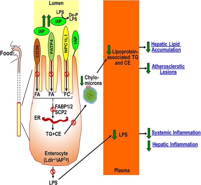
IAP is produced by enterocytes, and catalyzes removal of 1 of the 2 phosphate groups from the toxic lipid A moiety of LPS. This produces monophosphoryl-LPS, and results in attenuation of the downstream TLR (Toll-like receptor)-4–dependent inflammatory cascade.

IAP also:

  • Dephosphorylates other proinflammatory molecules such as flagellin and ATP, resulting in their detoxification;
  • Regulates expression of key gap junction proteins (zonula occludens, claudin, and occludin) and their cellular localization, which directly modulates intestinal barrier function;
  • Promotes growth of various commensal bacteria in the gut by decreasing luminal concentrations of nucleotide triphosphates via dephosphorylation; and
  • Translocates from the apical surface of enterocytes during fat absorption. Increased serum IAP accompanies fat absorption, which is consistent with observed increased levels of circulating LPS in WD-fed mice, providing one more likely mechanism by which WD affects intestinal barrier function via IAP.

Nutrients and food components/supplements that increase IAP include galacto- or chito- oligosaccharides, glucomannan, and vitamin D3. These provide a novel opportunity to develop simple strategies for modulation of diet/nutrition to target metabolic diseases including diabetes, fatty liver disease, atherosclerosis, and heart disease.”

https://www.ahajournals.org/doi/10.1161/CIRCRESAHA.120.317144 “Over-Expression of Intestinal Alkaline Phosphatase Attenuates Atherosclerosis”


Previously curated IAP studies were:

PXL_20210710_093310161.NIGHT

Gut and brain health

This 2021 human review subject was interactions of gut health and disease with brain health and disease:

“Actions of microbial metabolites are key for appropriate gut-brain communication in humans. Among these metabolites, short-chain fatty acids (SCFAs), tryptophan, and bile acid metabolites / pathways show strong preclinical evidence for involvement in various aspects of brain function and behaviour.

Dietary fibres, proteins, and fats ingested by the host contain components which are metabolized by microbiota. SCFAs are produced from fermentation of fibres, and tryptophan-kynurenine (TRP-KYN) metabolites from dietary proteins. Primary bile acids derived from liver metabolism aid in lipid digestion, but can be deconjugated and bio-transformed into secondary bile acids.

1-s2.0-S0149763421001032-gr1

One of the greatest challenges with human microbiota studies is making inferences about composition of colonic microbiota from faeces. There are known differences between faecal and caecal microbiota composition in humans along with spatial variation across the gastrointestinal tract.

It is difficult to interpret microbiome-host associations without identifying the driving influence in such an interaction. Large cohort studies may require thousands of participants on order to reach 20 % explanatory power for a certain host-trait with specific microbiota-associated metrics (Shannon diversity, relative microbial abundance). Collection of metadata is important to allow for a better comparison between studies, and to identify differentially abundant microbes arising from confounding variables.”

https://www.sciencedirect.com/science/article/pii/S0149763421001032 “Mining Microbes for Mental Health: Determining the Role of Microbial Metabolic Pathways in Human Brain Health and Disease”


Don’t understand why these researchers handcuffed themselves by only using PubMed searches. For example, two papers were cited for:

“Conjugated and unconjugated bile acids, as well as taurine or glycine alone, are potential neuroactive ligands in humans.”

Compare scientific coverage of PubMed with Scopus:

  • 2017 paper: PubMed citations 39; Scopus citations 69.
  • 2019 paper: PubMed citations 69; Scopus citations 102.

Large numbers of papers intentionally missing from PubMed probably influenced this review’s findings, such as:

  1. “There are too few fibromyalgia and migraine microbiome-related studies to make definitive conclusions. However, one fibromyalgia study found altered microbial species associated with SCFA and tryptophan metabolism, as well as changes in serum levels of SCFAs. Similarly, the sole migraine-microbiota study reported an increased abundance of the kynurenine synthesis GBM (gut-brain module).
  2. Due to heterogeneity of stroke and vascular disease conditions, it is difficult to make substantial comparisons between studies. There is convincing evidence for involvement of specific microbial genera / species and a neurovascular condition in humans. However, taxa were linked to LPS biosynthesis rather than SCFA production.
  3. Several studies suggest lasting microbial changes in response to prenatal or postnatal stress, though these do not provide evidence for involvement of SCFA, tryptophan, or bile-acid modifying bacteria. Similar to stress, there are very few studies assessing impact of post-traumatic stress disorder on microbiota.”

These researchers took on a difficult task. Their study design could have been better.


PXL_20210628_095746132

Wildlife

PXL_20210710_100826663

Improving epigenetic clocks’ signal-to-noise ratio

This 2021 computational study investigated several methods of improving epigenetic clock reliability:

“Epigenetic clocks are widely used aging biomarkers calculated from DNA methylation data. Unfortunately, measurements for individual CpGs can be surprisingly unreliable due to technical noise, and this may limit the utility of epigenetic clocks.

Noise produces deviations up to 3 to 9 years between technical replicates for six major epigenetic clocks. Elimination of low-reliability CpGs does not ameliorate this issue.

We present a novel computational multi-step solution to address this noise, involving performing principal component analysis (PCA) on the CpG-level data followed by biological age prediction using principal components as input. This method extracts shared systematic variation in DNAm while minimizing random noise from individual CpGs.

Our novel principal-component versions of six clocks show agreement between most technical replicates within 0 to 1.5 years, equivalent or improved prediction of outcomes, and more stable trajectories in longitudinal studies and cell culture. This method entails only one additional step compared to traditional clocks, does not require prior knowledge of CpG reliabilities, and can improve the reliability of any existing or future epigenetic biomarker.

PC-based clocks showed greatly improved agreement between technical replicates, with 90+% agreeing within 1-1.5 years. The median deviation ranged from 0.3 to 0.8 years, whereas CpG clocks ranged from 0.9-2.4 years.

PCPhenoAge vs. PhenoAge

The most dramatic improvement was in PhenoAge. CpG-trained PhenoAge has a median deviation of 2.4 years, 3rd quartile of 5 years, and maximum of 8.6 years. In contrast, PCPhenoAge has a median deviation of 0.6 years, 3rd quartile of 0.9 years, and maximum of 1.6 years. PCPhenoAge was trained directly on phenotypic age based on clinical biomarkers rather than DNAm.

Correlations between different PC clocks was stronger than between CpG clocks. This may be partly due to the shared set of CpGs used to train PCs, or due to the reduction of noise that would have biased correlations towards the null. Correlations between PC clocks and CpG clocks tended to be stronger compared to correlations between CpG clocks and CpG clocks, consistent with a reduction of noise.

PC clocks preserve relevant aging signals unique to each of their CpG counterparts. They reduce technical variance but maintain relevant biological variance.

PCA is a commonly used tool and does not require specialized knowledge. High reliability of principal component-based epigenetic clocks will make them particularly useful for applications in personalized medicine and clinical trials evaluating novel aging interventions.”

https://doi.org/10.1093/geroni/igab046.015 “A Computational Solution to Bolster Epigenetic Clock Reliability for Clinical Trials and Longitudinal Tracking”


I appreciate that a coauthor – who is the originator of PhenoAge – is open to evidence and improvements. There’s a fun do-it-yourself demo of PCA at https://setosa.io/ev/principal-component-analysis/.

I found this study from it citing a 2021 review:

https://www.sciencedirect.com/science/article/abs/pii/S1084952121000094 “Aging biomarkers and the brain” (not freely available)

I found that review from it citing a 2020 study:

https://www.cell.com/iscience/fulltext/S2589-0042(20)30384-9 “Human Gut Microbiome Aging Clock Based on Taxonomic Profiling and Deep Learning”

Maybe this last study could be improved from its “mean absolute error of 5.91 years” with PCA? See Part 2 for another view.


PXL_20210704_092829847

Take acetyl-L-carnitine for early-life trauma

This 2021 rodent study traumatized female mice during their last 20% of pregnancy, with effects that included:

  • Prenatally stressed pups raised by stressed mothers had normal cognitive function, but depressive-like behavior and social impairment;
  • Prenatally stressed pups raised by control mothers did not reverse behavioral deficits; and
  • Control pups raised by stressed mothers displayed prenatally stressed pups’ behavioral phenotypes.

Acetyl-L-carnitine (ALCAR) protected against and reversed depressive-like behavior induced by prenatal trauma:

alcar regime

ALCAR was supplemented in drinking water of s → S mice either from weaning to adulthood (3–8 weeks), or for one week in adulthood (7–8 weeks). ALCAR supplementation from weaning rendered s → S mice resistant to developing depressive-like behavior.

ALCAR supplementation for 1 week during adulthood rescued depressive-like behavior. One week after ALCAR cessation, however, the anti-depressant effect of ALCAR was diminished.

Intergenerational trauma induces social deficits and depressive-like behavior through divergent and convergent mechanisms of both in utero and early-life parenting environments:

  • We establish 2-HG [2-hydroxyglutaric acid, a hypoxia and mitochondrial dysfunction marker, and an epigenetic modifier] as an early predictive biomarker for trauma-induced behavioral deficits; and
  • Demonstrate that early pharmacological correction of mitochondria metabolism dysfunction by ALCAR can permanently reverse behavioral deficits.”

https://www.nature.com/articles/s42003-021-02255-2 “Intergenerational trauma transmission is associated with brain metabotranscriptome remodeling and mitochondrial dysfunction”


This study had an effusive endorsement of acetyl-L-carnitine in its Discussion section, ending with:

“This has the potential to change lives of millions of people who suffer from major depression or have risk of developing this disabling disorder, particularly those in which depression arose from prenatal traumatic stress.”

I take a gram daily. Don’t know about prenatal trauma, but I’m certain what happened during my early childhood.

I asked both these researchers and those of Reference 70 for their estimates of a human equivalent to “0.3% ALCAR in drinking water.” Will update with their replies.


No word from those researchers, so here’s what I calculate:

  • (.003 x .081) x 70 kg = 1.7% human equivalent dose.
  • 1 liter water = 1 kg, so .017 x 1000 g = 17 g per liter of water.

We all drink at least a liter of water every day. A 17 gram/liter dose is way too high for humans, considering:

I downgraded this study to Required further work. It’s likely these researchers overdosed mice to ensure their treatment produced an effect. That’s counterproductive to the purpose of animal studies: to help humans.


PXL_20210704_095621886

Vitamin K2 forms and effects

Two human studies using two forms of Vitamin K2. The first published in 2021 was with premenopausal women taking the MK-7 form:

“The aim of this 6-month randomised, controlled trial was to examine effects on bone metabolism of a nutritional supplement in women aged 25 to 44. The nutritional supplement was a protein-rich beverage powder fortified with multi-micronutrients including calcium (600 mg), vitamin D (400 IU), and vitamin K (55 mcg) per daily serving.

Co-primary outcome variables were the changes from baseline after 6 months of treatment in:

  • Bone resorption marker serum C-terminal cross-linking telopeptide of type I collagen (s-CTX-I); and
  • Bone formation marker expressed as ratio of carboxylated osteocalcin to under carboxylated-osteocalcin (c-OC/uc-OC).

The ratio of carboxylated to undercarboxylated OC is a marker of vitamin K status:

c-OC to uc-OC ratio

A meta-analysis of randomised controlled trials indicated that vitamin K2 administration reduces uc-OC and increases c-OC, and is associated with reduced bone loss and possibly a reduction in risk of fractures. The MK-7 dose of 55 mcg given in this study had a significant benefit (increase) at 3 months, but not at 6 months.

This randomised controlled 6-month trial of a nutritional supplement showed favorable changes in bone turnover markers (decreased) and calcium homeostasis. Such changes in older adults have been associated with slowing of bone loss and reduced fracture risk.”

https://www.mdpi.com/2072-6643/13/2/364/htm “Randomised Controlled Trial of Nutritional Supplement on Bone Turnover Markers in Indian Premenopausal Women”


A second study published in 2019 was with postmenopausal women taking the MK-4 form:

“This study assessed improvement in carboxylation of osteocalcin (OC) in response to escalating doses of MK-4 supplementation. A nine-week, open-labeled, prospective cohort study was conducted in 29 postmenopausal women who suffered hip or vertebral compression fractures. Mean ± SD age of participants was 69 ± 9 years.

Participants took:

  • Low-dose MK-4 (0.5 mg) for 3 weeks; then
  • Medium-dose MK-4 (5 mg) for 3 weeks; then
  • High-dose MK-4 (45 mg) for 3 weeks.

MK-4 dose, but neither age nor other relevant medications (e.g. bisphosphonates), correlated with improvement in %ucOC. Compared with baseline concentrations of 16.8 ± 2.4:

  • 0.5 mg supplementation halved %ucOC to 8.7 ± 2.2; and
  • 5-mg dose halved %ucOC again to 3.9 ± 2.2.

However, compared to 5 mg/day, there was no additional benefit of 45 mg/day (%ucOC 4.6).

MK-4 supplementation resulted in borderline increases in γ-carboxylated osteocalcin (glaOC; p = 0.07). There were no major side effects of MK-4 supplementation.

In postmenopausal women with osteoporotic fractures, supplementation with either 5 or 45 mg/day of MK-4 reduced ucOC to concentrations typical of healthy premenopausal women.”

https://econtent.hogrefe.com/doi/10.1024/0300-9831/a000554 “Maximal dose-response of vitamin K2 (menaquinone-4) on undercarboxylated osteocalcin in women with osteoporosis” (not freely available)


I asked coauthors of the first study for an estimate of MK-7 trans isomer content. Will update with their answer.

PXL_20210628_091432077.NIGHT

Eat broccoli sprouts every day

This 2020 rodent study demonstrated benefits from daily cooked broccoli intake, even when it contained no myrosinase enzyme and no sulforaphane:

“Broccoli consumption by rats influenced several metabolic pathways that impact liver health. Plasma metabolite changes are potential biomarkers of liver health, and also monitor broccoli benefits.

Rats fed a broccoli diet exhibited an enhanced Nrf2-Nqo1 pathway by day 4:

nrf2-nq01 pathway activation

Amino acid synthesis and glutathione (GSH) synthesis pathways were upregulated by Day 7. Fatty acid synthesis pathways, specifically α-linoleic acid synthesis pathways, were downregulated by Day 14.

Glucosinolate (GSL) metabolite sulforaphane alters liver GSH metabolism. It might be that consumption of any brassica, since all have GSLs, may lead to plasma glutamine and S-methyl-L-cysteine (SMC) as biomarkers. Future studies are needed to confirm whether glutamine and SMC are broccoli-specific or GSL-specific biomarkers.

Dietary broccoli caused plasma metabolite changes that correlate with:

  • Improved GSH status, suggesting protection from oxidative stress; and
  • Diversity and abundance of gut microbiota, suggesting that changes in gut microbiome may contribute to health benefits caused by dietary broccoli.”

https://www.mdpi.com/2072-6643/12/9/2514/htm “Biomarkers of Broccoli Consumption: Implications for Glutathione Metabolism and Liver Health”


I came across this study as a result of it citing the second study of A pair of broccoli sprout studies:

“A human clinical study reported changes in plasma fatty acids and GSH/GSH component levels after even a single meal of broccoli sprouts, similar to pathways we report here for rat plasma. We saw levels drop initially, then rise.

In that 2-day study, levels dropped like ours, but it was not sufficiently long to see the recovery and overshoot that we saw by 14 days when glutamine abundance and liver Nrf2 and NQO1 expression were all increased, suggesting increased GSH production, which might provide protection of liver from reactive oxygen species.”

Maybe a better comparison would have been against 0, 1-day, and 2-day rodent measurements, since the human study sampled at 0, 3, 6, 12, 24, and 48-hour intervals? People ate fresh broccoli sprouts only at time 0, though, whereas rodents ate a 10% cooked broccoli diet (0.11 mg/g glucoraphanin) ad libitum.


PXL_20210628_092632681

Vitamin K-dependent proteins

This 2020 review focused on three Vitamin K-dependent proteins (VKDPs):

“We summarize three important emerging VKDPs: Growth arrest‑specific protein 6 (Gas 6), Gla‑rich protein (GRP) and periostin in terms of their functions in physiological and pathological conditions. As examples:

  • Carboxylated Gas 6 and GRP effectively protect blood vessels from calcification;
  • Gas 6 protects from acute kidney injury and is involved in chronic kidney disease;
  • GRP contributes to bone homeostasis and delays progression of osteoarthritis; and
  • Periostin is involved in all phases of fracture healing and assists myocardial regeneration in the early stages of myocardial infarction.

IJMM-47-03-4835-g00

The ‘+’ refers to promotion and ‘-‘ refers to inhibition. Green represents Gas 6 physiological effects and red represents its pathological effects.

  • Gas 6 resists vascular calcification: i) Gas 6 promotes proliferation and migration of endothelial progenitor cells (EPCs); ii) Gas 6 inhibits apoptosis and senescence of vascular smooth muscle cells (VSMCs) by binding Tyro3, Axl and Mer (TAM) receptors; iii) Gas 6 decreases expression of inflammatory factors, including TNF-α and ICAM-1.
  • Gas 6 protects from acute kidney injury: i) Gas 6 significantly reduces creatinine and blood urea nitrogen; ii) Gas 6 enhances macrophages to uptake apoptotic cells; iii) Gas 6 reduces the expression of pro-inflammatory cytokines, such as IL-1β.
  • Gas 6 assists tumor progression: i) Gas 6 is necessary for survival, proliferation and growth of tumor cells; ii) Gas 6 contributes to drug resistance and tumor angiogenesis; iii) Gas 6 negatively regulates tumor immunity.

Numerous physiological benefits of vitamin K2 have been identified, such as anti-vascular calcification, glycemic control, and lipid-lowering effects. However, some questions about relationships between vitamin K2 and cancers remain unsolved. VKDPs are expected to be biomarkers for many diseases.”

https://www.spandidos-publications.com/10.3892/ijmm.2020.4835?text=fulltext “Role of emerging vitamin K‑dependent proteins: Growth arrest‑specific protein 6, Gla‑rich protein and periostin (Review)”


This review’s VKPD biomarkers included:

  • Vascular calcification;
  • Asthma;
  • Bronchial obstruction;
  • Diabetic nephropathy; and
  • Fracture risk.

Elaborating on this last item:

“In a cohort of 607 postmenopausal women from France that were followed up for 7 years, a positive correlation between serum periostin and fracture risk was observed. The association was independent of bone mineral density and prior fractures, indicating that periostin is an independent predictive marker of fracture risk.”

As pointed out in Chronological age by itself is an outdated clinical measurement, bone mineral density is one of several historical measurements that were selected for their relative convenience instead of chosen for their efficacy. We’re in a different century now.

PXL_20210628_094545802

Part 2 of Week 63 of Changing to a youthful phenotype with sprouts

To follow up Part 1, received Thursday’s lab results yesterday. Downloaded the workbook at https://michaellustgarten.com/2019/09/09/quantifying-biological-age/ and filled it in. Went to http://aging.ai/, selected 3.0, and entered values.

My starting point’s calculated values were:

biological age 1

A biological age snapshot from a year ago‘s video included optimal ranges:

optimizing biological age

Values in these optimal ranges were:

  • Albumin: 46;
  • Creatinine: 1.07;
  • high-sensitivity C-reactive protein: 0.24;
  • Red cell distribution width: 11.8; and
  • White blood cell count: 4.6.

I have some work to do on the other four. Good health while aging seldom happens on its own.

Reading more about Phenotypic age and its biological relationships. It definitely doesn’t mean I can do things I did 9.5 years ago like play golf and Frisbee football on the weekends.

I’d probably use DNAm PhenoAge to compare with other epigenetic clocks. Not sure how to use Aging.ai 3.0 calculations.

Sometime over the past year, Labcorp changed their adult alkaline phosphatase reference range from 39-117 to 48-121. Don’t know whether alkaline phosphatase’s optimal range will change with Labcorp’s new range, since < 48 was based on all-cause-mortality data.

PXL_20210622_093759263

Eat whole oats for your gut microbiota

Two papers on whole grains, with the first a 2021 review:

“Whole grains are more complex than refined grains and are promoted as part of a healthy and sustainable diet, mainly because the contribution of indigestible carbohydrates, and their co-passenger nutrients, is significantly higher. Changing composition and availability of grain carbohydrates and phytochemicals during processing may positively affect gut microbiota and improve health.

Processing is required for virtually all cereals that humans consume. However, eliminating bran has resulted in grain-based products that contribute to a lower-quality diet.

Currently, there are no specific recommendations on relative proportions of different dietary fiber types (based on variability in fermentability or degree of solubility). Switching from refined grain to whole grain will deliver more dietary fiber and nutrients associated with bran and germ, and improve diet quality.

crf312728-fig-0001-m

Carbohydrate-rich foods that are higher in slowly digested starches, resistant starch, oligosaccharides with prebiotic potential, and dietary fiber are considered to have a higher quality. Foods can be awarded an overall carbohydrate quality index (CQI). The optimum ratio of total carbohydrate (CHO) to dietary fiber should be ≤10:1.

Mostly only oligosaccharides and polysaccharides reach the colon. Even though larger molecules were fermented slower, they were still fermented within the proximal colon.

It is not surprising that there are conflicting reports with respect to effects of whole grains on gut microbiota. Part of this is due to whole grains comprising a diverse group of staple cereal foods, including wheat, corn, rice, oats, barley and rye, and hence different effects on gut microbiota are expected. Differences in study design, with respect to dose, duration, and study populations make it difficult to compare between studies and distill overarching similarities.

Enzymes can modify less fermentable dietary fiber to improve its fermentability by microbiota. Using different enzymes, dietary fibers can contribute to fermentation throughout the colon.”

https://onlinelibrary.wiley.com/doi/10.1111/1541-4337.12728 “Health benefits of whole grain: effects on dietary carbohydrate quality, the gut microbiome, and consequences of processing”


This review cited a 2019 paper as “an elegant study where oat bran (including co-passengers) was shown to be effective in increasing Bifidobacterium populations in the gut, whereas purified bioactive β-glucans did not show a bifidogenic effect”:

“Whole grain oats are known to modulate human gut microbiota and have prebiotic properties. Research todate mainly attributes these effects to fibre content. However, oats are also a rich dietary source of polyphenols, which may contribute to positive modulation of gut microbiota.

We found that oats increased bifidobacteria, acetic acid and propionic acid. This was mediated by synergy of all oat compounds within the complex food matrix, rather than its main bioactive β-glucan or polyphenols.

While human digestive enzymes cannot degrade plant cell wall polysaccharides, gut xylanolytic bacteria can, producing SCFA with health-beneficial effects. Certain strains down-regulate gene and protein expression of pro-inflammatory cytokines, notably isoform of nitric oxide synthase and PPAR-γ and interferon-γ, resulting in reduced inflammatory status, suggesting that oat β-glucan have beneficial effects on human health.

Oats as a whole food led to the greatest impact on microbiota.”

https://www.cambridge.org/core/journals/british-journal-of-nutrition/article/oat-bran-but-not-its-isolated-bioactive-glucans-or-polyphenols-have-a-bifidogenic-effect-in-an-in-vitro-fermentation-model-of-the-gut-microbiota/B23FAE2C7EED702132FC72F1C9CE990E “Oat bran, but not its isolated bioactive β-glucans or polyphenols, have a bifidogenic effect in an in vitro fermentation model of the gut microbiota”


The Avena nuda oats I eat for breakfast start out as 81.0 grams (1/2 cup). The only processing I do from an Illinois farmer is soaking them for 16 hours, draining then changing out to 1 1/2 cups water, then cooking for 20 minutes in a 1000W microwave at 80% power. They end up weighing 154.7 g.

I eat 51.9 g of 3-day-old sprouted Avena sativa oats from a Montana farmer at the same time, and concurrently take 2.5 g inulin. Pretty sure this 154.7 + 51.9 + 2.5 = 209.1 g combination meets an “optimum ratio of total carbohydrate to dietary fiber ≤10:1.”

Also pretty sure sprouted Avena sativa oats supply enzymes that facilitate breaking down Avena nuda complex molecules. Haven’t experienced any complaints over the past 3+ months. 🙂

Week 63 of Changing to a youthful phenotype with sprouts

Finally got around to getting an annual physical this morning. Two indicators so far, with more expected in five days. They came in early, so here’s Part 2.

1. HbA1C – glycated hemoglobin – was 4.8 on a scale of 4.8 to 5.6%. That’s down from 5.1 in June 2020. HbA1C shows a two-month average blood glucose level.

I’ve eaten advanced glycation end product (AGE)-less chicken vegetable soup almost every day since July 2019. Upcoming instantaneous blood glucose measurements may be informative, but it seems that with what I’m doing, there’s little impetus to glycate that glucose. Which satisfies my intention to avoid glycative stress.

2. BMI for a normal weight is 18.5-24.9 kg/m2. Measurements over the past two years:

  • June 2019 24.8, 0.1 below range high;
  • June 2020 22.4, 2.5 below range high and 3.9 above low; and
  • June 2021 21.0, 3.9 below range high and 2.5 above low.

Annual BMI trend is going in the right direction, but it’s too squishy to be a biomarker. I usually don’t curate studies that rely on BMI.

I eat a lot of food every day! Not going to turn my kitchen into a laboratory to quantify, though. See Switch on your Nrf2 signaling pathway for what intake was on 1/1/2021.

Once or twice a week lately I’ve backed off and skipped one of a daily two (fresh and leftovers) AGE-less chicken vegetable meals when it’s been too much food. Haven’t skipped:

  • Twice-daily combined broccoli-red cabbage-mustard sprouts; or
  • Twice-daily Avena sativa oat sprouts; or
  • My Avena nuda whole oats breakfast.

Lost 11 lbs. over 12 months without trying to lose weight. Maximal food intake didn’t result in weight gain when much of its purpose was to:

  • Reduce inflammation; and
  • Make my gut microbiota happy.

PXL_20210618_094601372

Go for the win-win with taurine

This 2021 rodent study investigated taurine effects on colitis:

“Taurine plays an important role in various essential biological processes. Health beneficial effects of taurine have been generally attributable to its antioxidant and anti-inflammatory effects.

Taurine chloramine (TauCl) is an endogenous anti-inflammatory substance derived from taurine. In fighting exogenous pathogens, neutrophils utilize one powerful weapon in their arsenal: generating the strong oxidant hypochlorous acid (HOCl), which is nature’s germ killer.

Taurine can act as a trap for HOCl forming the long-lived oxidant TauCl, which is more stable and less toxic than HOCl. TauCl (20 mg/kg) was given on daily basis by gavage for 10 days before and for 3 days after intrarectal administration of 2.5% TNBS:

antioxidants-10-00479-g008-550

TauCl inhibits generation of proinflammatory mediators by phagocytic cells. Taurine exerts an anti-inflammatory as well as antioxidant action by preventing cytolytic damage caused by HOCl generated by inflammatory cells, particularly neutrophils.

These results suggest that TauCl exerts a protective effect against colitis through upregulation of Nrf2-dependent cytoprotective gene expression, while blocking proinflammatory signaling mediated by NFκB and STAT3.”

https://www.mdpi.com/2076-3921/10/3/479/htm “Protective Effects of Taurine Chloramine on Experimentally Induced Colitis: NFκB, STAT3, and Nrf2 as Potential Targets”


Train your gut microbiota with taurine showed how taurine supplementation can be a win-win for both our brain and gut microbiota.

Smoke and die early, while your twin lives on

A 2021 human twin study investigated epigenetic clocks:

“This study showed that accelerated epigenetic aging is associated with increased mortality, and smoking plays a role in explaining this association. Present findings suggest that DNAm GrimAge is a strong predictor of mortality independent of genetic influences among female twin pairs.

An invitation to participate in the study was sent to 414 female twin pairs, aged 63–76 years. Of 199 twin pairs, at least one twin died in 112 pairs during follow-up:

twins

This epigenetic age estimate that measures biological age and runs alongside, but not always in parallel with chronological age, may inform life expectancy predictions. Further research is needed to determine whether results apply to men, and the extent to which DNA methylation age can be used as a clinical biomarker of lifespan.”

https://clinicalepigeneticsjournal.biomedcentral.com/articles/10.1186/s13148-021-01112-7 “Does the epigenetic clock GrimAge predict mortality independent of genetic influences: an 18 year follow-up study in older female twin pairs”


If you don’t have a twin, substitute yourself as an analogous entity who has opposite behaviors. Don’t assume that smoking cannabis will produce different results from tobacco.

Meanwhile, Dr. Steven Horvath, who has a twin, continued his 2021 torrent of coauthored studies last week with Epigenetic clock and methylation studies in elephants. Amazing epigenetic clock information being published this year. Dr. Horvath is completely open to evidence, IAW, a real scientist.

Take Vitamin K2 if you take statins

This 2021 human study related Vitamin K2 status and statin usage:

“We examined the connection between statin exposure, coronary artery calcification (CAC), and vitamin K-dependent proteins (VKDPs) in patients with cardiovascular (CV) conditions. VKDPs measured in plasma included undercarboxylated (ucOC), and carboxylated osteocalcin (cOC).

CAC score (CACS) was determined by multislice computed tomography:

statins and vitamin k2 deficiency

CACS was more pronounced in statin users compared to non-users. The same was also found among CVD patients and among controls. Both ucOC and ucOC / cOC ratio were significantly elevated in statin users, indicating vitamin K deficiency.

Our results are in agreement with existing evidence about positive associations between statins and vascular calcification. They enlighten possible mechanisms through which statins may enhance calcium accumulation in arterial wall, by inhibiting VKDPs and functions involved in vascular protection.”

https://onlinelibrary.wiley.com/doi/10.1002/kjm2.12373 “Statins, vascular calcification, and vitamin K-dependent proteins: Is there a relation?”


All of this study’s measurements were done outside the liver, so Vitamin K deficiency ≈ Vitamin K2 deficiency. The uncited third paper of Vitamin K2 – What can it do? provided evidence for findings of the current study.

Per the third paper, I should have reached a blood serum level Vitamin K2 MK-7 plateau by supplementing for three weeks. We’ll see later this week if an increasing cOC / ucOC ratio had any effect on hypertension.

PXL_20210606_093626698