Signaling pathways and disordered proteins

This 2022 review explored the title subject:

“Cell signaling imposes many demands on proteins that comprise these pathways, including abilities to form active and inactive states, and to engage in multiple protein interactions. Signaling often requires amplifying signals, regulating or tuning responses to signals, combining information sourced from multiple pathways, all while ensuring process fidelity.

Sensitivity, adaptability, and tunability are possible, in part, due to inclusion of intrinsically disordered regions in many proteins involved in cell signaling.  This review highlights the critical role of intrinsically disordered proteins for signaling:

  • In widely diverse organisms (animals, plants, bacteria, fungi);
  • In every category of cell signaling pathway (autocrine, juxtacrine, intracrine, paracrine, and endocrine); and
  • At each stage (ligand, receptor, transducer, effector, terminator) in the cell signaling process.

Function of the glucocorticoid receptor is regulated in part by its intrinsically disordered C-terminal tail. Prior to activation, the glucocorticoid receptor resides in cytosol:

glucocorticoid receptor

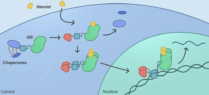
Intrinsic disorder in the glucocorticoid receptor not only enables multiple allosteric regulatory interactions to impact function, but also allows deployment of different surfaces of the protein to enable binding to many different sets of macromolecules, and regulation of these interactions via mRNA splicing and phosphorylation.

Combinations of alternative translation initiation and alternative mRNA splicing result in production of multiple glucocorticoid receptor isoforms from one gene. Various isoforms exhibit distinctive tissue distribution patterns and altered transcriptional regulatory profiles.

Greater than 90% of transcription factors either contain intrinsically disordered regions of proteins or are entirely intrinsically disordered. The many advantages conferred by disorder to cell signaling cascades means that:

  1. Understanding signaling required definition of roles disorder plays in each pathway;
  2. Many more examples of disordered proteins in cell signaling pathways are likely to be discovered; and
  3. More mechanisms by which disorder functions remain to be elucidated.”

https://biosignaling.biomedcentral.com/articles/10.1186/s12964-022-00821-7 “Intrinsically disordered proteins play diverse roles in cell signaling”


Cells in vivo seldom act on their own impetus. I would have liked discussion – or at least mention – of bidirectional signals between genes / cells / tissues / organs / organism / environment. This review’s topic of cell signaling pathways excluded “interactions of complex, interconnected systems spanning hierarchical levels” as explored in An environmental signaling paradigm of aging.

Vitamin K forms

Two papers on Vitamin K, beginning with a 2021 review:

“Vitamin K is involved in many biological processes. Menaquinones (MK) [Vitamin K2] and phylloquinone [Vitamin K1] vary in biological activity, showing different bioavailability, half-life, and transport mechanisms.

The effective dose to decrease uncarboxylated osteocalcin was six times lower for MK-7 than for MK-4. Similarly, MK-7 affected blood coagulation system at dose three to four times lower than vitamin K1.

Both vitamin K1 and MK-7 inhibited decline in bone mineral density. However, benefits for occurrence of cardiovascular diseases have been observed only for long-chain menaquinones. There are currently no guidelines for recommended doses and forms of vitamin K in prevention of osteoporosis, atherosclerosis, and other cardiovascular disorders.”

https://www.mdpi.com/2304-8158/10/12/3136/htm “Relationship between Structure and Biological Activity of Various Vitamin K Forms”


This first paper cited a 2019 meta-analysis for:

“Vitamin K2 supplementation is a preventative measure rather than an osteoporosis treatment.

Meta-analysis of various interventions for improving BMD revealed that vitamin K2 can increase lumbar spine BMD. It ranked sixth among eighteen different single or combined interventions including Ca, vitamin D, estrogen, isoflavone and exercise.

Effect size for change in bone mineral density (BMD) using forest plots. LS, lumbar spine; D, vitamin D; Est, oestrogen; Ex, exercise; K, vitamin K; Iso, isoflavone; FN, femoral neck.

urn cambridge.org id binary-alt 20211204100437-73338-optimisedImage-S0007114519002290_fig3g

Lumbar spine:

  • Ca, vitamin D, vitamin K, oestrogen, exercise, Ca + vitamin D, vitamin D + vitamin K, and vitamin D + oestrogen were associated with significantly beneficial effects relative to no treatment.
  • Ca, vitamin D, oestrogen, and Ca + vitamin D were associated with beneficial effects compared with placebo.
  • Vitamin D + vitamin K was associated with positive effect with Ca.
  • Oestrogen, vitamin D + vitamin K, and vitamin D + oestrogen were associated with beneficial effect compared with vitamin D.
  • Ca + vitamin D + exercise had a beneficial effect compared with Ca + vitamin D.
  • Ca + oestrogen, and isoflavone + exercise were related to negative effects relative to oestrogen.

Femoral neck:

  • Ca, exercise, and vitamin D + oestrogen were associated with significant beneficial intervention effects relative to no treatment.

The present study demonstrated that many interventions were valuable for improving BMD in the LS and FN of postmenopausal women. It confirmed the need for postmenopausal women to improve BMD through preventive measures such as nutrients or oestrogen.

It also confirmed that different single or combined preventions can affect BMD at different sites in different orders. This reveals to medical and health workers and postmenopausal women which methods can be selected preferentially to prevent bone loss.”

https://doi.org/10.1017/S0007114519002290 “Impact of calcium, vitamin D, vitamin K, oestrogen, isoflavone and exercise on bone mineral density for osteoporosis prevention in postmenopausal women: a network meta-analysis”


Amazingly oblivious that this freely-available second 2019 paper has been cited only by this first paper. What recent literature is more relevant to postmenopausal women’s health?

Eat broccoli sprouts for depression, Part 2

Here are three papers that cited last year’s Part 1. First is a 2021 rodent study investigating a microRNA’s pro-depressive effects:

“Depressive rat models were established via chronic unpredicted mild stress (CUMS) treatment. Cognitive function of rats was assessed by a series of behavioral tests.

Nrf2 CUMS

Nrf2 was weakly expressed in CUMS-treated rats, whereas Nrf2 upregulation alleviated cognitive dysfunction and brain inflammatory injury.

Nrf2 inhibited miR-17-5p expression via binding to the miR-17-5p promoter. miR-17-5p was also found to limit wolfram syndrome 1 (Wfs1) transcription.

We found that Nrf2 inhibited miR-17-5p expression and promoted Wfs1 transcription, thereby alleviating cognitive dysfunction and inflammatory injury in rats with depression-like behaviors. We didn’t investigate the role of Nrf2 in other depression models (chronic social stress model and chronic restraint stress model) and important brain regions other than hippocampus, such as prefrontal cortex and nucleus accumbens. Accordingly, other depression models and brain regions need to be designed and explored to further validate the role of Nrf2 in depression in future studies.”

https://link.springer.com/article/10.1007/s10753-021-01554-4 “Nrf2 Alleviates Cognitive Dysfunction and Brain Inflammatory Injury via Mediating Wfs1 in Rats with Depression‑Like Behaviors” (not freely available)

This study demonstrated that activating the Nrf2 pathway inhibited brain inflammation, cognitive dysfunction, and depression. Would modulating one microRNA and one gene in vivo without Nrf2 activation achieve similar results?


A 2021 review focused on the immune system’s role in depression:

“Major depressive disorder is one of the most common psychiatric illnesses. The mean age of patients with this disorder is 30.4 years, and the prevalence is twice higher in women than in men.

Activation of inflammatory pathways in the brain is considered to be an important producer of excitotoxicity and oxidative stress inducer that contributes to neuronal damage seen in the disorder. This activation is mainly due to pro-inflammatory cytokines activating the tryptophan-kynurenine (KP) pathway in microglial cells and astrocytes.

Elevated levels of cortisol exert an inhibitory feedback mechanism on its receptors in the hippocampus and hypothalamus, stopping stimulation of these structures to restore balance. When this balance is disrupted, hypercortisolemia directly stimulates extrahepatic enzyme 2,3-indolimine dioxygenase (IDO) located in various tissues (intestine, placenta, liver, and brain) and immune system macrophages and dendritic cells.

Elevation of IDO activities causes metabolism of 99% of available tryptophan in the KP pathway, substantially reducing serotonin synthesis, and producing reactive oxygen species and nitrogen radicals. The excitotoxicity generated produces tissue lesions, and activates the inflammatory response.”

https://academic.oup.com/ijnp/article/25/1/46/6415265 “Inflammatory Process and Immune System in Major Depressive Disorder”

This review highlighted that stress via cortisol and IDO may affect the brain and other parts of the body.


A 2022 review elaborated on Part 1’s findings of MeCP2 as a BDNF inhibitor:

“Methyl-CpG-binding protein 2 (MeCP2) is a transcriptional regulator that is highly abundant in the brain. It binds to methylated genomic DNA to regulate a range of physiological functions implicated in neuronal development and adult synaptic plasticity.

Ability to cope with stressors relies upon activation of the hypothalamic–pituitary–adrenal (HPA) axis. MeCP2 has been shown to contribute to early life stress-dependent epigenetic programming of genes that enhance HPA-axis activity.

We describe known functions of MeCP2 as an epigenetic regulator, and provide evidence for its role in modulating synaptic plasticity via transcriptional regulation of BDNF or other proteins involved in synaptogenesis and synaptic strength like reelin. We conclude that MeCP2 is a promising target for development of novel, more efficacious therapeutics for treatment of stress-related disorders such as depression.”

https://www.mdpi.com/2073-4409/11/4/748/htm “The Role of MeCP2 in Regulating Synaptic Plasticity in the Context of Stress and Depression”


Osprey lunch

PXL_20220221_192924474

Broccoli sprouts and your gut barrier

This 2021 human cell study investigated sulforaphane’s gut barrier protective effects:

“Intestinal epithelial cells (IECs) are an important component of the epithelial barrier, which helps prevent passage of pathogens, toxins, and allergens from gastrointestinal lumen into the circulatory system. Destruction of the intestinal barrier increases intestinal permeability, destroys homeostasis of the immune system, and induces inflammatory responses and oxidative stress.

Interconnections in each cellular network maintain homeostasis. AMPK is a highly conserved serine/threonine protein kinase that helps regulate levels of ROS in mitochondria.

AMPK acts together with its downstream target SIRT1 to upregulate PGC-1α and help control mitochondrial biosynthesis, energy metabolism, and oxidative stress as a homeostasis-sensing network. It induces intracellular NAD+ which can show biological effects.

kbie_a_1952368_uf0001_oc

Sulforaphane:

  • Increased cell viability and reduced lactate dehydrogenase activity in a concentration-dependent manner.
  • Weakened LPS-induced increases in intestinal epithelial cell permeability and oxidative stress.
  • Increased levels of antioxidants.
  • Weakened the ability of LPS to induce production of inflammatory cytokines and pro-apoptotic caspases.

We showed that sulforaphane exerts these effects by activating the AMPK/SIRT1/PGC-1α cascade.”

https://www.tandfonline.com/doi/full/10.1080/21655979.2021.1952368 “Sulforaphane protects intestinal epithelial cells against lipopolysaccharide-induced injury by activating the AMPK/SIRT1/PGC-1ɑ pathway”


PXL_20220217_201213685

Beneficially stressing sprouts with light

This 2021 study investigated effects of light on red cabbage sprouts and microgreens. I’ll highlight its 3-day-old sprout findings:

“Periodic ultraviolet UV-B (280–320 nm) pulses at low doses improved morphological development of red cabbage sprouts, and probably will have the same effect on other sprouts. Similarly, total phenolic content, total flavonoid content, and total antioxidant capacity presented a UV-B dose-dependence response.

Although UV-B radiation may cause damage to plant tissues, an optimum dose can promote accumulation of antioxidant and UV-protective molecules that enhance nutraceutical biosynthesis in plant foods without altering sensory quality. Such an increase in concentration of bioactive compounds is mainly due to environmental stress generated by UV-B light, which leads to changes in morphology, physiology, and molecular conformation of DNA, RNA, and proteins.

red cabbage sprout phenolics

Total phenolic content of control (0, CTRL) or UV-B-treated (5, 10, and 15 kJ m−2) red cabbage sprouts after 10 days growth at 20 °C. Different capital letters indicate significant differences among treatments at < 0.05. Different lowercase letters indicate significant differences among time of analysis of the same treatment at p < 0.05.

Our results demonstrated that application of UV-B light during germination induces a positive effect on growth of red cabbage sprouts, as well as on secondary metabolite content related to nutritional quality. Analysed bioactive compounds (phenolics, flavonoids, and carotenoids) increased during germination, and tended to remain constant throughout a refrigerated shelf life.”

https://www.mdpi.com/2311-7524/7/12/567/htm “UV-B Radiation as Abiotic Elicitor to Enhance Phytochemicals and Development of Red Cabbage Sprouts”


I came across this study after lead author Dr. Lorena Martínez-Zamora provided an earlier study, Postharvest UV-B and UV-C radiation enhanced the biosynthesis of glucosinolates and isothiocyanates in Brassicaceae sprouts (not freely available). Its focus was also a worthwhile commercialization of cruciferous microgreens.

Nearby researchers also published studies such as Red cabbage effects on gut microbiota and Sprout bioaccessibility last year. Let’s hope a push for cruciferous sprouts and microgreens continues, although consumer acceptance is limited by products not being sweet.

I think these research efforts will succeed. Take a look at oat milk’s quick rise, for example. I had trouble getting delivery of Avena sativa seeds last month because oat milk producers bought up last year’s supplies and futures on this year’s crops.

For me, it’s been 98 weeks of spending at least 45 minutes a day growing 3-day-old broccoli, red cabbage, mustard, and oat sprouts at home. I’m satisfied with results, and won’t turn my kitchen into a laboratory to eke out extra effects with light and other elicitors.

Here’s a photo of this study’s sponsoring institution’s harbor from the latest of two visits:

IMG_20170611_125837

I’d like to return whenever we individually stop being herded and recover our sanity.

Your thymus and calories

This 2022 paper studied caloric restriction in humans followed up by rodent experiments:

“Extension of lifespan driven by 40% caloric restriction (CR) in rodents causes trade-offs in growth, reproduction, and immune defense that make it difficult to identify therapeutically relevant CR-mimetic targets. We report that about 14% CR for 2 years in healthy humans improved thymopoiesis.

thymus cr

Expression of the gene PLA2G7 is inhibited in humans undergoing CR. Deletion of Pla2g7 in mice showed decreased thymic lipoatrophy, protection against age-related inflammation, lowered NLRP3 inflammasome activation, and improved metabolic health.

Twenty-four-month-old PLA2G7-deficient mice (analogous to ~70-year-old humans) had larger thymi and higher thymocyte abundance, and were protected from age-related thymic involution.

thymus mice

We propose that reduction of PLA2G7 caused by CR in humans might contribute to better adipose tissue metabolism, lower inflammation, and reduced thymic lipoatrophy.”

https://www.science.org/doi/10.1126/science.abg7292 “Caloric restriction in humans reveals immunometabolic regulators of health span” (not freely available). Thanks to Dr. Alexander Predeus for providing a copy.


I would have liked rodent experiments to continue another year or so to determine the control group’s and PLA2G7-deficient group’s healthspans and lifespans. These researchers could have strengthened their findings if increased healthspans also increased lifespans.

CD38 and balance

I’ll highlight this 2022 review’s relationships between inflammation and cluster of differentiation 38:

“We review the nicotinamide adenine dinucleotide (NAD) catabolizing enzyme CD38, which plays critical roles in pathogenesis of diseases related to infection, inflammation, fibrosis, metabolism, and aging.

NAD is a cofactor of paramount importance for an array of cellular processes related to mitochondrial function and metabolism, redox reactions, signaling, cell division, inflammation, and DNA repair. Dysregulation of NAD is associated with multiple diseases. Since CD38 is the main NADase in mammalian tissues, its contribution to pathological processes has been explored in multiple disease models.

CD38 is upregulated in a cell-dependent manner by several stimuli in the presence of pro-inflammatory or secreted senescence factors or in response to a bacterial infection, retinoic acid, or gonadal steroids. CD38 is stimulated in a cell-specific manner by lipopolysaccharide, tumor necrosis factor alpha, interleukin-6, and interferon-γ.

dysregulated inflammation

CD38 plays a critical role in inflammation, migration, and immunometabolism, but equally important is resolution of the inflammatory response which left unchecked leads to loss of self-tolerance, tissue infiltration of lymphocytes, and circulation of autoantibodies.

  • Depending upon context, CD38 can either promote or protect against an autoimmune response.
  • Chronic mucosal inflammation and tissue damage characteristic of inflammatory bowel disease predisposes IBD patients to development of colorectal cancer, and the risks increase with duration, extent, and severity of inflammation.
  • Pulmonary fibrosis occurs in the presence of unresolved inflammation and dysregulated tissue repair, and results from an array of injurious stimuli including infection, toxicant exposure, adverse effects of drugs, and autoimmune response.
  • Modulating CD38 and NAD levels in kidney disease may provide therapeutic approaches for prevention of inflammatory conditions of the kidney.
  • Inflammation as well as evidence of senescence are present in pathophysiology of chronic liver diseases that progress to cirrhosis.
  • Inflammation-associated metabolic diseases impair vascular function. Chronic inflammation can lead to vascular senescence and dysfunction.

One cause of NAD decline during aging is due to increase of NAD breakdown in the presence of increased CD38 expression and activity on immune cells, thus linking inflammaging with tissue NAD decline. Other sources of NAD decline include increased DNA-damage requiring PARP1 activation, and decreased NAMPT levels leading to diminished NAD synthesis through the salvage pathway.

Inflammation is among the major risk factors that predispose organisms to age-associated diseases. During aging, accumulation of senescent cells creates an environment rich in proinflammatory signals, leading to ‘inflammaging.’ Metabolically active cells lose their replicative capacity by entering an irreversible quiescent state, and are considered both a cause and a consequence of inflammaging.

Recent findings uncover a major role of CD38 in inflammation and senescence, showing that age-related NAD+ decline and the sterile inflammation of aging are partially mediated by a senescence / senescence associated secretory phenotype (SASP)-induced accumulation of CD38+ inflammatory cells in tissues. Given the clear association between the phenomenon of inflammaging, senescence, and CD38, as well as the impact of CD38 on degradation of NAD and the NAD precursor NMN, future studies should focus on CD38 as a druggable target in viral illnesses.”

https://journals.physiology.org/doi/abs/10.1152/ajpcell.00451.2021 “The CD38 glycohydrolase and the NAD sink: implications for pathological conditions”


We extend good-vs.-bad thinking to nature. Does that paradigm explain much, though?

All pieces of a puzzle are important. Otherwise, evolution would have eliminated what wasn’t necessary for its purposes.

Restoring balance to an earlier phenotype suits my purposes. Don’t want to eliminate inflammatory responses, but instead, calm them down so that they’re evoked appropriately.

Studying AGEs and neurodegeneration

This 2022 review suggested more effective ways to conduct in vitro studies of advanced glycation end products (AGEs) and neurodegenerative diseases:

“The main goal of this review was to present and discuss in vitro models that were applied or have the potential to be used in research on AGEs and ND.

  • We introduced and explained current knowledge on AGEs regarding their formation and accumulation in humans.
  • We presented existing evidence linking involvement of AGEs in ND and explained basic concepts of brain physiology and immunology affected by AGEs.
  • We presented and discussed available in vitro models to study AGE-mediated neurodegeneration by dividing them into sections from simple models. These have been applied to more complex models that have not been yet applied in the field of AGEs, but offer opportunities.
  • We gathered advisable in vitro tools based on their relevance to three primary endpoints that AGEs can impact brain pathophysiology and their characteristics and suitability to mimic ND pathophysiology.

nutrients-14-00363-g002

Several studies have indicated intracellular formation of AGEs by microglia or neurons, but identification of intracellular AGEs in those cases is made by immunoassays, which have received much criticism regarding their reliability to identify and quantify AGEs. Concerns about these techniques are mostly related to undefined specificity and affinity of anti-AGE antibodies.

The source of observed AGE accumulation in the brain of patients (dietary or endogenous) is not yet fully understood. For that reason, studies on AGE digestion and absorption (i.e., in vitro digestion models) are crucial to understanding the type of dietary AGEs that will circulate and cross the BBB to reach the brain.

On the other hand, endogenous AGEs can also be formed due to increased glucose levels derived from a high glycemic diet. Highly reactive molecules in the brain can contribute to locally produced AGEs extracellularly or intracellularly.

Clinical studies mainly focus on the fate and metabolism of dietary AGEs. Exposure based on consumption of certain foods is difficult to translate to a concentration that cells are going to be exposed to. The complexity and multiple sources of protein glycation require application of in vitro models to understand potential contribution to neurodegeneration.”

https://www.mdpi.com/2072-6643/14/2/363/htmIn Vitro Methodologies to Study the Role of Advanced Glycation End Products (AGEs) in Neurodegeneration”


While we’re waiting for research to catch up, we can hedge neurodegenerative disease bets by:

  • Not spiking our blood glucose levels;
  • Avoiding foods with medium and high levels of AGEs;
  • Giving our gut microbiota the intake they need instead of what our unconscious programming dictates; and
  • Maintaining youthful activities.

PXL_20220212_194256506

Optimizing glucosinolate analysis

I’ll highlight microwave findings of this 2021 study:

“Glucosinolates (GSLs) are important precursor compounds with anticancer activities in Brassicaceae vegetables and are readily hydrolyzed by myrosinase. Given the diversity of these species, establishing an accurate and universal method to quantify intact GSLs in different plant tissues is necessary.

We compared and optimized three tissue disruption methods for sample preparation:

  • Recoveries of GSLs in a Chinese cabbage sample were significantly lower than 100% after microwave treatment for 60 s, due to insufficient inactivation of myrosinase.
  • After microwave treatment for 90 s, recoveries of 13 GSLs were in the range of 73–124%, indicating that this condition could inactivate myrosinase completely.
  • The increase in GSL recoveries with microwave treatment for 120 s might be due to increased extractability of GSLs.
  • A limitation of this method was that different tissues could not be processed under the same microwave conditions.

molecules-27-00231-g001

SIN, Sinigrin; NAP, Gluconapin; GBN, Glucobrassicanapin; PRO, Progoitrin; ERU, Glucoerucin; RAE, Glucoraphenin; RAA, Glucoraphanin; ALY, Glucoalyssin; GBC, Glucobrassicin; 4ME, 4-Methoxyglucobrassicin; NEO, Neoglucobrassicin; TRO, Glucotropaeolin; NAS, Gluconasturtiin.

Five GSLs without available standards were estimated using calibration curves of structurally similar compounds. Specifically, the pair glucoberteroin (GOB) and glucoerucin (ERU), glucoiberin (GIB) and glucoraphanin (RAA), gluconapoleiferin (GNL) and PRO, and 4OH and 4ME are homologs that differ in structure by one -CH2 group, with glucoraphasatin (GRH) containing an alkenyl group in its molecular structure, which is two hydrogen atoms less than ERU.

The verified method of intact GSLs by UHPLC-MS/MS established in this study was more accurate and time-saving than the commonly used ISO method for desulfo-GSLs.”

https://www.mdpi.com/1420-3049/27/1/231/htm “Determination of 18 Intact Glucosinolates in Brassicaceae Vegetables by UHPLC-MS/MS: Comparing Tissue Disruption Methods for Sample Preparation”


This study was in line with other studies that increased GLS amounts by microwaving. For example, most of the above graphic’s Chinese cabbage GLS measurements at 90 seconds were greater than raw samples, which kept going:

“The increase in GSL recoveries with microwave treatment for 120 s might be due to increased extractability of GSLs.”

Unlike this study, my goal is to optimize glucosinolate hydrolysis products such as sulforaphane. I increase myrosinase enzyme activity rather than decrease it, and want to have less GLS amounts than what I started with after processing.

I facilitate myrosinase activity by:

  • Adjusting immersion water to pH 5; and
  • Stopping at 60°C (140°F) to avoid a myrosinase deactivation cliff between 60°C and 65°C.

This study used a 900W microwave to process 30-gram broccoli floret samples at Figure S1 different times (20, 40, 60, 90, 120, 180 seconds). I use a 1000W microwave to process a 65-gram broccoli / red cabbage / mustard sprouts mix in 100 ml water for 40 seconds.

Kickstarting endogenous regenerative pathways

A 2022 amphibian study by the Electroceuticals team investigated limb regeneration:

“Organisms such as Xenopus laevis – whose limited regenerative capacities in adulthood mirror those of humans – are important models with which to test interventions that can restore form and function. We demonstrate long-term (18 months) regrowth, marked tissue repatterning, and functional restoration of an amputated X. laevis hindlimb following a 24-hour exposure to a multidrug, pro-regenerative treatment delivered by a wearable bioreactor.

  • Regenerated multidrug treatment (MDT) hindlimbs were longer than the no added factors (BD) and no device (ND) groups by 2.5 mpa, as indicated by growth beyond resection site (red dashed line).
  • At 4 mpa, vascularized structures developed at the distal extension of MDT (yellow arrow), but not BD or ND regenerates.
  • At 9 mpa, digit-like projections appeared (blue arrow), contrasting hypomorphic spikes of BD and ND regenerates (pink arrows).

limb regeneration

We suggest that the overall strategy of providing wound cells with an aqueous, amniotic-like environment, which is uniquely given through our bioreactor, that contains pro-regenerative signals is likely to be an effective method for kickstarting biomedically relevant growth and patterning cascades that are too complex to directly implement. One additional direction that may present new opportunities for enhanced regeneration is to assess immune function in relation to tissue remodeling.”

https://www.science.org/doi/10.1126/sciadv.abj2164 “Acute multidrug delivery via a wearable bioreactor facilitates long-term limb regeneration and functional recovery in adult Xenopus laevis”

Sulforaphane and hair loss

This 2021 human clinical trial evaluated sulforaphane’s cosmetic effects:

“We demonstrated that sulforaphane has the potential to become a highly effective functional hair cosmetic to relieve hair loss with androgen alopecia. Sulforaphane increases expression of the dihydrotestosterone (DHT)-degrading enzyme 3α-hydroxysteroid dehydrogenase (3α-HSD) in the liver, which accelerates DHT degradation, thereby inhibiting hair loss.

We performed a visual evaluation of parietal and frontal lines of 23 men and women from 18 to 54 years old before and after using the product, and then calculated total number of hairs. This clinical study showed that parietal lines and bangs visually improved, and the number of hairs increased by 6.71% from before using the test product to 18 weeks after using the test product.

hair growth

We tested expression levels of Ak1c21 and Dhrs9 isoforms of 3α-HSD in the in vitro cell culture experiment where Hepa1c1c7 cells were treated with sulforaphane or a mixture of biotin, dexpanthenol, and l-menthol. This study showed that sulforaphane alone achieved a hair loss-relieving effect in our experimental cell culture conditions.

Our finding that sulforaphane induces Akr1c2 in a dose-dependent manner is consistent with previous studies. Sulforaphane treatment induced expression of Dhrs9, which has several sites in the promoter region that bind to Nrf2, which is induced by sulforaphane.

It is highly likely that sulforaphane might enhance degradation of DHT, not only via the induction of degrading enzymes 3α-HSD, but also by functional activation of these enzymes. Further studies remain to test this possibility.”

https://www.mdpi.com/2079-9284/8/3/63/htm “Sulforaphane, L-Menthol, and Dexpanthenol as a Novel Active Cosmetic Ingredient Composition for Relieving Hair Loss Symptoms”

Gut signals

I’ll highlight signaling pathway aspects of this 2022 review:

“The gut bacterial community plays an important role in regulation of multiple aspects of metabolic disorders. This regulation depends, among other things, on production of a wide variety of metabolites by microbiota and on their interactions with receptors on host cells that can activate or inhibit signalling pathways, and either be beneficial and detrimental to the host’s health.

Colonocytes and endocrine cells express a variety of receptors able to sense and transmit signals from the microbial environment:

gutjnl-2021-326789-F4.large

  • TLRs cover a wide range of both external stimuli (PAMPs) and internal signals derived from tissue damage. Their activation induces antigen-presenting cell activation, thereby bridging innate and adaptive immune responses, and stimulates signalling cascades as an attempt to fend off microbial invaders or repair damaged tissue.
  • The endocannabinoid signalling system appears to play a key role in regulating energy, glucose, and lipid metabolism but also in immunity, inflammation, and more recently in microbiota-host interactions.
  • Although the primary function of bile acids (BAs) is to regulate digestion and absorption of cholesterol, triglycerides, and fat-soluble vitamins, it has been recently recognised that BAs also serve an endocrine function as they act as signalling molecules. BAs have been shown to modulate epithelial cell proliferation, gene expression, lipid, glucose, and energy metabolism by activating several receptors. Because of their signalling capacities and the fact that BAs are chemically transformed by gut microbiota, BAs can be considered as microbiota-derived signalling metabolites.
  • Numerous AhR ligands exist including environmental triggers, nutrition-derived signals, various phytochemicals, and bacterial metabolites such as tryptophan.

Most signalling metabolites can be produced by large numbers of different gut bacteria, and hence have limited specificity.”

https://gut.bmj.com/content/early/2022/01/31/gutjnl-2021-326789.long “Gut microbiome and health: mechanistic insights”

Predicting atherosclerosis

Starting this blog’s eighth year with a 2022 epigenetic clock study that assessed young people’s common blood tests fifteen and twenty years later:

GrimAge acceleration (GAA), an epigenetic marker that represents physiologic aging, is associated with atherosclerotic cardiovascular disease. We used multivariable regression models to examine associations of Y15 and Y20 GAA estimates with plasma lipid levels measured at prior examination years (Y0, Y5, and Y10) and concurrently: triglycerides (TG), low-density lipoprotein cholesterol (LDL-C), and high-density lipoprotein cholesterol (HDL-C) levels:

  • Each 1-SD higher cumulative TG level was associated with an average 0.73 ± 0.12 years older GAA;
  • Each 1-SD higher cumulative HDL-C level was associated with an average 0.57 ± 0.17 years younger GAA;
  • Associations between TG and GAA were stronger among female and Black participants; and
  • Associations between HDL-C and GAA were stronger among female and White participants.

lipid-profiles-and-GrimAge-acceleration

We observed that elevated TG and low HDL-C levels in young adulthood are associated with accelerated midlife epigenetic aging, and epigenetic aging mediates some of the well-described associations between elevated TG levels in early life and subclinical atherosclerosis in middle age. These findings suggest that maintaining optimal lipid levels in early adulthood may help to slow epigenetic aging, which reflects delays in the onset of age-related diseases like atherosclerosis.”

https://clinicalepigeneticsjournal.biomedcentral.com/articles/10.1186/s13148-021-01222-2 “Plasma lipid profiles in early adulthood are associated with epigenetic aging in the Coronary Artery Risk Development in Young Adults (CARDIA) Study”


Which is better for resolving a health situation?

  • Hope for luck / providence before subclinical symptoms become clinical problems?
  • Do nothing constructive, and depend on interventions after problems occur?
  • Take responsibility for your own one precious life?

PXL_20220131_201832917

An epigenetic regulator of vascular aging

This 2022 rodent and human cell study investigated the smooth muscle cell mineralocorticoid receptor:

“Vascular stiffness increases with age and independently predicts cardiovascular disease risk. Epigenetic changes, including histone modifications, accumulate with age, but the global pattern has not been elucidated nor are the regulators known.

Rising mineralocorticoid receptor (MR) in aging vascular smooth muscle cells downregulates EZH2 to globally shift to a more open chromatin thereby allowing MR to be recruited to promoters to transcriptionally upregulate target genes involved in vascular stiffness. This mechanism provides multiple potential targets to prevent vascular stiffness in aging humans.

cvac007f8

We demonstrate for the first time that:

  1. MR expression increases with age in primary, low passage, human aortic smooth muscle cell (SMC) and correlates with age in whole aortic tissue from aging humans;
  2. The global proteomic profile of histone modifications in mouse vessels changes profoundly with aging with a significant overall decrease in H3K27 methylation;
  3. Expression of H3K27 methyltransferase EZH2 decreases with age in mouse vessels and in human SMCs in a MR-dependent manner and negatively correlates with MR expression in whole human aortic tissue;
  4. The aging-induced decline in EZH2 associates with reduced H3K27 methylation and increased H3K27 acetylation in vitro and in vivo;
  5. These epigenetic changes in aging human SMC and mouse vessels correspond with increased expression of the vascular stiffness genes, CTGF and integrin-α5, previously identified vascular MR target genes;
  6. Induction of an aging phenotype in human SMC associates with increased MR enrichment and H3K27 acetylation at these stiffness gene promoters; and
  7. Inhibition of MR in aged mice and aged human aortic SMCs reverses the entire process; increasing EZH2 and H3K27 methylation, increasing locus-specific EZH2 enrichment and decreasing H3K27 acetylation at stiffness gene promoters, decreasing vascular expression of CTGF and integrin-α5, and decreasing the stiffness and adhesiveness of aged human SMC in vitro and mouse aortic stiffness and fibrosis in vivo.”

https://academic.oup.com/cardiovascres/advance-article-abstract/doi/10.1093/cvr/cvac007/6502304 “Smooth muscle mineralocorticoid receptor as an epigenetic regulator of vascular ageing” (not freely available) Thanks to Dr. Seung Kyum Kim for providing a copy.