Prenatal stress produces offspring who as adults have cognitive, emotional, and memory deficiencies

This 2018 French/Italian/Swiss rodent study used a prenatally restraint stressed (PRS) model to create problems that could be resolved by various chemicals:

“S 47445 is a positive modulator of glutamate AMPA-type receptors, possessing neurotrophic and enhancing synaptic plasticity effects as well as pro-cognitive and anti-stress properties.

Most of studies examining the antidepressant effects of new molecules are carried out using behavioral tests performed in unstressed animals.

Corticosterone-treated mice and rats exposed to chronic stress are models that do not recapitulate the early programming of stress-related disorders, which likely originates in the perinatal period. The PRS rat model is characterized by a prolonged corticosterone response to stress and by abnormal behavior.

All the behavioral alterations induced by PRS were corrected by chronic S 47445 administration at both doses.”


The paper included a section comparing S 47445 to ketamine:

“Ketamine, however, causes severe cognitive impairment and psychotomimetic [mimics the symptoms of psychosis, reference not freely available] effects that are direct consequences of the prolonged inhibition of NMDA receptors in cortical and hippocampal interneurons, and seriously limit the chronic administration of the drug in the clinical setting. [reference not freely available]

S 47445 by inducing a direct activation of AMPARs displayed an antidepressant activity without the adverse effect of ketamine. Indeed, contrary to ketamine, S 47445 presented no psychotomimetic effects and induced no occurrence of spontaneous epileptic seizures. [reference freely available] Moreover, S 47445 also presented pro-cognitive properties.”

Compare the above with this April 2018 Chicago Tribune story that had opinions with no linked references:

“ketamine, an anesthetic used to sedate both people and animals before surgery. It’s also a notorious street drug, abused by clubgoers seeking a trancelike, hallucinatory high. But in recent years, numerous studies have found that ketamine can be an effective and speedy treatment for people with depression.”

Which coverage better informed us?


Treating prenatal stress-related disorders with an oxytocin receptor agonist was performed by several of this paper’s coauthors. One references to it was:

“We have already reported that depolarization-evoked glutamate release in the ventral hippocampus is negatively correlated with risk-taking behavior of PRS rats, and that such correlation can be corrected by chronic treatment with monoaminergic/ melatoninergic antidepressants or oxytocin receptor agonist. Thus, an impairment of glutamatergic transmission in the ventral hippocampus lies at the core of the pathological phenotype of PRS rats.”

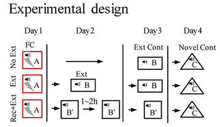
Looking at the above graphic of the experimental design, I’m not sure why the term perinatal (occurring during or pertaining to the phase surrounding the time of birth) was used in the paper’s title and content to describe the stress period. The pregnant females were stressed three times every day during the second half of pregnancy up until delivery, so the prenatal (previous to birth) term was more applicable.


So, how does this study help humans?

One takeaway is to avoid stressing pregnant mothers-to-be if her children will be expected to become adults without cognitive, emotional, and behavioral problems.

The study demonstrated one way prenatal events cause lifelong effects. The PRS model provides another example of why it’s useless to ask adult humans to self-report causes of epigenetic problems in their lives when these originated before birth, during infancy, or in early childhood, well before humans develop sufficient cognitive capability to recognize such situations. It’s incomprehensible that this unreliable paradigm is still given significant weight in stress studies, especially when experimental designs:

“Do not recapitulate the early programming of stress-related disorders, which likely originates in the perinatal period.”

Also, a relevant difference between humans and PRS rats is that we can ourselves individually change our responses to experiential causes of ongoing adverse effects. Standard methodologies can only apply external treatments such as those mentioned above.

https://www.sciencedirect.com/science/article/pii/S0028390818301291 “The reduction in glutamate release is predictive of cognitive and emotional alterations that are corrected by the positive modulator of AMPA receptors S 47445 in perinatal stressed rats” (not freely available) Thanks to coauthors Stefania Maccari and Dr. Jerome Mairesse for providing a copy.

The role of DNMT3a in fear memories

This 2018 Chinese rodent study found:

“Elevated Dnmt3a [a DNA methyltransferase] level in the dorsal dentate gyrus (dDG) of hippocampus was associated with the absence of fear renewal in an altered context after extinction training. Overexpression and knockdown of Dnmt3a in the dDG regulated the occurrence of fear renewal in a bi-directional manner.

We found that renewal of remote fear memory can be prevented, and the absence of renewal was concurrent with an elevated Dnmt3a level.

Our results indicate that Dnmt3a in the dDG is a key regulator of fear renewal after extinction, and Dnmt3a may play a critical role in controlling fear memory return and thus has therapeutic values.”


The study was a collection of five experiments investigating causes and effects of biology and behavior. The researchers used different techniques to achieve their goals. I’ve quoted extensively below to show some background and results.

“Alterations in histone acetylation and DNA methylation are involved in the formation and extinction of long-term memory. DNMTs catalyze the cytosine methylation and are required to establish and maintain genomic methylation.

Dnmt3a and Dnmt3b are de novo DNA methyltransferases. Dnmt1 is the maintenance DNA methyltransferase.

  1. Dnmt3a expression was elevated in the dDG after extinction training followed by a brief memory retrieval (Rec+Ext), which was associated with the absence of fear renewal when tested in an altered context.
  2. Increasing Dnmt3a expression in the dDG using AAV [recombinant adeno-associated virus] expression led to the prevention of fear renewal following a standard extinction training protocol. 
  3. Knockdown of Dnmt3a in the dDG using CRISPR/Cas9 resulted in fear renewal following Rec+Ext protocol.
  4. Renewal of remote fear memory can be prevented using the Rec+Ext protocol.
  5. The absence of renewal was concurrent with an elevated Dnmt3a level.

Current exposure therapy, although effective in many patients, suffers from the inability to generalize its efficacy over time, or is limited by the potential return of adverse memory in the new/novel contexts. These limitations are caused by the context-dependent nature of extinction which is widely viewed as the biological basis of exposure therapy.

Achieving a context-independent extinction may significantly reduce fear renewal to improve the efficacy of exposure therapy. Our current study suggests that the effectiveness of these approaches, and ultimately the occurrence of fear renewal, is determined by the level of Dnmt3a after extinction training, especially in the dDG.

There are two potential mechanisms underlying extinction, one is erasure or updating of the formed memory, and the other is the formation of a new extinction memory which suppresses or competes with the existing memory in a context-dependent manner. While most studies favor the suppression mechanism in the adult, limited studies do suggest that erasure occurs in the immature animals.

We propose that if Dnmt3a level is elevated with extinction training (such as with Rec+Ext protocol), modification to the existing memory occurs and as a consequence extinction does not act as a separate mechanism or form a new memory; but if Dnmt3a level is unaltered with extinction training, a separate extinction memory is formed which acts to suppress or compete with the existing memory.”


The relevant difference between humans and lab rats is that we can ourselves individually change our responses to experiential causes of ongoing adverse effects. Standard methodologies can only apply external treatments such as exposure therapy and manipulating Dnmt3a levels.

https://www.nature.com/articles/s41598-018-23533-w “Dnmt3a in the dorsal dentate gyrus is a key regulator of fear renewal”

This dietary supplement is better for depression symptoms than placebo

This 2018 Italy/UK meta-analysis subject was the use of dietary supplement acetyl-L-carnitine to treat depression symptoms:

“Deficiency of acetyl-L-carnitine (ALC) appears to play a role in the risk of developing depression, indicating dysregulation of fatty acids transport across the inner membrane of mitochondria. However, the data regarding ALC supplementation in humans are limited. We thus conducted a systematic review and meta-analysis investigating the effect of ALC on depressive symptoms across randomized controlled trials (RCTs).

Pooled data across nine RCTs (231 treated with ALC versus 216 treated with placebo and 20 no intervention) showed that ALC significantly reduced depressive symptoms.

In these nine RCTs, the majority of the studies used 3 grams of ALC as intervention.

In three RCTs comparing ALC versus antidepressants (162 for each group), ALC demonstrated similar effectiveness compared with established antidepressants [fluoxetine (Prozac), duloxetine (Cymbalta), amisulpride (Solian) respectively below] in reducing depressive symptoms. In these latter RCTs, the incidence of adverse effects was significantly lower in the ALC group [79%] than in the antidepressant group.

Subgroup analyses suggested that ALC was most efficacious in older adults. Future large scale trials are required to confirm/refute these findings.”

From the Methods section:

“Studies were excluded if:

  1. did not include humans;
  2. did not include a control group;
  3. did not use validated scales for assessing depression;
  4. did not report data at follow-up evaluation regarding tests assessing depression;
  5. included the use of ALC with another agent vs. placebo/no intervention.”

The Discussion section was informative regarding possible mechanisms of ALC affecting depression, pain, and linked symptoms. Several citations were of a review rather than of the original studies, however.


Research needs to proceed on to investigate therapies that address ultimate causes for depression and pain. Researchers and sponsors shouldn’t stop at just symptoms and symptom relief, notwithstanding the requirement from a statistical point of view for “future large scale trials.”

Here are other acetyl-L-carnitine topics I’ve curated:

https://journals.lww.com/psychosomaticmedicine/Citation/2018/02000/Acetyl_L_Carnitine_Supplementation_and_the.4.aspx “Acetyl-L-Carnitine Supplementation and the Treatment of Depressive Symptoms: A Systematic Review and Meta-Analysis” (not freely available)


This post has somehow become a target for spammers, and I’ve disabled comments. Readers can comment on other posts and indicate that they want their comment to apply here, and I’ll re-enable comments.

RNA and neurodegenerative diseases

This 2018 Chinese paper reviewed the associations among long non-coding RNA and four neurodegenerative diseases:

“lncRNAs are widely implicated in various physiological and pathological processes, such as epigenetic regulation, cell cycle regulation, cell differentiation regulation, cancer, and neurodegenerative diseases, through their interactions with chromatin, protein, and other RNAs. Numerous studies have suggested that lncRNAs are closely linked with the occurrence and development of a variety of diseases, especially neurodegenerative diseases, of which the etiologies are complicated and the underlying mechanisms remain elusive.

We focus on how lncRNA dysfunctions are involved in the pathogenesis of Alzheimer’s disease, Parkinson’s disease, Huntington’s disease, and amyotrophic lateral sclerosis.”


Table 1 showed specific lncRNAs that acted as “bodyguards” in inherited Huntington’s disease, “culprits” in Alzheimer’s disease, and as both in Parkinson’s disease. The table didn’t include lncRNAs associated with amyotrophic lateral sclerosis although the review text mentioned several.

https://www.sciencedirect.com/science/article/pii/S2162253117303104 “Long Non-coding RNAs, Novel Culprits, or Bodyguards in Neurodegenerative Diseases”

Sleep and adult brain neurogenesis

This 2018 Japan/Detroit review subject was the impact of sleep and epigenetic modifications on adult dentate gyrus neurogenesis:

“We discuss the functions of adult‐born DG neurons, describe the epigenetic regulation of adult DG neurogenesis, identify overlaps in how sleep and epigenetic modifications impact adult DG neurogenesis and memory consolidation..

Whereas the rate of DG neurogenesis declines exponentially with age in most mammals, humans appear to exhibit a more modest age‐related reduction in DG neurogenesis. Evidence of adult neurogenesis has also been observed in other regions of the mammalian brain such as the subventricular zone, neocortex, hypothalamus, amygdala, and striatum.

Adult‐born DG neurons functionally integrate into hippocampal circuitry and play a special role in cognition during a period of heightened excitability and synaptic plasticity occurring 4–6 weeks after mitosis. Adult DG neurogenesis is regulated by a myriad of intrinsic and extrinsic factors, including:

  • drugs,
  • diet,
  • inflammation,
  • physical activity,
  • environmental enrichment,
  • stress, and
  • trauma.”


Some of what the review stated was contradicted by other evidence. For example, arguments for sleep were based on the memory consolidation paradigm, but evidence against memory consolidation wasn’t cited for balanced consideration.

It reminded me of A review that inadvertently showed how memory paradigms prevented relevant research. That review’s citations included a study led by one of those reviewers where:

“The researchers elected to pursue a workaround of the memory reconsolidation paradigm when the need for a new paradigm of enduring memories directly confronted them!”

Some of what this review stated was speculation. I didn’t quote any sections after:

 “We go one step further and propose..”

The review also had a narrative directed toward:

“Employing sleep interventions and epigenetic drugs..”

It’s storytelling rather than pursuing the scientific method when reviewers approach a topic as these reviewers did.

Instead of reading a directed narrative, read this informative blog post from a Canadian researcher. The post provided scientific contexts to summarize what was and wasn’t known in 2018 about human neurogenesis.

http://onlinelibrary.wiley.com/doi/10.1002/stem.2815/epdf “Regulatory Influence of Sleep and Epigenetics on Adult Hippocampal Neurogenesis and Cognitive and Emotional Function”

Sex-specific impacts of childhood trauma

This 2018 Canadian paper reviewed evidence for potential sex-specific differences in the lasting impacts of childhood trauma:

“This paper will provide a contextualized summary of neuroendocrine, neuroimaging, and behavioral epigenetic studies on biological sex differences contributing to internalizing psychopathology, specifically posttraumatic stress disorder and depression, among adults with a history of childhood abuse.

Given the breadth of this review, we limit our definition [of] trauma to intentional and interpersonal experiences (i.e., childhood abuse and neglect) in childhood. Psychopathological outcomes within this review will be limited to commonly explored internalizing disorders, specifically PTSD and depression.

Despite the inconsistent and limited findings in this review, a critical future consideration will be whether the biological effects of early life stress can be reversed in the face of evidence-based behavioral interventions, and furthermore, whether these changes may relate to potentially concurrent reductions in susceptibility to negative mental health outcomes.”


It was refreshing to read a paper where the reviewers often interrupted the reader’s train of thought to interject contradictory evidence, and display the scientific method. For example, immediately after citing a trio of well-respected studies that found:

“Psychobiological research on relationships linking impaired HPA axis functioning and adult internalizing disorders are suggestive of lower basal and afternoon levels of plasma cortisol in PTSD phenotype.”

the reviewers stated:

“However, a recent meta-analysis suggests no association between basal cortisol with PTSD.”

and effectively ended the cortisol discussion with:

“Findings are dependent upon variance in extenuating factors, including but not limited to, different measurements of:

  • early adversity,
  • age of onset,
  • basal cortisol levels, as well as
  • trauma forms and subtypes, and
  • presence and severity of psychopathology symptomology.”

The reviewers also provided good summaries of aspects of the reviewed subject. For example, the “Serotonergic system genetic research, childhood trauma and risk of psychopathology” subsection ended with:

“Going forward, studies must explore the longitudinal effects of early trauma on methylation as well as comparisons of multiple loci methylation patterns and interactions to determine the greatest factors contributing to health outcomes. Only then, can we start to consider the role of sex in moderating risk.”


I didn’t agree with the cause-ignoring approach of the behavior therapy mentioned in the review. Does it make sense to approach one category of symptoms:

“the biological effects of early life stress”

by treating another category of symptoms?

“can be reversed in the face of evidence-based behavioral interventions.”

But addressing symptoms instead of the sometimes-common causes that generate both biological and behavioral effects continues to be the direction.

After receiving short-term symptom relief, wouldn’t people prefer treatments of originating causes so that their various symptoms don’t keep bubbling up? Why wouldn’t research paradigms be aligned accordingly?

I was encouraged by the intergenerational and transgenerational focus of one of the reviewer’s research:

“Dr. Gonzalez’s current research focus is to understand the mechanisms by which early experiences are transmitted across generations and how preventive interventions may affect this transmission.”

This line of hypotheses requires detailed histories, and should uncover causes for many effects that researchers may otherwise shrug off as unexplainable individual differences. Its aims include the preconception through prenatal periods when both the largest and the largest number of epigenetic changes occur, and is when our susceptibility and sensitivity to our environment is greatest. There are fewer opportunities for effective “preventive interventions” in later life compared with these early periods.

Unlike lab rats, women and men can reach some degree of honesty about our early lives’ experiential causes of ongoing adverse effects. Experiential therapies that allow humans to potentially change their responses to these causes deserve more investigation than do therapies that apply external “interventions.”

https://www.sciencedirect.com/science/article/pii/S0272735817302647 “Biological alterations affecting risk of adult psychopathology following childhood trauma: A review of sex differences” (not freely available) Thanks to lead author Dr. Ashwini Tiwari for providing a copy.

Non-CpG DNA methylation

This 2017 Korean review compared and contrasted CpG and non-CpG DNA methylation:

“Non-CpG methylation is restricted to specific cell types, such as pluripotent stem cells, oocytes, neurons, and glial cells. Accumulation of methylation at non-CpG sites and CpG sites in neurons seems to be involved in development and disease etiology.

Non-CpG methylation is established during postnatal development of the hippocampus and its levels increase over time. Similarly, non-CpG methylation is scarcely detected in human fetal frontal cortex, but is dramatically increased in later life. This increase in non-CpG methylation occurs simultaneously with synaptic development and increases in synaptic density.

In contrast, CpG methylation occurs during early development and does not increase over time.

Neurons have considerably higher levels of non-CpG methylation than glial cells. The human male ES [embryonic stem] cell line (H1) is more highly methylated than the female ES cell line (H9).

Among the different types of non-CpG methylation (CpA [adenosine], CpT [thymine], and CpC [another cytosine]), methylation is most common at CpA sites. For instance, in human iPS [induced pluripotent stem] cells, 5mCs are found in approximately 68.31%, 7.81%, 1.99%, and 1.05% of CpG, CpA, CpT, and CpC sites, respectively.”


The reviewers’ referenced statement:

“CpG methylation occurs during early development and does not increase over time.”

was presented outside of its context. The 2013 cited source’s statement was restricted to “selected loci” in the rodent hippocampus:

“Consistent with a recent study of the cortex, time-course analyses revealed that CpH [non-CpG] methylation at the selected loci was established during postnatal development of the hippocampus and was then present throughout life, whereas CpG methylation was established during early development.”

Epigenetic study methodologies improved in 2017 had more information on CpA methylation.

http://www.mdpi.com/2073-4425/8/6/148/htm “CpG and Non-CpG Methylation in Epigenetic Gene Regulation and Brain Function”

Can researchers make a difference in their fields?

The purpose and finding of this 2017 UK meta-analysis of human epigenetics and cognitive abilities was:

“A meta-analysis of the relationship between blood-based DNA methylation and cognitive function.

We identified [two] methylation sites that are linked to an aspect of executive function and global cognitive ability. The latter finding relied on a relatively crude cognitive test..which is commonly used to identify individuals at risk of dementia.

One of the two CpG sites identified was under modest genetic control..there are relatively modest methylation signatures for cognitive function.”

The review’s stated limitations included:

“It is, of course, possible that a reliable blood-based epigenetic marker of cognitive function may be several degrees of separation away from the biological processes that drive cognitive skills.

There are additional limitations of this study:

  • A varying number of participants with cognitive data available for each test;
  • Heterogeneity in relation to the ethnicity and geographical location of the participants across cohorts; and
  • Relating a blood-based methylation signature to a brain-based outcome.

A 6-year window [between ages 70 and 76] is possibly too narrow to observe substantial changes in the CpG levels.”

All of these limitations were known before the meta-analysis was planned and performed. Other “possible” limitations already known by the 47 coauthors include those from Genetic statistics don’t necessarily predict the effects of an individual’s genes.

The paper referenced studies to justify the efforts, such as one (cited twice) coauthored by the lead author of A problematic study of DNA methylation in frontal cortex development and schizophrenia:

“Epigenome-wide studies of other brain-related outcomes, such as schizophrenia, have identified putative blood-based methylation signatures.”


Was this weak-sauce meta-analysis done just to plump up 47 CVs? Why can’t researchers investigate conditions that could make a difference in their fields?

Was this meta-analysis done mainly because the funding was available? I’ve heard that the primary reason there are papers like the doubly-cited one above is that the US NIMH funds few other types of research outside of their biomarker dogma.

The opportunity costs of this genre of research are staggering. Were there no more productive topics that these 47 scientists could have investigated?

Here are a few more-promising research areas where epigenetic effects can be observed in human behavior and physiology:

I hope that the researchers value their professions enough to make a difference with these or other areas of their expertise. And that sponsors won’t thwart researchers’ desires for difference-making science by putting them into endless funding queues.

https://www.nature.com/articles/s41380-017-0008-y “Meta-analysis of epigenome-wide association studies of cognitive abilities”

Epigenetic study methodologies improved in 2017

Let’s start out 2018 paying more attention to advancements in science that provide sound empirical data and methodology. Let’s ignore and de-emphasize studies and reviews that aren’t much more than beliefs couched in models and memes, whatever their presumed authority.

Let sponsors direct researchers to focus on ultimate causes of diseases. Let’s put research of treatments affecting causes ahead of those that only address symptoms.

Here are two areas of epigenetic research that improved in 2017.


Improved methodologies enabled DNA methylation studies of adenine, one of the four bases of DNA, to advance, such as this 2017 Wisconsin/Minnesota study N6-methyladenine is an epigenetic marker of mammalian early life stress:

“6 mA is present in the mammalian brain, is altered within the Htr2a gene promoter by early life stress and biological sex, and increased 6 mA is associated with gene repression. These data suggest that methylation of adenosine within mammalian DNA may be used as an additional epigenetic biomarker for investigating the development of stress-induced neuropathology.”

Most DNA methylation research is performed on the cytosine and guanine bases.


Other examples of improved methodologies were discussed in this 2017 Japanese study Genome-wide identification of inter-individually variable DNA methylation sites improves the efficacy of epigenetic association studies:

“A strategy focusing on CpG sites with high DNA methylation level variability may attain an improved efficacy..estimated to be 3.7-fold higher than that of the most frequently used strategy.

With ~90% coverage of human CpGs, whole-genome bisulfite sequencing (WGBS) provides the highest coverage among the currently available DNAm [DNA methylation] profiling technologies. However, because of its high cost, it is presently infeasible to apply WGBS to large-scale EWASs [epigenome-wide association studies], which require DNAm profiling of hundreds or thousands of subjects. Therefore, microarrays and targeted bisulfite sequencing are currently practicable for large-scale EWASs and thus, effective strategies to select target regions are essentially needed to improve the efficacy of epigenetic association studies.

DNAm levels measured with microarrays are invariable for most CpG sites in the study populations. As invariable DNAm signatures cannot be associated with exposures, intermediate phenotypes, or diseases, current designs of probe sets are inefficient for blood-based EWASs.”

Differing approaches to a life wasted on beliefs

Let’s start by observing that people structure their lives around beliefs. As time goes on, what actions would a person have taken to ward off non-confirming evidence?

One response may be that they would engage in ever-increasing efforts to develop new beliefs that justified how they spent their one precious life’s time so far.

Such was my take on beliefs embedded in https://www.ncbi.nlm.nih.gov/pmc/articles/PMC5684598/pdf/PSYCHIATRY2017-5491812.pdf “Epigenetic and Neural Circuitry Landscape of Psychotherapeutic Interventions”:

“Animal models have shown the benefits of continued environmental enrichment (EE) on psychopathological phenotypes, which carries exciting translational value.

This paper posits that psychotherapy serves as a positive environmental input (something akin to EE).”

The author conveyed his belief that wonderful interventions were going to happen in the future. However, when scrutinized, most human studies have demonstrated NULL effects of psychotherapeutic interventions on causes. Without sound evidence that treatments affect causes, his belief seemed driven by something else.

The author cited findings of research like A problematic study of oxytocin receptor gene methylation, childhood abuse, and psychiatric symptoms as supporting external interventions to tamp down symptoms of patients’ presenting problems. Did any of the 300+ cited references concern treatments where patients instead therapeutically addressed their problems’ root causes?


For an analogous religious example, a person’s belief caused him to spend years of his life trying to convince men to act so that they could get their own planet after death, and trying to convince women to latch onto men who had this belief. A new and apparently newsworthy belief developed from his underlying causes:

“The founder and CEO of neuroscience company Kernel wants “to expand the bounds of human intelligence.” He is planning to do this with neuroprosthetics; brain augmentations that can improve mental function and treat disorders. Put simply, Kernel hopes to place a chip in your brain.

He was raised as a Mormon in Utah and it was while carrying out two years of missionary work in Ecuador that he was struck by what he describes as an “overwhelming desire to improve the lives of others.”

He suffered from chronic depression from the ages of 24 to 34, and has seen his father and stepfather face huge mental health struggles.”

https://www.theguardian.com/small-business-network/2017/dec/14/humans-20-meet-the-entrepreneur-who-wants-to-put-a-chip-in-your-brain “Humans 2.0: meet the entrepreneur who wants to put a chip in your brain”

The article stated that he had given up Mormonism. There was nothing to suggest, though, that he had therapeutically addressed any underlying causes for his misdirected thoughts, feelings, and behavior.

So he developed other beliefs instead.


What can people do to keep their lives from being wasted on beliefs? As mentioned in What was not, is not, and will never be:

“The problem is that spending our time and efforts on these ideas, beliefs, and behaviors won’t ameliorate their motivating causes. Our efforts only push us further away from our truths, with real consequences: a wasted life.

The goal of the therapeutic approach advocated by Dr. Arthur Janov’s Primal Therapy is to remove the force of presenting problems’ motivating causes. Success in reaching this goal is realized when patients become better able to live their own lives.

Do you have your family’s detailed medical histories?

Imagine that you were a parent who puzzled over the mystery of your pre-teen daughter’s hyperactive behavior. Without detailed family medical histories, would anyone recognize this as a preprogammed phenotype?

Could anyone trace the daughter’s behavior back to her maternal great-grandmother being treated with glucocorticoids near the end of the second trimester of carrying her grandfather?

Such was a finding of a 2017 Canadian guinea pig study that was undertaken to better inform physicians of the transgenerationally inherited epigenetic effects of glucocorticoid treatments commonly prescribed during human pregnancies:

“This study presents the first evidence that prenatal treatment with sGC [synthetic glucocorticoid] results in transgenerational paternal transmission of hyperactivity and altered hypothalamic gene expression through three generations of young offspring. Female offspring appear to be more sensitive than male offspring to the programming effects of sGC, which suggests an interaction between sGC and sex hormones or sex-linked genes. Paternal transmission to F3 strongly implicates epigenetic mechanisms in the process of transmission, and small noncoding RNAs likely play a major role.”


Some details of the study included:

Veh[icle] was the control group initially treated with saline.

The study was informative and conclusive for the aspects studied. From the Methods section:

“Data from same-sex littermates were meaned to prevent litter bias. Sample sizes (N) correspond to independent litters, and not to the total number of offspring across all litters.

Power analyses based on previous studies determined N ≥ 8 sufficient to account for inter-litter variability and detect effects in the tests performed.”

https://www.nature.com/articles/s41598-017-11635-w “Prenatal Glucocorticoid Exposure Modifies Endocrine Function and Behaviour for 3 Generations Following Maternal and Paternal Transmission”

“Transgenerationally” inherited epigenetic effects of fetal alcohol exposure

The fourth paper of Transgenerational epigenetic inheritance week was a 2016 German rodent study of of improperly-termed “transgenerational” epigenetic effects of alcohol:

“We investigated 2 generations of offspring born to alcohol-treated mothers. Here, we show that memory impairment and reduced synthesis of acetylcholine occurs in both F1 (exposed to ethanol in utero) and F2 generation (never been exposed to ethanol). Effects in the F2 generation are most likely consequences of transgenerationally transmitted epigenetic modifications in stem cells induced by alcohol.

The results further suggest an epigenetic trait for an anticholinergic endophenotype associated with cognitive dysfunction which might be relevant to our understanding of mental impairment in neurodegenerative disorders such as Alzheimer’s disease and related disorders.”

F0 generation mothers modeled human fetal alcohol syndrome. They were exposed to ethanol gradually up to 20%, then mated. The 20% ethanol intake level was maintained until the F1 generation pups were born, then gradually diminished to 0%. After a ten-day wait, an eight-week handling and shaping period started, followed by five weeks of behavioral testing.

The F1 children and F2 grandchildren started an eight-week handling and shaping period after young adulthood, followed by five weeks of behavioral testing. The F1 children were mated after behavioral testing.

The F0 parents showed no significant differences in working memory and reference memory compared with controls. Both the F1 children and F2 grandchildren were significantly impaired in the same tests compared with controls, with the F1 children performing worse than the F2 grandchildren. No sex-dependent differences were noted.

After behavioral impairments due to intergenerational epigenetic modifications were established, the F2 grandchildren received treatments to ascertain the contribution of cholinergic dysfunction in their behavioral impairments. It was confirmed, as an acetylcholine esterase inhibitor that crosses the blood-brain barrier almost completely erased working-memory and reference-memory performance deficits.

Items in the Discussion section included:

  • A dozen studies from 2014-2016 were cited for epigenetic mechanisms of inheritance stemming from parental alcohol consumption; and
  • Transgenerational inheritance of alcohol-induced neurodevelopmental deficits may involve epigenetic mechanisms that are resistant to developmental clearance.

As argued in Transgenerational effects of early environmental insults on aging and disease and A review of epigenetic transgenerational inheritance of reproductive disease, testing of F3 great-grandchildren was needed in order to establish transgenerational vs. intergenerational results. A F3 generation necessarily controls for the variable of F2 direct germline exposure.

http://www.neurobiologyofaging.org/article/S0197-4580(16)30303-7/pdf “Transgenerational transmission of an anticholinergic endophenotype with memory dysfunction” (not freely available)

Experience-induced transgenerational programming of neuronal structure and functions

The second paper of Transgenerational epigenetic inheritance week was a 2017 German/Israeli review focused on:

“The inter- and transgenerational effects of stress experience prior to and during gestation..the concept of stress-induced (re-)programming in more detail by highlighting epigenetic mechanisms and particularly those affecting the development of monoaminergic transmitter systems, which constitute the brain’s reward system.

We offer some perspectives on the development of protective and therapeutic interventions in cognitive and emotional disturbances resulting from preconception and prenatal stress.”

The reviewers noted that human studies have difficulties predicting adult responses to stress that are based on gene expression and early life experience. Clinical studies that experimentally manipulate the type, level and timing of the stressful exposure aren’t possible. Clinical studies are also predicated on the symptoms being recognized as disorders and/or diseases.

The researchers noted difficulties in human interventions and treatments. Before and during pregnancy, and perinatal periods are where stress effects are largest. But current human research hasn’t gathered sufficient findings to develop practical guidelines for early intervention programs.


I’m not persuaded by arguments that cite the difficulties of performing human research on transgenerational epigenetic inheritance. There are overwhelming numbers of people who have obvious stress symptoms: these didn’t develop in a vacuum.

Researchers:

  • Design human studies to test what’s known from transgenerational epigenetic inheritance animal studies that will include documenting the subjects’ detailed histories with sufficient biometric samples and data obtained from their lineage.
  • Induce pregnant subjects to at least temporarily avoid what’s harmful for them and/or the offspring, in favor of what’s beneficial.
  • Document the subjects’ actions with history and samples.

I acknowledge that economic incentives may not be enough to get people to participate. I’m familiar with a juvenile sickle-cell study that didn’t get enough subjects despite offering free transportation and hundreds of dollars to the caregivers per visit. The main problem seemed to be that the additional income would be reported and threaten the caregivers’ welfare benefits.

Stop whining that your jobs are difficult, researchers. Society doesn’t owe you a job. EARN IT – get yourself and the people in your organization motivated to advance science!

http://www.sciencedirect.com/science/article/pii/S014976341630731X “Experience-induced transgenerational (re-)programming of neuronal structure and functions: Impact of stress prior and during pregnancy” (not freely available)

Transgenerational effects of early environmental insults on aging and disease

The first paper of Transgenerational epigenetic inheritance week was a 2017 Canadian/Netherlands review that’s organized as follows:

“First, we address mechanisms of developmental and transgenerational programming of disease and inheritance. Second, we discuss experimental and clinical findings linking early environmental determinants to adverse aging trajectories in association with possible parental contributions and sex-specific effects. Third, we outline the main mechanisms of age-related functional decline and suggest potential interventions to reverse negative effects of transgenerational programming.”

A transgenerational phenotype was defined as an epigenetic modification that was maintained at least either to F2 grandchildren in the paternal lineage, or to F3 great-grandchildren in the maternal lineage.

The reviewers noted that mechanisms of transgenerational programming are complex and multivariate.  Severity, timing, and type of exposure; lineage of transmission; germ cell exposure; and gender of an organism were the main factors that may determine consequences. Mechanisms reviewed were:

  1. Parental exposure to an adverse environment;
  2. Altered maternal behavior and care of offspring; and
  3. Experience-dependent modifications of the epigenome.

There was a long list of diseases and impaired functionalities that were consequences of ancestral experiences and exposures. Most studies were of animals, but a few were human, such as those done on effects of extended power outages during a Quebec ice storm of January 1998.


One intervention that was effective in reversing a transgenerational phenotype induced by deficient rodent maternal care was to place pups with a caring foster female soon after birth. It’s probably unacceptable in human societies to preemptively recognize all poor-care human mothers and remove the infant to caring foster mothers. But researchers could probably find enough instances to develop studies of the effectiveness of such placements in reversing a transgenerational phenotype.

The review didn’t have suggestions for reversing human transgenerational phenotypes, just “potential interventions to reverse negative effects of transgenerational programming.” Interventions suggested for humans – exercise, enriched lifestyle, cognitive training, dietary regimens, and expressive art and writing therapies – only reduced impacts of transgenerational epigenetic effects.

Tricky wording of “reverse negative effects of transgenerational programming” showed that research paradigms weren’t aimed at resolving causes. The review was insufficient for the same reasons mentioned in How one person’s paradigms regarding stress and epigenetics impedes relevant research, prompting my same comment:

Aren’t people interested in human treatments of originating causes so that their various symptoms don’t keep bubbling up? Why wouldn’t research paradigms be aligned accordingly?

When reversals of human phenotypes aren’t researched, problems may compound by being transmitted to the next generations.

http://www.sciencedirect.com/science/article/pii/S014976341630714X “Transgenerational effects of early environmental insults on aging and disease incidence” (not freely available)

Does living near a forest keep your amygdala healthier?

A thought-provoking post from A Paper a Day Keeps the Scientist Okay entitled “Living Near a Forest Keeps Your Amygdala Healthier” referenced a 2017 German human study which found:

“..a relationship between place of residence and brain health: those city dwellers living close to a forest were more likely to show indications of a physiologically healthy amygdala structure and were therefore presumably better able to cope with stress.”

The researchers accomplished the imperative of meeting the study’s stated objective:

“We set out to identify and characterize the geographical elements of a city that are associated with these brain structures following a suggestion by Kennedy and Adolph that studies should begin to derive recommendations for urban planning and architecture.

The results of our study may suggest that forests in and around the cities are a valuable resource that should be promoted. However future longitudinal studies are needed to investigate the causal directionality of the effect in order to disentangle whether more forest in ones habitat facilitates brain structural integrity or potentially those people with better brain structural integrity choose to live closer to forests. Moreover we need to investigate whether living close to the forest is associated with an absence of risk factors such as noise, air pollution or stress and thereby has beneficial effects or whether the forest itself constitutes a salutary factor that promotes well-being.”

https://www.nature.com/articles/s41598-017-12046-7 “In search of features that constitute an “enriched environment” in humans: Associations between geographical properties and brain structure”


A major limitation of this study’s methodology was intentional non-use of an available data source. Referring to Do we need to study the brain to understand the mind? posted earlier this week:

“Self-report is still the gold standard for assessing emotional experience and the contents of thought. Isn’t it easier just to ask?”

These researchers put the forest before the trees, and designed a study that didn’t ask subjects important questions such as why they lived where they lived. The researchers inferred sketchy fMRI-geography associations because they didn’t solicit relevant primary information via individual self-reports.


I don’t live in Berlin, and I’m not part of the selected cohort, but I otherwise generally meet this study’s subject parameters. Something in my past causes me to actively select housing that isn’t in a noisy environment. If I were asked why I lived where I lived, my answer would have included:

  • A deciding factor in why I sold my second house was traffic noise in wintertime;
  • A deciding factor in why I bought my fourth house was its location in the housing development’s center, away from street noise; and
  • A deciding factor in why I live where I now live is the house’s orientation away from both direct and reflective traffic noise sources.

Processing my hypothetical fMRI data with my self-reported historical housing choices may or may not have found:

“Geographical features in the proximal participants’ habitat are associated with brain integrity.”

Using better-quality information of self-reports, though, it’s unlikely that an association this study would have found to be significant – a chance fact that I live within one kilometer of a forest – would have been deemed significant.