A fast-paced 2020 presentation on studies underlying the plasma portion of Levine’s PhenoAge epigenetic clock:
Watch it on YouTube to read comments and replies.
A fast-paced 2020 presentation on studies underlying the plasma portion of Levine’s PhenoAge epigenetic clock:
Watch it on YouTube to read comments and replies.
This 2021 rodent study investigated effects of four different types of dietary fiber on two different types of aged human microbiota:
“Individual differences in gut microbiota may influence host metabolic responses to dietary fiber in humans. Dietary fibers are edible carbohydrates resistant to host digestive enzymes, and not broken down or absorbed in the small intestine.
We colonized genetically identical germ-free mice with two distinct human fecal communities and fed them isocaloric diets containing different types of fiber. We used fecal specimens from a cohort of previously analyzed samples obtained from adults in their mid-seventies.
We used 10% dietary fiber and 35% kcal derived from fat as comparable to the intake level of dietary fiber in US adults:
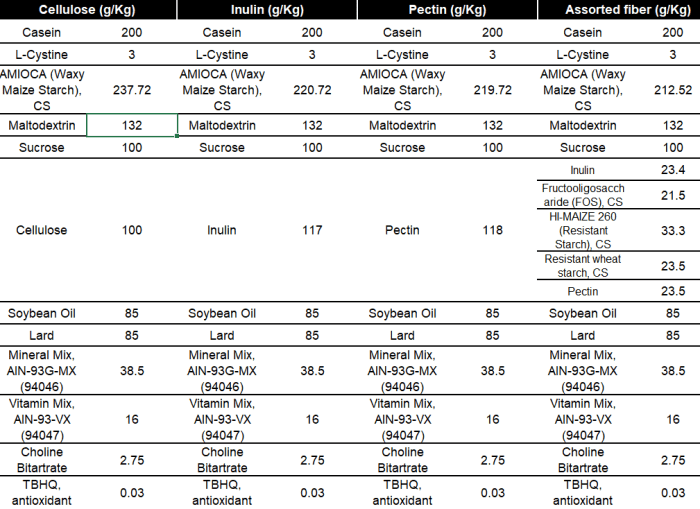
All mice had the same assorted fiber diet for two weeks. Mice were then switched to one of four diets described above: cellulose, inulin, pectin, and assorted fiber, and maintained in these diets for another 4 weeks.
There was a ~ 4-fold range in levels of cecal butyrate among the eight groups despite all animals consuming the same diet [before switching]. Butyrate is known to vary widely among humans and has been linked with beneficial health effects on the host:

We chose inulin and pectin as the former is commonly used as a prebiotic, while the latter has been proven to support growth of a wide variety of gut microbes, and it is commonly used as a dietary supplement. We also chose these two dietary fibers due to their distinct structures, including differences in basic units, linkages, and degree of polymerization.
Assorted fiber diet had the same total amount of dietary fiber as treatment groups used in this study, but with more diversity [FOS and two resistant starches] in fermentable substrates, which we reasoned would support engraftment of taxa relevant to all dietary treatments. Inclusion of this group in the experimental phase also served as a control to inform whether this diet used during colonization drove major differences.
Diet and its interaction with gut community showed a significant effect on serum glucose levels. While pectin diet had an overall beneficial effect on metabolic phenotypes relative to non-fermentable cellulose for SubA-colonized mice, this diet was less favorable for SubB-colonized animals, which showed the strongest benefits on inulin fiber.
In inulin diet, mice inoculated with SubB showed decreased adiposity, decreased liver triglycerides (TG) and lower serum levels of fasting glucose relative to animals colonized with SubA. In contrast, pectin-fed mice colonized with SubB accumulated more fat mass relative to SubA-colonized counterparts, whereas serum glucose and liver TG were comparable between the two community groups.
Mice colonized with SubB showed significantly lower levels of adiposity than those colonized with SubA in the assorted fiber diet, whereas serum glucose and liver TG were comparable.
We found that these two transplanted communities elicited divergent metabolic epigenetic and transcriptional responses to the same dietary fiber. Furthermore, differences between mice colonized with these two communities varied depending on type of fiber consumed.
Populations contain a significant amount of genetic variation derived from their largely individual associated microbiomes. Dissecting effects of gut microbial vs. host genetic variation while controlling environmental exposure is practically impossible to achieve in human studies.
One-size-fits-all approaches to promote health are unlikely to elicit consistent effects across individuals. Identifying gut microbial biomarkers associated with beneficial responses to common interventions may help to stratify subjects into more effective personalized treatments.”
https://microbiomejournal.biomedcentral.com/articles/10.1186/s40168-021-01061-6 “Gut microbiome variation modulates the effects of dietary fiber on host metabolism”
1. This study nailed it! You are what you eat, and The future of your brain is in your gut right now.
2. Group differences in cecal butyrate in the second graphic were instructive. But what really needed to be analyzed was each individual subject’s responses within the eight groups, and each individual’s characteristics.
What did or didn’t matter to each individual could then be applied and analyzed to what did or didn’t matter to its group. Researchers need to flip from a top-down statistics-package approach, to a bottoms-up individual paradigm for evidence.
3. Haven’t mentioned Increasing soluble fiber intake with inulin recently. I eat the labeled 2.5 grams serving. More than that runs into a 10 g “Over this dose would induce mild gastrointestinal symptoms” threshold.
I eat a half-dozen cloves of garlic in daily AGE-less chicken vegetable soup. Garlic contains ≈ 16% inulin, contributing 4-5 g inulin.
4. My dietary fiber intake of current practices is well beyond this study’s 10%. Several times more than our human ancestors’ estimated 100 g/day if Switch on your Nrf2 signaling pathway measurements are correct?
Trying to make my gut microbiota happy, expecting that they’ll reciprocally respond. Dietary fat content is < 10 %.
This 2019 review focused on one Vitamin K-deficiency biomarker. All parts I’ve quoted are outside the liver, so Vitamin K deficiency ≈ Vitamin K2 deficiency.
This is a hard read with many technical details, but sometimes that’s how researchers do it:
“Active MGP (matrix Gla protein), once released into extracellular space, acts as a local inhibitor of calcification. Widespread expression of MGP points to a role of MGP that by far exceeds its well-known function as local inhibitor of calcification.
Recent research confirmed this concept, usually by measuring plasma dp-ucMGP (desphospho-uncarboxylated MGP), a biomarker reflecting poor vitamin K status:

Vitamin K plays a pivotal role in maintaining bone health. Increasing evidence also implicates MGP in maintaining bone health.
In the Health, Aging and Body Composition study, 791 older community-dwelling adults underwent magnetic resonance imaging to measure bilateral knee structural features. The highest [25%] compared with the lowest fourth of the dp-ucMGP distribution had higher odds of having:
- Meniscus damage;
- Osteophytes;
- Bone marrow lesions; and
- Subarticular cysts.
Regarding Vitamin K supplementation:
- Studies showed a dose-dependent decrease in circulating dp-ucMGP with an 86% decrease already observed after 4 weeks of substitution by 360 μg menaquinone-7 [in 50 hemodialysis patients];
- In a randomized double-blind trial of 244 postmenopausal women followed up for 3 years, arterial stiffness as captured by aortic pulse wave velocity or stiffness index β, decreased in intervention compared with control group.
These results should be considered as hypothesis-generating in view of small sample size, and because there were no between-group differences in vitamin K–induced changes in elastic properties of the carotid artery.
Plasma dp-ucMGP levels ranging from 1.4 to 4.6 μg/L were optimal in terms of risk of mortality and macrovascular cardiovascular illness (4.6 μg/L threshold corresponding to the 65th percentile of dp-ucMGP distribution).
Vitamin K supplementation before irreversible organ damage sets in might find its application in prevention of a wide range of disabling diseases. Circulating dp-ucMGP levels might be measured over time to track risk of vascular complications.”
https://www.ahajournals.org/doi/10.1161/HYPERTENSIONAHA.119.12412 “Vitamin K–Dependent Matrix Gla Protein as Multifaceted Protector of Vascular and Tissue Integrity”
I usually don’t give 5+ stars to reviews. This one was different.
Yes, there could be factors other than this one Vitamin K deficiency biomarker involved in study findings. Sure, these coauthors cited their own studies. Its overall purpose, though, was to inform readers.
I’ll summarize this paper as providing evidence for a biomarker of Vitamin K2 deficiency being implicated in the development and progression of many diseases.
Two papers on Vitamin K2, and an online database to continue Part 1:
“Precise quantitative assessments of vitamin K bioavailability in humans is challenging due to unquantified tissue conversion of PK [phylloquinone, Vitamin K1] to MK [menaquinone, Vitamin K2]-4, and contributions of gut microbiota. Absorption of long-chain MKs (MK-7, MK-8 and MK-9) from natto, cheese and egg yolk is close to 100%.
Long-chain MKs have a longer half-life. Long half-life may not necessarily indicate increased bioavailability, but instead non-preferential utilisation by tissues compared to PK and MK-4. A long half-life may also indicate that long-chain MKs may be of particular importance for extrahepatic tissues.
12 databases list vitamin K content of individual food items, which is required to more accurately determine vitamin K intake. The Dutch database is the most comprehensive, and includes PK and several types of MKs, ranging from MK-4 to MK-10.”
https://pubs.rsc.org/en/content/articlelanding/2020/FO/C9FO02321F “Quantifying dietary vitamin K and its link to cardiovascular health: a narrative review” (not freely available)
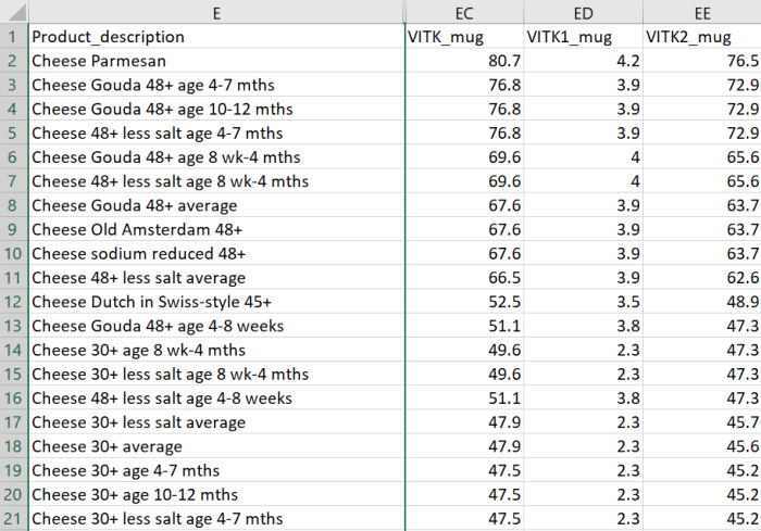
One online database mentioned is at https://www.rivm.nl/en/dutch-food-composition-database:
“The Dutch Food Composition Database (NEVO) contains data on the composition of foods eaten frequently by a large part of the Dutch population. These foods contribute significantly to the intake of energy and nutrients. Foods of importance for specific groups of the Dutch population are also included.
The NEVO online website contains data on 133 nutrients of 2152 food items. The most recent version of NEVO online dates from November 2019.”
I downloaded a copy in Excel format, selected all, and sorted by column EE “VITK2_mug” in descending order. There were 263 food items with Vitamin K2 measurements.
A second paper detailed a 2021 double-blind, placebo-controlled, feasibility study:
“INTRICATE will assess the influence of combined vitamin K2 and vitamin D3 supplementation on micro-calcification in carotid artery disease. Considering recent advancements in medical imaging, ultrasound, PET/MRI, and computed tomography can be used for selection and stratification of patients with atherosclerosis.
Subjects will be randomized (1:1) to a vitamin K2 (400 µg MK-7/day) and vitamin D3 (80 µg [3200 IU]/day) dose or to placebo. Primary endpoint is change in Na[18F]F PET/MRI (baseline vs. after 3 months) in treatment group as compared to placebo arm. Secondary endpoints are changes in plaque composition and in blood-biomarkers.
Studies suggest positive effects of vitamin D on vitamin K-dependent metabolism. The MGP-gene promoter contains a vitamin D response element, capable of a two to threefold enhanced MGP expression after vitamin D binding.
Upregulation of MGP due to vitamin D needs vitamin K to ensure full activation of MGP for optimal functioning. This implies that the combination of both vitamin K and vitamin D could provide enhanced protection against progressive vascular calcification, cardiovascular disease, and mortality.”
https://www.mdpi.com/2072-6643/13/3/994/htm “Effects of Combined Vitamin K2 and Vitamin D3 Supplementation on Na[18F]F PET/MRI in Patients with Carotid Artery Disease: The INTRICATE Rationale and Trial Design”
The second study was somewhat of a tell in that after two decades, researchers are still testing Vitamin K2 dose efficacy. Researchers don’t consider it proper science to not use a statistics package to lump subjects into groups.
Someday researchers will thoroughly analyze each individual, and relate measurements to each individual’s causal and symptomatic characteristics. Then we’ll find out whether what did or didn’t matter to each individual, will or won’t matter to a group.
Until then they’ll focus on one dimension of health like Vitamin K2 foods per their sponsor’s directions. Nevermind that Vitamin K2-rich foods like cheeses are full of advanced glycation end products (AGEs) that humans can’t adequately metabolize, to our detriment.
A trio of papers on Vitamin K2, the first being a 2021 review that emphasized dual effects:
“Osteoporosis (OP) is the most common bone disease that affects elderly men and women. It is a metabolic skeletal disorder caused by an imbalance between bone formation and resorption, leading to a loss of bone mass and quality, skeletal structure deterioration, and an increased risk of fractures.
Vascular calcification is defined as ectopic deposition of mineral matrix in vessel wall. It occurs prevalently in aging and primary chronic conditions (hypertension, diabetes mellitus, and chronic kidney disease), representing an important risk factor for cardiovascular morbidity and mortality.
Studies have provided support for a close link between bone and vascular health. Findings suggest that bone loss in OP may promote and increase the risk of cardiovascular events and vascular atherosclerosis.
Vitamin K2 is involved in a phenomenon in which a low calcium deposition in bone tends to be associated with a parallel increase of calcium deposition in vessel wall as a consequence of impaired calcium metabolism. Most production of Vitamin K2 in humans takes place in intestines. However, the amount derived from intestinal bacteria is poorly absorbed, and is not able to reach concentrations required to exert physiological functions.
Vitamin K2‘s ability to reduce loss of bone mineral density and fracture risk, as well as to improve bone quality, has been described by several clinical studies, which have confirmed that osteocalcin (OC) γ-carboxylation is the main mechanism of action through which this natural compound is able to improve bone health. Clinical evidence suggests an analogous protective role of Vitamin K2 at the vascular level, emphasizing a strict association between:
- Vitamin serum level;
- Matrix gla protein (MGP) γ-carboxylation levels;
- Reduction of vascular smooth muscle cells osteogenic trans-differentiation; and
- Possible risk of cardiovascular events.”
https://www.mdpi.com/2072-6643/13/4/1222/htm “The Dual Role of Vitamin K2 in ‘Bone-Vascular Crosstalk’: Opposite Effects on Bone Loss and Vascular Calcification”
A second 2021 review emphasized aging:
“Vitamin K can:
- Carboxylate OC (a protein capable of transporting and fixing calcium in bone);
- Activate MGP (an inhibitor of vascular calcification and cardiovascular events); and
- Carboxylate Gas6 protein (involved in brain physiology and a cognitive decline and neurodegenerative disease inhibitor).
By improving insulin sensitivity, Vitamin K lowers diabetes risk. It also exerts antiproliferative, proapoptotic, autophagic effects, and has been associated with a reduced risk of cancer.
The most common [Vitamin K2] subtypes in humans are the short-chain MK[menaquinone]-4, which is the only MK produced by systemic conversion of phylloquinone [Vitamin K1] to menaquinone, and MK-7 through MK-10, which are synthesized by bacteria. The main sources of Vitamin K2 are fermented foods, cheeses, eggs, and meats.”
https://www.mdpi.com/2076-3921/10/4/566/htm “The Role of Vitamin K in Humans: Implication in Aging and Age-Associated Diseases”
The third paper – somehow not cited by these two reviews – was a 2006 human study that performed four experiments:
“The synthetic short-chain vitamin K1 is commonly used in food supplements, but recently the natural long-chain MK-7 has also become available as an over-the-counter supplement. The purpose of this paper was to compare in healthy volunteers absorption and efficacy of K1 and MK-7.
Serum vitamin K species were used as a marker for absorption and OC carboxylation as a marker for activity. Both K1 and MK-7 were absorbed well, with peak serum concentrations at 4 hours after intake.
A major difference was:
- Very long half-life time of MK-7, resulting in much more stable serum levels; and
- Accumulation of MK-7 to higher levels (7- to 8-fold) during prolonged intake.
MK-7 induced more complete carboxylation of OC.

Accumulation and efficacy of K vitamins during long-term daily administration. Participants received in a crossover design either K1 (○) or MK-7 (•) or placebo; in the latter case only K1 (▴) could be detected.
- (A) Circulating levels of vitamin K; baseline levels for K1 were subtracted; no MK-7 could be detected at baseline.
- (B) Ratio between circulating carboxylated and undercarboxylated osteocalcin (cOC/ucOC); at baseline the ratio was 1.74 for MK-7, 1.8 for K1, and 1.7 for the placebo group.
MK-7 accumulated during the first 2 weeks until it reached a plateau level of about 10 nM (6 μg/L), whereas K1 remained slightly above placebo values during the entire study period. Efficacy of both K vitamins for OC carboxylation was monitored using the ratio between circulating cOC and ucOC, and it turned out that within 3 days both vitamins had induced increased cOC.
But only by taking MK-7 did the effect continue to increase during the entire study period.
Taken together, these data demonstrate considerable differences between MK-7 and K1:
- Higher and more stable serum levels are reached with MK-7; and
- MK-7 has a higher efficacy in both hepatic and extrahepatic protein carboxylation.”
https://ashpublications.org/blood/article/109/8/3279/23729/Vitamin-K-containing-dietary-supplements “Vitamin K–containing dietary supplements: comparison of synthetic vitamin K1 and natto-derived menaquinone-7″
I’ve tried various things over the years to address hypertension. I stopped high blood pressure medications briefly to see if each intervention worked. They all haven’t, presumably because I didn’t address causes.
More recently, I broke my left big toe on furniture while walking around in the dark last month, and haven’t recovered. No pictures from walking on the beach at sunrise because it still isn’t possible. 😦
A link between these two health conditions could be Vitamin K2. I don’t eat fermented foods because of their high sodium, or dairy products, and haven’t supplemented Vitamin K2.
Next week I’ll start a 300 μg MK-7 daily dose. Current Vitamin D3 dose is 3800 IU, compared to the second paper of Part 2 of Vitamin K2 – What can it do? which is 400 μg MK-7 and 3200 Vitamin D3.
I partially read more than a dozen studies this week of overdosed rodents producing p < .05 significant results. Net effect was to thwart the purpose of rodent studies – to help humans.
The latest came from search term “SIRT1” “DHEA” 2021 after I read a 2021 study Dehydroepiandrosterone protects against acetaminophen-induced liver damage in rats by upregulation of Bcl-2 and activation of sirt signalling that found:
“The study examined protective effect of exogenous administration of dehydroepiandrosterone (DHEA) against acetaminophen (APAP) -induced liver damage in rats, and tested underlying mechanisms. DHEA prevents APAP-induced liver damage by concomitant upregulation of Bcl-2 and SIRT1-dependent effect.”
The daily DHEA dose was 50 mg/kg, which is a (.162 x 50 mg) x 70 kg = 567 mg human equivalent. Eleven times the most frequent human dose of Take responsibility for your one precious life – DHEA. Anyone who took this study’s DHEA amount would hurt themself.
The one-time acetaminophen dose was 800 mg/kg which is a (.162 x 800 mg) x 70 kg = 9 grams human equivalent. Someone who took 18 pills of the most frequent 500 mg acetaminophen dose would be attempting suicide.
How does this help humans?

See posts like Problematic rodent sulforaphane studies and Human relevance of rodent sulforaphane studies for further evidence and observations.
This 2020 cell study investigated antibiotic effects of broccoli sprout compounds:
“In this work, we asked whether isothiocyanates (ITCs) could act synergistically with each other to increase antibacterial effect. A set of aliphatic ITCs, such as iberin, iberverin, alyssin, erucin, sulforaphene, erysolin, and cheirolin was tested in combination with sulforaphane against E. coli.
All tested ITCs exhibit strong antimicrobial effect individually. Synergistic action observed for iberin, iberverin, and alyssin led to minimal inhibitory concentration necessary for antibacterial effect four- to eight-fold lower than for individual ITCs.
Effectiveness of antimicrobial effect is correlated with both type of ITC used and bacterial growth conditions. The combination of several fold lower concentration of ITCs gives a similar effect as much higher amounts of individual ITCs.
Antimicrobial action of sulforaphane analogs was impaired by specific amino acids. Antibacterial effect of ITC treatment is related to stringent response induction, which is triggered by amino acid starvation.
The use of ITCs as antibacterial agents can be advantageous, as there are very few examples of bacterial resistance to these compounds.”
https://www.frontiersin.org/articles/10.3389/fmicb.2020.591802/full “Induction of the Stringent Response Underlies the Antimicrobial Action of Aliphatic Isothiocyanates”
One of this study’s references was the 2016 Relationship between Chemical Structure and Antimicrobial Activities of Isothiocyanates from Cruciferous Vegetables against Oral Pathogens which found that broccoli and red cabbage compound indole-3-carbinol and mustard compound benzyl isothiocyanate were even more potent antibiotics than half of the aliphatic isothiocyanates in this study:

Our ancestors evolved to deal with everyday bacteria, viruses, and other pathogens. Not sure about the current virus developed to herd humans into an agenda.
Train your immune system every day! disclosed that I was in Milan, Italy on the same February 22-23, 2020 weekend that ten towns were closed south of Milan. I’ve never experienced any symptoms.
Treat your gut microbiota well. Give them what they want – including cruciferous sprouts – instead of prescription antibiotics, and expect reciprocity.
This 2018 human study found:
“The objective of this study was to determine whether daily broccoli consumption alters absorption and metabolism of isothiocyanates derived from broccoli glucosinolates. We conducted a randomised cross-over human study (n = 18) balanced for BMI and glutathione S-transferase μ 1 (GSTM1) genotype in which subjects consumed a control diet with no broccoli (NB) for 16 d or the same diet with 200 g of cooked broccoli and 20 g of raw daikon radish daily for 15 d (daily broccoli, DB) and 100 g of broccoli and 10 g of daikon radish on day 16.
On day 17, all subjects consumed a meal of 200 g of broccoli and 20 g of daikon radish. Plasma and urine were collected for 24 h and analysed for sulphoraphane (SF) and metabolites of SF and erucin (ER). (a) BMI < 26 (b) BMI > 26.


Plasma AUC [area under the curve] and urinary excretion rates were higher on DB diet than on NB diet. Daily consumption of broccoli interacted with BMI to affect plasma concentrations and urinary excretion of glucosinolate-derived compounds.
Plasma and urinary levels of SF and mercapturic acid pathway products of SF and ER following a broccoli challenge meal were altered when preceded by 16 d of daily broccoli ingestion, and the effect depended on BMI.”
https://www.cambridge.org/core/journals/british-journal-of-nutrition/article/absorption-and-metabolism-of-isothiocyanates-formed-from-broccoli-glucosinolates-effects-of-bmi-and-daily-consumption-in-a-randomised-clinical-trial/ “Absorption and metabolism of isothiocyanates formed from broccoli glucosinolates: effects of BMI and daily consumption in a randomised clinical trial”
Humans are the same, yet we’re each individually unique. These researchers could have explored individual differences, but that wasn’t part of this study’s design.
So we’re left with BMI as a discriminator. I don’t think that’s evidentiarily sufficient.
Eat broccoli sprouts every day. You’ll figure it out.
A tremendous 2021 study involving the group who published Our model clinical trial for Changing to a youthful phenotype with broccoli sprouts:
“The aim was to evaluate the influence of red cabbage extracts on bioaccessibility of their isothiocyanates, and their effect on intestinal microbiota using a dynamic model of human digestion treated with the gut microbiome of obese adults.
Plant plasma membrane vesicles as delivery systems for bioactive compounds has been studied. Diverse types of plant membrane vesicles could be good candidates for this purpose, such as extracellular vesicles, which are spheroids of cytosolic material surrounded by a lipid bilayer, or extracted plasma membrane from fresh plant tissue.
As an example of the latter, we used cauliflower plasma membrane vesicles, which are proteoliposomes with a high proportion of unsaturated fatty acids. There could be an interaction between plant aquaporins found in our vesicles and isothiocyanates present in red cabbage aqueous extract, which could have increased stability.
Plasma membrane vesicles may act as stabilizing carriers and feeding agents for enzymes and bile salts rather than an encapsulating agent per se. However, this aspect should be further studied.

In the transversal colon reactor, butyric acid production by gut microbiota had a 3-fold increase after 14-day treatment for free red cabbage aqueous extract when compared to stabilization period. A 3.5-fold increase was observed when using nanonencapsulated extract.
Regarding the descending colon, a 2-fold increase in butyric acid was produced after 14 days of treatment with free red cabbage aqueous extract. A 4-fold increase was observed in production after treatment with nanoencapsulated extract.
Propionic and acetic acids were studied, but no changes were observed. The fact that encapsulated red cabbage extract provided a higher production of butyric acid pointed to future developments for design of a functional ingredient or food product for management of overweightness and obesity.”
https://www.mdpi.com/2304-8158/10/5/1038/htm “The Influence of Red Cabbage Extract Nanoencapsulated with Brassica Plasma Membrane Vesicles on the Gut Microbiome of Obese Volunteers”
This study demonstrated that iberin was initially the third highest isothiocyanate of red cabbage after glucosinolate hydrolysis. Iberin surpassed sulforaphane to become the predominant isothiocyanate – in both free and nanoencapsulated forms – when it reached the lower colon, where most of our gut microbiota reside.
These in vitro findings were after 14 days, though, which doesn’t happen in healthy humans in vivo. Also, if sulforaphane metabolites such as dithiocarbamates and I3C breakdown products such as DIM were measured, these findings may have changed.
As noted in Tailoring measurements for broccoli sprouts, study findings of mature plants don’t necessarily apply to their sprouts. Lab analyses of broccoli sprout compounds used 9-day-old red cabbage sprouts to measure iberin (3MSOP-ITC in Figure 5). Haven’t found recent studies on iberin’s effects on gut microbiota and intestinal epithelial cells.
This study showed “a 3 to 4-fold increase in production of butyric acid with encapsulated extract treatment.” Keep leading the way. 🙂
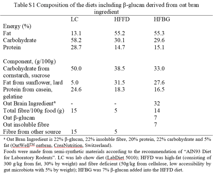
This 2020 rodent study investigated effects of different diets:
“The present study aimed to evaluate effects of β-glucan on the microbiota gut-brain axis and cognitive function in an obese mouse model induced by a high-fat and fiber-deficient diet (HFFD). After long-term supplementation for 15 weeks, β-glucan prevented HFFD-induced cognitive impairment, assessed behaviorally by object location, novel object recognition, and nesting building tests:
- Long-term β-glucan supplementation suppressed microglia activation and inflammation in hippocampus of HFFD-fed mice;
- β-glucan attenuated deleterious engulfment of synapses by activation of microglia seen in HFFD mice;
- β-glucan significantly prevented upregulation of TNF-α, IL-1β, and IL-6 mRNA expression in hippocampus; and
- A broad-spectrum antibiotic intervention abrogated β-glucan-induced improvement in cognitive function, highlighting the essential role of gut microbiota to mediate cognitive function and behavior.
We found that short-term β-glucan supplementation did not change cognitive behavior in HFFD fed mice. HFFD feeding for 7 days dramatically changed gut microbial profile, with β-glucan-fed mice clustered apart from HFFD-fed mice sample, suggesting:
- Quick changes in gut microbiota are induced by short-term β-glucan consumption and
- Possible causality of gut microbiota profile on cognition.
β-glucan supplementation increased place discrimination ratio in object location test compared with HFFD mice; however, there was no significant difference in total exploration time with objects during test phases between the two groups. Higher place discrimination index in β-glucan supplementation group was not due to better general performance, but increased recognition memory.
Results provide consistent evidence linking increased β-glucan intake to improved:
- Gut microbiota profile;
- Intestinal barrier function;
- Reduced endotoxemia; and
- Enhanced cognitive function via more optimized synaptic and signaling pathways in critical brain areas.
It is speculative that β-glucan improvement of gut microbiota composition, but not necessarily diversity per se, may be most critical for improved cognition. Enhanced consumption of β-glucan-rich foods is an easily implementable nutritional strategy to attenuate diet-induced cognitive decline.”
https://microbiomejournal.biomedcentral.com/articles/10.1186/s40168-020-00920-y “β-glucan attenuates cognitive impairment via the gut-brain axis in diet-induced obese mice”
This study did well by elaborating It’s the fiber, not the fat and Eat oats to prevent diabetes related findings. How many humans eat themselves into essentially the same situation as this HFFD group with no gut-microbiota-friendly dietary fiber?
Experiments were with β-glucan 1,3/1,4 found in oats. β-glucan 1,3/1,6 has separate effects, especially on innate immunity.
It’s a coin toss on whether observed cognitive improvement was due to 7% β-glucan soluble fiber, 7% indigestible fiber, or both since they were part of the same HFBG diet. I eat both fibers, beginning with Avena nuda oats for breakfast.
By request, research on astaxanthin bioavailability. I used a “astaxanthin” “bioavailability” “quinone reductase” 2021 search term, and read citing papers.
“The bioaccessibility, bioavailability, and antioxidative activities of three astaxanthin geometric isomers were investigated using an in vitro digestion model.
- 13Z-Astaxanthin showed higher bioaccessibility than 9Z- and all-E-astaxanthins during in vitro digestion, and
- 9Z-astaxanthin exhibited higher transport efficiency than all-E- and 13Z-astaxanthins.
These might explain why 13Z- and 9Z-astaxanthins are found at higher concentrations in human plasma than all-E-astaxanthin.
9Z- and 13Z- astaxanthins exhibited a higher protective effect than all-E-astaxanthin against oxidative stress.”
https://pubs.acs.org/doi/10.1021/acs.jafc.7b04254 “Bioaccessibility, Cellular Uptake, and Transport of Astaxanthin Isomers and their Antioxidative Effects in Human Intestinal Epithelial Caco-2 Cells” (2017, not freely available)
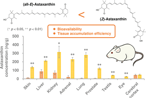
“Astaxanthin with a high proportion of Z-isomer (especially rich in 9Z- and 13Z-isomers) was prepared from (all-E)-astaxanthin by thermal treatment and solid–liquid separation. Z-isomer-rich astaxanthin diet resulted in higher levels of astaxanthin in blood and many tissues (in particular, skin, lung, prostate, and eye) compared to all-E-isomer-rich diet.
Z-isomer-rich diet enhanced the level of 13Z-isomer in blood and tissues rather than that of 9Z-isomer. (13Z)-astaxanthin would have higher bioavailability and tissue accumulation than other isomers.”
https://pubs.acs.org/doi/10.1021/acs.jafc.1c00087 “Z-Isomers of Astaxanthin Exhibit Greater Bioavailability and Tissue Accumulation Efficiency than the All-E-Isomer” (2021, not freely available)
“Astaxanthin is highly susceptible to light, oxygen, and heat stress degradation. In addition, poor water solubility and bioavailability limit its efficacy in vivo. Investigating novel astaxanthin delivery systems is necessary in order to solve these drawbacks.”
https://www.mdpi.com/1420-3049/24/14/2640/htm “The Neuroprotective Effects of Astaxanthin: Therapeutic Targets and Clinical Perspective” (2019)
“Astaxanthin Z-isomers potentially have greater bioavailability and biological activity than (all-E)-astaxanthin. However, stability of Z-isomers is lower than all-E-isomer, which is a serious problem affecting its practical use.
In this study, we investigated impacts of different suspension media (oils and fats) and additives on astaxanthin isomer stability.
- Z-isomers of astaxanthin isomerized to all-E-isomer during storage.
- When soybean and sunflower oils were used as the suspension medium, astaxanthin isomers were hardly degraded. However the total Z-isomer ratio decreased from ~80% to ~50% during 6-week storage at 30 °C.
- (9Z)-astaxanthin showed higher stability than 13Z- and 15Z-isomers.”
https://www.sciencedirect.com/science/article/abs/pii/S0308814621003770 “Evaluation and improvement of storage stability of astaxanthin isomers in oils and fats” (2021, not freely available)
I looked for but didn’t find a graph similar to this one that comparatively plotted astaxanthin:

I also didn’t find recent human studies.
It seems that a special delivery system is required for taking astaxanthin as a supplement. It would require investigating manufacturers’ claims about isomer content and stability.
Eating colorful seafood is another way to get astaxanthin. Don’t know about eating raw or dried algae.
This 2021 cell study investigated a dietary supplement’s role in preventing nerve disease:
“A loss of epigenetic control has been implicated in development of neurodegenerative diseases. Previous studies have implicated aberrant DNA and histone methylation in multiple sclerosis (MS) disease pathogenesis.
We have previously reported that methyl donor betaine is depleted in MS and is linked to changes in histone H3 trimethylation (H3K4me3) in neurons. We have also shown that betaine increases histone methyltransferase activity by activating chromatin bound betaine homocysteine S-methyltransferase (BHMT).
A hallmark of MS is the death of oligodendrocytes, the cells responsible for wrapping axons in myelin in the central nervous system and maintaining a healthy sheath. In demyelinating diseases like MS, oligodendrocyte progenitor cells (OPCs) fail to differentiate and make more myelin, resulting in sclerotic lesions.
Promoting differentiation of OPCs and generation of myelin is of great interest as a novel MS therapy. Waves of gene regulation (repression and activation) need to occur to promote myelination.
This BHMT-betaine methylation pathway ensures availability of S-adenosylmethionine (SAM) for a variety of DNA and histone methylation processes. OPC survival and differentiation are dependent upon DNA and histone methylation, and both processes require SAM.
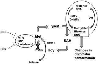
BHMT uses betaine to remethylate homocysteine to methionine. Betaine can be taken in through the diet or synthesized through the oxidation of choline in mitochondria.
We demonstrated that oligodendrocyte gene expression can be modulated by betaine supplementation through the BHMT-betaine methylation pathway. Our study suggests that dietary betaine supplementation may prove to be a therapeutic agent for MS and other demyelinating disorders.”
https://journals.plos.org/plosone/article?id=10.1371/journal.pone.0250486 “The BHMT-betaine methylation pathway epigenetically modulates oligodendrocyte maturation”
I started taking betaine 16 years ago. Didn’t know of these effects until reading this study.
Treating psychopathological symptoms will somehow resolve causes? had more on betaine (aka trimethyl glycine). Current dose is 1.5 grams twice daily.
This 2020 stem cell review argued against rodent models of human neurodegenerative diseases:
“Neuronal loss is not caused solely by intrinsic degenerative processes but rather via impaired interactions with surrounding glia and other brain cells. Dysfunctional astrocytes do not provide sufficient nutrients and antioxidants to neurons, while dysfunctional microglia cannot efficiently clear pathogens and cell debris from extracellular space, resulting in chronic inflammatory processes in the brain.
Human glia, especially astrocytes, differ significantly in morphology and function from their mouse counterparts. Recent advances in stem cell technology make it possible to reprogram human patients’ somatic cells to induced pluripotent stem cells (iPSC) and differentiate them further into patient‐specific glia and neurons, thus providing a source of human brain cells.
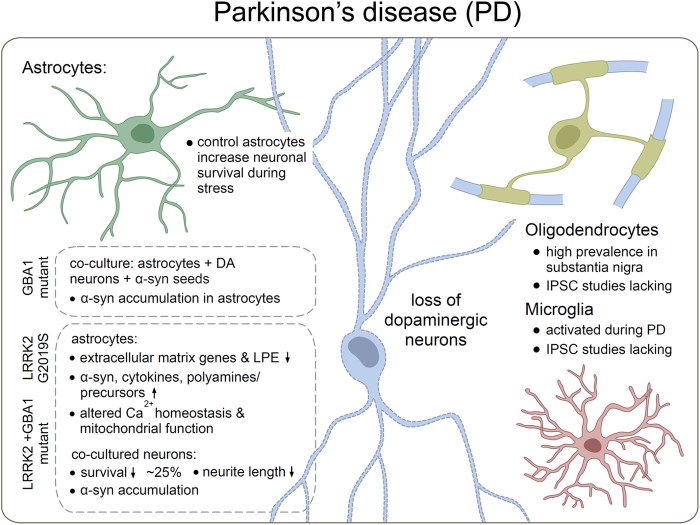
Astrocytes do not efficiently utilize energy resources and cannot provide adequate metabolic support to neurons. A coculture of healthy human neurons with diseased astrocytes impaired neuronal calcium responses to glutamate and γ‐aminobutyric acid (GABA) as compared to coculture with healthy human astrocytes.
Treatment with sulforaphane:
- Normalized basal level glycolysis;
- Decreased basal level Aβ42 secretion; as well as
- Ameliorated inflammatory response to pro‐inflammatory cytokines TNF-α and IL1-β in PSEN1 mutant iPSC astrocytes.
It is essential to make sure that what we see in the dish is the real patient‐specific phenotype. Transplantation of human brain organoids containing microglia into mice could provide a novel tool for drug screening in vivo.”
https://stemcellsjournals.onlinelibrary.wiley.com/doi/full/10.1002/stem.3309 “Metabolic and immune dysfunction of glia in neurodegenerative disorders: Focus on iPSC models”
This review’s thesis seems plausible. However, one problem with in vitro stem cell studies is that they often don’t have a control group.
This 2021 human study investigated development and persistence of allergies:
“Allergic rhinitis (AR) is a common IgE-mediated disorder involving troublesome symptoms of nasal congestion, nasal itch, sneezing, and associated eye symptoms. Like many chronic health conditions, AR stems from complex gene–environment interactions.
130 subjects with AR were recruited. Control population included 154 healthy children who underwent a regular physical examination in the same ear, nose and throat clinic as AR patients. Individuals with history of asthma or atopic dermatitis were excluded.
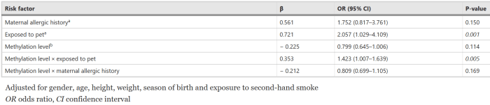
Plenty of contradictory associations exist as whether furred pet exposure (cats and dogs) may be a risk or a protective factor for AR development. Discrepancies are likely due to the ubiquitous nature of pet allergens, while pet owners are more concerned about sanitation and many other hygiene-related reasons.
Interaction of early-life pet exposure with methylation level of ADAM33 increased the risk for AR onset 1.423 times more in children. This study provides evidence that:
- Early-life pet exposure and low methylation level of ADAM33 increase AR risk in children; and
- The interaction between pet exposure and methylation level of ADAM33 may play an important role in development of AR.”
https://aacijournal.biomedcentral.com/articles/10.1186/s13223-021-00526-5 “Interaction between early-life pet exposure and methylation pattern of ADAM33 on allergic rhinitis among children aged 3–6 years in China”
There’s nothing children can do about who their parents were. Exposing them to pet allergens, though, may be another example of early-life experiences causing lifelong effects.
This 2021 rodent study investigated effects on offspring of maternal high-fat diet (HFD) during gestation and lactation, and offspring HFD during young adulthood:
“We found that gestation was the most sensitive period to induce obesity in late life, and there was no difference between sexes in chance of obesity. Furthermore, we found that lactation and administration of a HFD post‐weaning increased incidence of lipid metabolism disorders and obesity in offspring.

There are different windows of opportunity for programming epigenetically labile genes. Some studies support the alteration of epigenetic status during development as an important cause induced adult obesity.
Gestation is considered as the most sensitive period because high DNA synthesis and DNA methylation patterns are established for normal tissue development during the embryonic period. These two programming events are the times when the epigenetic state changes most widely in the life cycle.”
https://onlinelibrary.wiley.com/doi/10.1111/jcmm.16551 “Gestational high-fat diet impaired demethylation of Pparα and induced obesity of offspring”
Hey mothers! Do what you please. But don’t turn around and deny consequences of your behavior and choices on your descendants’ physiology and behavior, and possibly those of further descendants.
Gestation, birth, infancy, and early childhood are critical periods for humans. There’s no going back to correct errors and problems.
