Preventing human infections with dietary fibers

This 2020 review covered interactions of gut microbiota, intestinal mucus, and dietary fibers. I’ve outlined its headings and subheadings, and ended with its overview:

“I. Dietary fibers and human mucus-associated polysaccharides: can we make an analogy?

I.1 Brief overview of dietary fibers and mucus polysaccharides structures and properties

I.I.1 Dietary fibers

  • Dietary fiber intake and health effects

I.I.2 Intestinal mucus polysaccharides

  • Structure
  • Main functions

I.2 Similarities and differences between dietary fibers and mucus carbohydrates

  • Origin and metabolism
  • Structure

II. Interactions of dietary fibers and mucus-associated polysaccharides with human gut microbiota

II-1 Substrate accessibility and microbial niches

  • Dietary fibers
  • Mucus polysaccharides

II-2 Recognition and binding strategies

  • Dietary fibers
  • Mucus polysaccharides

II-3 Carbohydrate metabolism by human gut microbiota

II-3.1 Specialized carbohydrate-active enzymes

II-3.2 Vertical ecological relationships in carbohydrate degradation

  • Dietary fibers
  • Mucus polysaccharides

II-3.3 Horizontal ecological relationships in carbohydrate degradation

II.4 Effect of carbohydrates on gut microbiota composition and sources of variability

II.4.1 Well-known effect of dietary fibers on the gut microbiota

II.4.2 First evidences of a link between mucus polysaccharides and gut microbiota composition

III. Gut microbiota, dietary fibers and intestinal mucus: from health to diseases?

[no III.1]

III.2 Current evidences for the relationship between dietary fibers, mucus and intestinal-inflammatory related disorder

III.2.1 Obesity and metabolic-related disorders

  • Dietary fibers
  • Mucus polysaccharides

III.2.2 Inflammatory bowel diseases

  • Dietary fibers
  • Mucus polysaccharides

III.2.3 Colorectal cancer

  • Dietary fibers
  • Mucus polysaccharides

IV. How enteric pathogens can interact with mucus and dietary fibers in a complex microbial background?

IV.1 Mucus-associated polysaccharides: from interactions with enteric pathogens to a cue for their virulence?

IV.1.1 Pathogens binding to mucus

  • Binding structures
  • Sources of variations

IV.1.2 Mucus degradation by pathogens

  • Bacterial mucinases
  • Glycosyl hydrolases

IV.1.3 Mucus-based feeding of pathogens

  • Primary degraders or cross-feeding strategies
  • Importance of microbial background

IV.1.4 Pathogens and inflammation in a mucus-altered context

IV.1.5 Modulation of virulence genes by mucus degradation products

IV.2 How can dietary fiber modulate enteric pathogen virulence?

IV.2.1 Direct antagonistic effect of dietary fibers on pathogens

  • Bacteriostatic effect
  • Inhibition of cell adhesion
  • Inhibition of toxin binding and activity

IV.2.2 Indirect effect of dietary fibers through gut microbiota modulation

  • Modulation of microbiota composition
  • Modulation of gut microbiota activity

IV.2.3 Inhibition of pathogen interactions with mucus: a new mode of dietary fibers action?

  • Binding to mucus: dietary fibers acting as a decoy
  • Inhibition of mucus degradation by dietary fibers

V. Human in vitro gut models to decipher the role of dietary fibers and mucus in enteric infections: interest and limitations?

V.1 Main scientific challenges to be addressed

V.2 In vitro human gut models as a relevant alternative to in vivo studies

V.3 In vitro gut models to decipher key roles of digestive secretions, mucus and gut microbiota

V.4 Toward an integration of host responses

V.5 From health to disease conditions

dietary fibers prevent infections

Overview of the potential role of dietary fibers in preventing enteric infections. Reliable and converging data from scientific literature are represented with numbers in circles, while data more hypothetical needing further investigations are represented with numbers in squares.

  1. Some dietary fibers exhibit direct bacteriostatic effects against pathogens.
  2. Dietary fiber degradation leads to short-chain fatty acids (SCFAs) production that can modulate pathogens’ virulence.
  3. By presenting structure similarities with receptors, some dietary fibers can prevent pathogen adhesin binding to their receptors.
  4. By the same competition mechanism, dietary fibers can also prevent toxins binding to their receptors.
  5. Dietary fibers are able to promote gut microbiota diversity.
  6. Dietary fibers may promote growth of specific strains with probiotic properties and therefore exhibit anti-infectious properties.
  7. Suitable dietary fiber intake prevents microbiota’s switch to mucus consumption, limiting subsequent commensal microbiota encroachment and associated intestinal inflammation.
  8. Dietary fibers may prevent pathogen cross-feeding on mucus by limiting mucus degradation and/or by preserving diversity of competing bacterial species.
  9. By preventing mucus over-degradation by switcher microbes, dietary fibers can hamper pathogen progression close to the epithelial brush border, and further restrict subsequent inflammation.”

https://doi.org/10.1093/femsre/fuaa052 “Tripartite relationship between gut microbiota, intestinal mucus and dietary fibers: towards preventive strategies against enteric infections” (not freely available)


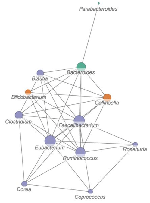
There were many links among gut microbiota studies previously curated. For example, Go with the Alzheimer’s Disease evidence found:

“Akkermansia cannot always be considered a potentially beneficial bacterium. It might be harmful for the gut–brain axis in the context of AD development in the elderly.”

The current review provided possible explanations:

“Akkermansia muciniphila could be considered as a species that fulfills a keystone function in mucin degradation. It is a good example of a mucus specialist.”

Points #7-9 of the above overview inferred that insufficient dietary fiber may disproportionately increase abundance of this species. But Gut microbiota strains also found that effects may be found only below species at species’ strain levels.

These reviewers provided copies in places other than what’s linked above. Feel free to contact them for a copy.


Moon bandit

PXL_20210822_100718644.NIGHT

No magic bullet, only magical thinking

Consider this a repost of Dr. Paul Clayton’s blog post The Drugs Don’t Work:

“The drug industry has enough funds to:

  • Rent politicians;
  • Subvert regulatory agencies;
  • Publish fake data in the most august peer-reviewed literature; and
  • Warp the output of medical schools everywhere.

Their products are a common cause of death. Every year, America’s aggressively modern approach to disease kills over 100,000 in-hospital patients, and twice that number of out-patients.

In 1900, a third of all deaths occurred in children under the age of 5. By 2000 this had fallen to 1.4%. The resulting 30-year increase in average life expectancy fed into the seductive and prevailing myth that we are all living longer; which is manifestly untrue. Improvements in sanitation were far more significant in pushing infections back than any medical developments.

There is currently no pharmaceutical cure for Alzheimer’s or Parkinsonism, nor can there be when these syndromes are in most cases driven by multiple metabolic distortions caused by today’s diet. The brain is so very complex, and it can go wrong in so many ways. The idea that we can find a magic bullet for either of these syndromes is ill-informed and philosophically mired in the past.

It is also dangerous. There is a significant sub-group of dementia sufferers whose conditions are driven and exacerbated by pharmaceuticals. Chronic use of a number of commonly prescribed drugs – and ironically, anti-Parkinson drugs – increases the risk of dementia by roughly 50%.

Big Pharma’s ability to subvert regulatory authorities is even more dangerous. The recent FDA approval of Biogen’s drug aducanumab is a scandal; not one member of the FDA Advisory Committee voted to approve this ineffective product, and three of them resigned in the aftermath of the FDA’s edict. This ‘anti-Alzheimer’s’ drug, which will earn Biogen $56,000 / patient / year, was licensed for financial reasons; it reduced amyloid plaque but was clinically ineffective.

So did the eagerly awaited gantenerumab and solanezumab. But they, too, failed to produce any significant clinical benefit.”


A knee-replacement patient enduring her daily workout

PXL_20210822_102713662

Eating sprouts prevents AGEs

This 2021 in vitro study found:

“Prolonged and chronic hyperglycemia is a leading factor in inducing formation of advanced glycation end-products (AGEs) generated by reaction of free amino groups of proteins and carbonyl groups in reducing sugars, especially glucose and fructose. Metabolism of glucose via the glycolysis pathway also produces the most reactive compounds such as methylglyoxal (MG), a potent precursor of AGEs.

Previous studies reported that red cabbage extract could decrease glycated hemoglobin concentration in streptozotocin-induced diabetic rats and oxidative stress makers including protein carbonyl content and malondialdehyde in red blood cells. Emerging evidence supports that inhibition of protein glycation and oxidative damage may be attributed to free radical scavenging activity of plant extracts.

three brassicae

Extracts of Brassica vegetables cauliflower, cabbage and Chinese cabbage:

  • Inhibited formation of AGEs;
  • Prevented loss of protein thiol group; and
  • May act as a MG-trapping and antioxidant agent.

Phenolic acids, particularly sinapic acid and p-hydroxybenzoic acid, were commonly found in Brassica vegetables. These findings suggest that Brassica vegetables may be promising antiglycation and antioxidant agents for preventing formation of AGEs.”

https://link.springer.com/article/10.1007/s11130-021-00903-w “Phytochemical Composition, Antiglycation, Antioxidant Activity and Methylglyoxal‑Trapping Action of Brassica Vegetables” (not freely available)


Regarding this study’s sinapic acid findings, Broccoli sprout compounds include sinapic acid derivatives found with 6-day-old broccoli sprouts:

“Sprouting in darkness results in overall decrease in total content of sinapic acid derivatives with growth time, but promotes replacement of relatively low active constituents, such as sinapine, by stronger antioxidants. These structural changes are beneficial for total antioxidant capacity of broccoli sprouts, and are correlated with their increasing ability to scavenge free radicals, reduce transition metal ions, and inhibit lipid peroxidation.”

Regarding this study’s p-hydroxybenzoic acid findings, Advantages of 3-day-old oat sprouts over oat grains found with 3-day-old oat sprouts:

“Six hydroxybenzoic acids were found in greater amounts in sprouts, whereas two were reduced or lost.”


Getting onboard before sunrise

PXL_20210822_100455999.NIGHT

Gut reaction

Two papers on broccoli compounds and gut microbiota relationships, with the first a 2021 article:

“We provide a supportive environment and a supply of nutrition and, in return, the microbiome delivers benefits to our health. What exactly are those benefits, and how can we maximise them?

Fibre component of food was thought to be completely indigestible roughage, but we now know that there is a digestible (a.k.a. soluble) component that can be fermented by bacteria resident in the large intestine, providing them with nutrition. There is also non-digestible fibre (a.k.a. insoluble fibre), which is not fermented by gut bacteria and includes plant cell walls formed from cellulose and lignin.

However, when cell walls remain intact, they encapsulate starch contained within cells and physically protect it from full digestion in the small intestine, ensuring that more passes into the large intestine where it can then be fermented by bacteria.

A bioactive is any chemical found in plant-based food that affects biological processes in the body, promoting better health or reducing risk of disease. Unlike macronutrients, such as carbohydrates and proteins, bioactive compounds are usually found in small amounts.

One class of bioactives where this has been known for some time is glucosinolates. For some compounds, including glucosinolates, we have identified particular bacteria that perform this task. For others, we still do not know which microbes are responsible.

S-methylcysteine sulphoxide (SMCSO) is found in brassicas but also in garlic and its relatives. Its metabolic breakdown products have been associated with protective effects against prostate and colon cancer, diabetes, and cardiovascular disease.

SMCSO-derived compounds are highly bioactive, so understanding how they affect the body’s central metabolic pathways could explain some of their health benefits. Only recently have we found clues to bacteria responsible.”

https://ifst.onlinelibrary.wiley.com/doi/10.1002/fsat.3501_6.x “Gut reaction”


The 2020 study cited for SMCSO was an in vitro 2020 study by their coworkers:

“We examined effects of a broccoli leachate (BL) on composition and function of human faecal microbiomes of five different participants under in vitro conditions. Bacterial isolates from these communities were then tested for their ability to metabolise glucosinolates and SMCSO.

We believe that this is the first study that shows reduction of dietary compound SMCSO by bacteria isolated from human faeces. Microbial communities cultured in vitro in BL media were observed to have enhanced growth of lactic acid bacteria, such as lactobacilli, with a corresponding increase in levels of lactate and short-chain fatty acids (SCFAs).

lactate

These results would have been strengthened by analysing soluble fibre content of BL media. As such, it is difficult to relate these results to in vivo SCFA production following consumption of broccoli.”

https://link.springer.com/article/10.1007/s00394-020-02405-y “Effects of in vitro metabolism of a broccoli leachate, glucosinolates and S-methylcysteine sulphoxide on the human faecal microbiome”


Which one of this pair is a male? I’ll guess on the right, as it subsequently turned to face me – a threat – when I walked passed them at a distance.

PXL_20210817_095617355.NIGHT

Gut microbiota functional relationships

This 2021 study investigated environmentally-organized gut microbiome functional relationships:

“There has been a substantial gap between understanding microbiome assemblage and how its functionality is organized. In this study, we demonstrated the usefulness of metaproteomics in gaining a system-level understanding of microbiome functionality.

Our current finding highlights the value of further investigation into functional hubs and hub functions in microbiome proteomic content networks. This will provide a unique and systematic insight for prediction of community functional responses, or manipulation of microbiome functioning.

Across all metaproteomics datasets, Eubacterium, Faecalibacterium, Ruminococcus, Bacteroides, Clostridium and Coprococcus were found to be the most frequent functional hubs.

functionally related genera

Taxon-function bipartite network based on functional distances between microbial genera. Size of a node corresponds to its degree.

Highly connected functions were enriched in metabolism of carbohydrates and amino acids, suggesting that microbial acquisition of nutrients from the environment and trophic interactions between microbes could be major factors that shape their active functional organization. Our result showing robustness of between-taxa functional distances across individual microbiomes implied a more fundamental mechanism that underlies selective organization of microbiome functionalities by environment.

We observed a universal pattern of between-taxa functional distances (dij) across all analyzed datasets. Notably, this pattern was fully shifted by a global increase in dij values, and subsequently a significant decrease of normalized taxonomic diversity in a subset of inflammatory bowel disease samples mostly obtained from inflamed areas.

This finding may support, from a functional angle, the hypothesis that there are alternative stable states (bi-stability or multi-stability) in the gut ecosystem. One frequently discussed mechanism behind these alternative states has been continuous exposure of the microbiome to a altered environmental parameter:

  • An inflamed area in the gut will have a reduced mucus layer and elevated host defense responses.
  • Host mucus layer is a nutritional source of cross-feeding in the gut microbiome.
  • Loss of this layer may firstly affect network hub functions of carbohydrate and amino acid metabolism, and subsequently affect functional interactions in the whole community.

In addition, host defense responses attenuate microbial oxidative stress responses, which have been associated to microbiome dysfunction. Decrease of within-sample functional redundancy has been associated with impaired microbiome stability and resilience.

Resilient microbiota resist external pressures and return to their original state. A non-resilient microbiome is likely to shift its composition permanently and stay at an altered new state instead of restoring to its original state of equilibrium.”

https://www.biorxiv.org/content/10.1101/2021.07.15.452564v1.full “Revealing Protein-Level Functional Redundancy in the Human Gut Microbiome using Ultra-deep Metaproteomics”


My top genus Faecalibacterium – a cross-feeding, acetate-consuming, butyrate-producing commensal – would be more than twice the size of this study’s Faecalibacterium network projection in the above graphic. In this year’s efforts to make my gut microbiota happy, I’ve apparently done much to express its relevant gene network.

my genera

I came across this study by it citing Gut microbiota guilds.

PXL_20210809_101612117

Gut microbiota guilds

This 2021 review investigated gut microbiota self-organizing units:

“We discuss how to use guilds as the aggregation unit to reduce dimensionality and sparsity in microbiome-wide association studies for identifying candidate gut bacteria that may causatively contribute to human health and diseases. Due to strain-level genetic complexity of gut microbiota, microbiome datasets are challenging to identify putative causative agents of a particular disease phenotype.

Members of an ecosystem seldomly live independently from each other. Instead, they develop local interactions and form inter-member organizations to influence higher-level patterns and functions of the ecosystem. In this context, members of a guild tend to exhibit co-abundance patterns by thriving or declining together without regard to their taxonomic positions whenever resources become available or depleted.

Genus-level results showed positive correlations between Bacteroides genus and disease phenotypes, giving the impression that all OTUs in this genus may play a detrimental role in host health. Guild-based analysis clustered these 13 Bacteroides OTUs into seven different guilds.

13073_2021_840_Fig4_HTML

a shows that correlations between clinical parameters and prevalent genera are significantly different among PCOS patients and non-obese controls. b and c show different abundance distributions of Bacteroides genus and 3 Bacteroides OTUs or Alistipes genus and 2 Alistipes OTUs in different patient groups (CN, non-obese control; CO, obese control; PN, non-obese PCOS; PO, obese PCOS):

  • Bacteroides OTU4 belonged to a guild that was positively correlated with disease phenotype, while Bacteroides OTU7 and Bacteroides OTU63 belonged to a negatively correlated guild.
  • Alistipes OTU200 belonged to a guild that was positively correlated with disease phenotype, and Alistipes OTU130 belonged to a guild that was negatively correlated with disease phenotype.

Aggregating microbial populations into guilds facilitates pattern recognition between microbiome and host phenotypes. Recognized patterns and isolates can help identify key functional gut bacteria contributing to human health and diseases causatively.”

https://genomemedicine.biomedcentral.com/articles/10.1186/s13073-021-00840-y “Guild-based analysis for understanding gut microbiome in human health and diseases”


PXL_20210809_094609983.NIGHT

Microwave your Brassica vegetables

This 2021 review evaluated effects on glucoraphanin and sulforaphane content of cooking broccoli and other Brassica vegetables:

“The amount of glucosinolates (GLS) in brassica vegetables can be affected significantly during processing and cooking, depending on their specific conditions and types:

  • Microwaving can retain or even increase content of glucoraphanin (GLR), and can increase production of sulforaphane (SLR) within a short time;
  • Fermentations generally decrease content of GLR;
  • Short-time steaming may promote formation of SLR; and
  • Short-time microwaving may promote formation of SLR from GLR better than fermentation and steaming.

Other processing and cooking effects include:

  • Packaging and freezing can reduce loss of GLR content. Freezing treatment promotes hydrolysis of GLS to form SLR, and freezing stress may lead to GLS degradation;
  • Boiling and blanching result in the largest loss of GLR from broccoli, as loss of GLR content is mainly due to its leakage into the water; and
  • Stir-frying may be a suitable and healthful cooking option to prevent loss of GLR, but contents of GLR and SLR were still influenced due to different factors.

It is better for consumers to microwave or steam brassica vegetables before consumption to obtain greater health benefits.”

https://www.sciencedirect.com/science/article/abs/pii/S030881462101013X “The effect of processing and cooking on glucoraphanin and sulforaphane in brassica vegetables” (not freely available). Thanks to Dr. Jing Sun for providing a copy.


This review found mainly negative effects of cooking Brassica vegetables with boiling, stir frying, blanching, or high pressure on glucoraphanin and sulforaphane content. Previously curated studies cited were:


PXL_20210809_101626026

Blue heron on its way to the breakfast buffet

Prevent your brain from shrinking

My 800th curation is a 2021 human diet and lifestyle study:

“Brain atrophy is correlated with risk of cognitive impairment, functional decline, and dementia. This study (a) examines the statistical association between brain volume (BV) and age for Tsimane, and (b) compares this association to that of 3 industrialized populations in the United States and Europe.

Tsimane forager-horticulturists of Bolivia have the lowest prevalence of coronary atherosclerosis of any studied population, and present few cardiovascular disease (CVD) risk factors. They have a high burden of infections and inflammation, reflected by biomarkers of chronic immune activation, including higher leukocytes counts, faster erythrocyte sedimentation rates, and higher levels of C-reactive protein, interleukin-6, and immunoglobulin-E than in Americans of all ages.

The Tsimane have endemic polyparasitism involving helminths and frequent gastrointestinal illness. Most morbidity and mortality in this population is due to infections.

brain volume

The Tsimane exhibit smaller age-related BV declines relative to industrialized populations, suggesting that their low CVD burden outweighs their high, infection-driven inflammatory risk. If:

  1. Cross-sectional data (which we believe are population-representative of Tsimane adults aged 40 and older) represent well the average life course of individuals; and
  2. The Tsimane are representative of the baseline case prior to urbanization;

these results suggest a ~70% increase in the rates of age-dependent BV decrease accompanying industrialized lifestyles.

Despite its limitations, this study suggests:

  • Brain atrophy may be slowed substantially by lifestyles associated with very low CVD risk; and
  • There is ample scope for interventions to improve brain health, even in the presence of chronically high systemic inflammation.

Lastly, the slow rate of age-dependent BV decrease in the Tsimane raises new questions about dementia, given the role of both infections and vascular factors in dementia risk.”

https://gurven.anth.ucsb.edu/sites/default/files/sitefiles/papers/irimiaetal2021.pdf “The indigenous South American Tsimane exhibit relatively modest decrease in brain volume with age despite high systemic inflammation”


I came across this study by its citation in Dr. Paul Clayton’s 2021 blog post We’ve got to get ourselves back to the garden.

Part 3 of Make your gut microbiota happy

Continuing from Part 2, my 7/15/2021 sample found that no bad bacteria needed work. Top three reasons why this may be are:

1. I’ve eaten microwaved broccoli sprouts every day for 68 weeks now. Relevant research:

helicobacter 0

2. This is the 17th year of training my immune system every day with yeast cell wall β-glucan.

acinetobacter

3. Basic hygiene practices such as brushing my teeth twice a day.

aggregatibacter 0


PXL_20210731_094258419.NIGHT

Part 2 of Make your gut microbiota happy

Continuing from Part 1, 7/15/2021 test results received 7/27 showed I was putatively below average in four gut bacteria. The most relatively deficient (percentage-wise) were populations in genus Bifidobacterium:

bifido level

Looking through Thryve’s recommended foods, eating all but one (green lentils) of twenty legumes increased genus Bifidobacterium. Here’s a sample:

legumes

I already had dried garbanzo and Adzuki beans in my pantry. One serving (35 grams, 1/4 cup) of each are soaking overnight.

Adzuki beans would be expected to improve genus Bifidobacterium populations through resistant starch 2. Garbanzo beans would be expected to improve genus Bifidobacterium populations primarily through resistant starch 3, while also improving relatively-deficient Akkermansia and Lactobacillus bacteria.

Resistant starch was curated in studies such as:

Resistant starch types and their effects were summarized in https://www.sciencedirect.com/topics/agricultural-and-biological-sciences/resistant-starch.


PXL_20210726_100958622

Advantages of 3-day-old oat sprouts over oat grains

This 2021 in vitro study investigated different composition and resultant effects of oat grain and sprouts:

“The aim of this study was to:

  • Evaluate the effect of polyphenols and avenanthramides extracted from oat grains and sprouts on glucose and lipid metabolisms in 3T3 L1 adipocytes; and
  • Identify compounds associated with their beneficial effects through a chemometric approach.

Oat (Avena sativa var. Turquesa) seeds soaked in distilled water at 1:6 w/v ratio for 12 hr. Seeds were then placed in trays covered with a wet filter paper, then into a germination chamber for 3 days at 25°C and 60% relative humidity.

Both polyphenol and avenanthramide extracts from oat sprouts showed a greater beneficial effect than those from oat grains:

Effect of oat grain and sprouts on glucose (a) and lipid (b) metabolism

Effect of oat grain and sprouts on glucose (a) and lipid (b) metabolism. PE-OG Polyphenol extract from oat grain; PE-OS polyphenol extract from oat sprout; AE-OG avenanthramide extract from oat grain; AE-OS avenanthramide extract from oat sprout. Glucose metabolism (a) Glut4, glucose transporter-4; Irs1, insulin receptor substrate-1; Pi3k, phosphoinositide 3-kinase. Lipid metabolism (b) Fasn, fatty acid synthase; Acaca, acetyl-CoA carboxylase; Cpt1, carnitine palmitoyltransferase 1a; Acadm, acyl-CoA dehydrogenase.

Flavonoids:

  • Twelve major and minor flavonols were found in greater amount in sprouts, whereas two were lost; and
  • Two flavones were found in greater amounts in sprouts, whereas seven were reduced or lost. This is the first study that reports the profile of flavone derivatives in oat grains and sprouts.

Phenolic acids:

  • Six hydroxybenzoic acids were found in greater amounts in sprouts, whereas two were reduced or lost.
  • Fifteen hydroxycinnamic acids were found in greater amounts in sprouts, whereas four were reduced or unchanged or lost. Hydroxycinnamic acids esterified with quinic acid such as sinapoylquinic, coumaroylquinic, and feruloylquinic acids, as well as other derivatives, were identified in this study for the first time in oat grain and oat sprouts.

Avenanthramides: all avenanthramides were significantly increased during sprouting (1.7 to 9.0-fold).

Health beneficial effects of oat grains and sprouts were mainly related to their high content of:

  • Avenanthramides A (2p), B (2f), and C (2c);
  • Flavonols quercetin 3-O-rutinoside and kaempferol;
  • Hydroxycinnamic acid sinapoylquinic acid; and
  • Flavones apigenin and luteolin derivatives.

Polyphenol and avenanthramide extracts from oat grains and oat sprouts increased expression of genes involved in glucose uptake and fatty acids β-oxidation, and decreased expression of genes involved in fatty acids de novo synthesis (Fasn and Acaca) in 3T3 L1 adipocytes. Oat sprout extracts exerted an overall greater beneficial effect as compared to oat grain extracts.

This is the first study that demonstrates that oat avenanthramides and polyphenols modulate expression of key genes involved in glucose and lipid metabolisms in adipocytes. Further studies are necessary to validate these results using an in vivo approach.”

https://onlinelibrary.wiley.com/doi/10.1111/jfbc.13738 “Polyphenols and avenanthramides extracted from oat (Avena sativa L.) grains and sprouts modulate genes involved in glucose and lipid metabolisms in 3T3 L1 adipocytes” (not freely available) Thanks to Dr. Iza F. Pérez-Ramírez for providing a copy.


PXL_20210714_100126693

Blood pressure and pain

A trio of papers, with the second and third citing a 2013 review:

“The relationship between pain and hypertension is potentially of great pathophysiological and clinical interest, but is poorly understood. Perception of acute pain initially plays an adaptive role, which results in prevention of tissue damage.

The consequence of ascending nociception is recruitment of segmental spinal reflexes through physiological neuronal connections:

  • In proportion to magnitude and duration of the stimulus, these spinal reflexes cause sympathetic nervous system activation, which increases peripheral resistances, heart rate, and stroke volume; and
  • The response also involves the neuroendocrine system, in particular, the hypothalamic-pituitary-adrenal axis, in addition to further activation of the sympathetic system by adrenal glands.

Persistent pain tends to become chronic and to increase BP values. After a long time, dysfunction of release of endogenous opioids results in a reduction of their analgesic effect. A vicious circle is established, where further pain leads to a reduction in pain tolerance, associated with decreased analgesia mediated by baroreceptors, in a kind of process of exhaustion.”

https://onlinelibrary.wiley.com/doi/epdf/10.1111/jch.12145 “The Relationship Between Blood Pressure and Pain”


A second paper was a 2021 human experimental pain study:

“We investigated the effectiveness of physiological signals for automatic pain intensity estimation that can either substitute for, or complement patients’ self-reported information. Results indicate that for both subject-independent and subject-dependent scenarios, electrodermal activity (EDA) – which is also referred to as skin conductance (SC) or galvanic skin response – was the best signal for pain intensity estimation.

EDA gave mean absolute error (MAE) = 0.93 using only 3 time-series features:

  1. Time intervals between successive extreme events above the mean;
  2. Time intervals between successive extreme events below the mean; and
  3. Exponential fit to successive distances in 2-dimensional embedding space.

Although we obtained good results using 22 EDA features, we further explored to see if we could reach similar or better results with fewer EDA features. This plot highlights that by considering only the top 3 features, we obtained the same level of performance given by all 22 features together.

journal.pone.0254108.g002

This is the first study that achieved less than 1-unit error for continuous pain intensity estimation using only one physiological sensor’s 3 time-series feature, and a Support Vector Regression machine learning model. Considering that this is an encouraging result, we can estimate objective pain using only the EDA sensor, which needs neither a complex setup nor a complex computationally intense machine learning algorithm.

This study paves the way for developing a smart pain measurement wearable device that can change the quality of pain management significantly.”

https://doi.org/10.1371/journal.pone.0254108 “Exploration of physiological sensors, features, and machine learning models for pain intensity estimation”


A third paper was a 2020 human rotator cuff surgery study:

“Results of our study demonstrated that:

  • Pain during the early postoperative period;
  • Time until occurrence of a retear; and
  • Existence of hypertension

were correlated with severity of pain in patients with a retorn rotator cuff.

Pain was selected as the sole outcome parameter of this study because:

  • Pain is an important factor that compels patients to seek treatment for rotator cuff tears, along with functional disability;
  • Pain and subjective functional deficits are important factors that influence a surgeon’s decision to continue with treatment in cases of retearing; and
  • Analyzing pain severity can be a good way to determine patients’ overall satisfaction after rotator cuff repair.

However, pain is not always correlated with disease severity or tear size and vice versa. A lack of pain does not necessarily depend on integrity of the repaired tendon or constitute a good prognosis. In fact, patients with partial-thickness rotator cuff tears showed more pain than did those with full-thickness tears.

Existence of hypertension had a proportional relationship with pain at 12 months postoperatively in patients with retears. This can be interpreted as a suggestion that pain in patients with retears is not acute, but rather chronic, and may be connected to pain in the early postoperative period at 3 months. However, results of this study cannot explain benefits of controlling hypertension in alleviating pain in patients with retears.”

https://journals.sagepub.com/doi/10.1177/2325967120947414 “Factors Related to Pain in Patients With Retorn Rotator Cuffs: Early Postoperative Pain Predicts Pain at 12 Months Postoperatively”


PXL_20210722_100353787

Fat-soluble vitamin competition

This 2015 rodent study investigated interactions of Vitamins A, D, E, and K:

“Significant competitive interactions for uptake were elucidated among vitamin D, E and K, supporting the hypothesis of common absorption pathways:

  • Vitamin A – Neither vitamin D nor K impacted vitamin A uptake. Vitamin E significantly improved vitamin A uptake at medium and high concentrations (up to 40%);
  • Vitamin D – Uptake was significantly reduced by vitamin E at medium and high concentrations (15% and 17% respectively), as well as by vitamin A at high concentration (30%);
  • Vitamin E – Vitamins A and D significantly reduced vitamin E uptake in a dose-dependent manner, while vitamin K had a negative effect only at the highest concentration; and
  • Vitamin K – Vitamins A, D, and E significantly decreased vitamin K uptake (from 34% to 58%).

FSV competition

Our data show that vitamin A was mostly absorbed in the mouse proximal intestine, while vitamin D was absorbed in the median intestine, and vitamins E and K in the distal intestine. These results should be taken into account, especially for supplement formulation.”

https://www.sciencedirect.com/science/article/abs/pii/S0308814614013880 “Fat-soluble vitamin intestinal absorption: Absorption sites in the intestine and interactions for absorption” (not freely available)


Subsequent studies have tested this study’s absorption pathway hypothesis, and whether there actually is competition. This study used Vitamin K1, and I haven’t seen more recent research using K2 for similar fat-soluble-vitamin pathway analysis.

Regardless, I reserved a late morning time slot an hour after yeast cell wall β-glucan intake and an hour before AGE-less chicken vegetable soup where I only eat walnuts and Vitamin K2. Current dose is 600 μg of this:

PXL_20210706_181522044


PXL_20210717_100230612

Back pain and advanced glycation end products (AGEs)

Two 2020 rodent studies investigated intervertebral disk degeneration, with the first on AGEs’ role:

“This study evaluated the role of AGEs and RAGE in driving early intervertebral disk (IVD) degeneration processes in mice. Aging and diabetes are associated with increased low-back pain and IVD degeneration, yet causal mechanisms remain uncertain. AGEs:

  • Accumulate in IVDs from aging;
  • Are implicated in diabetes-related disorders;
  • Alter collagen; and
  • Induce proinflammatory conditions.

A mixed population of 23 male and female wild type AC57BL/6J mice were each assigned to two isocaloric diet groups after weaning. They received either low-AGE chow containing 7.6 μg/mg AGE, or high-AGE chow containing 40.9 μg/mg AGE generated via high-temperature heating (NIH-31 open formula chow autoclaved for 30 minutes at 120°C [248° F]). This in vivo dietary model was previously shown to increase IVD AGE accumulation without systemic obesity or diabetes.

disc AGE damage

AGE accumulation leads to RAGE-dependent collagen disruption in the annulus fibrosus, and can initiate molecular and tissue level collagen disruption. Second harmonic generation (SHG) and collagen-hybridizing peptide (CHP) analyzes were sensitive to collagenous alterations at multiple hierarchical levels due to AGE.

These methods may be useful in identifying additional contributors to collagen damage in IVD degeneration processes.”

https://onlinelibrary.wiley.com/doi/10.1002/jsp2.1126 “Advanced glycation end products cause RAGE-dependent annulus fibrosus collagen disruption and loss identified using in situ second harmonic generation imaging in mice intervertebral disk in vivo and in organ culture models”

Other human studies found degenerative spine disorders start at detectable levels during adolescence. Those study designs didn’t trace disc degeneration to diet, though.


A second study was summarized in a conference I’m sure researchers would like to reconvene:

“Kritschil et al investigated the role of insulin-like growth factor 1 (IGF-1) signaling on progression of disc degeneration in aging mice. They showed that diminished IGF-1 bioavailability confers both beneficial effects of decreased disc cell senescence and extracellular matrix catabolism, whilst at the same time negatively impacting proteoglycan production.”

jsp21134-fig-0001-m

https://onlinelibrary.wiley.com/doi/10.1002/jsp2.1134 “Advancing basic and preclinical spine research: Highlights from the ORS PSRS 5th International Spine Research Symposium”

https://onlinelibrary.wiley.com/doi/10.1002/jsp2.1112 “Effects of suppressing bioavailability of insulin-like growth factor on age-associated intervertebral disc degeneration”

This study asserted:

“Despite some inconsistent findings on the role of IGF-1 among human centenarian and animal model studies, there is overwhelming evidence to support that disruptions to the IGF-1 signaling pathway promotes healthy longevity.”

See Take responsibility for your one precious life – DHEA for other evidence on IGF-1.


Spent a large part of this weekend reading abstracts and studies concerning diet interactions with spinal disc degeneration. This AGE study provided more evidence than others on these relationships.

I’ve eaten AGE-less chicken vegetable soup almost every day for two years:

  • 237 g chicken breast cubes, 179 g celery, and 262 g carrots in 1 cup Savignon Blanc get up to 100° C around 9 minutes initially, then again about 6 minutes after I add 1 quart chicken broth, then I turn off the Instant Pot.
  • I stir in 340 g mushrooms, 31 g garlic, and 387 g Roma tomatoes five minutes later at about 85° C, and they cool the soup down to around 70° C. I let it stew for another 15 minutes before eating half (1.5 quarts).
  • A 1.5 quart leftover heated the next day for six minutes in a 1000W microwave reaches 55° C.

I do stretches every day to accommodate a L5-S1 disc replacement with a titanium-cage-and-rods apparatus done ten years ago, and a C5-C6-C7 similar operation done eleven years ago. Can’t say whether recent diet, last decades’ disc replacement surgeries, daily stretches and exercises, or other factors are responsible for absence of spine pain.

PXL_20210717_093614850.NIGHT