Plasmalogens Week #6 – Health and Diseases, Part 2

Continuing Part 1 with three 2025 papers, starting with a rodent study of dietary mussel plasmalogens’ effects on atherosclerosis:

“The purpose of this study was to clarify the underlying mechanisms of Mytilus edulis-derived plasmalogens (Pls) against atherosclerosis (AS) in ApoE−/− mice induced by a high-fat diet (HFD), through a comprehensive analysis of hepatic metabolomics and aortic transcriptomics data. Besides Pls role as the storage pool of n-3 PUFAs, the structural feature of vinyl ether bond at sn-1 position confers multiple advantages upon Pls compared to their diacyl counterparts, including enhanced antioxidant capacity, increased membrane fluidity, as well as improved stability and stability of biomembranes.

The C57BL/6 mouse strain is susceptible to high-fat diet (HFD)-induced AS lesions, and ApoE knockout accelerates AS development. Molecular mechanisms by which Pls ameliorate AS were investigated through a comprehensive analysis of hepatic metabolomics and aortic transcriptome profiles, focusing on changes in gene related to the p38 mitogen-activated protein kinase (MAPK) signaling pathway and the downstream inflammatory response.

The concentration of Pls in mussel tissues is 32 μgmg−1 (dry weight), and the obtained Pls contains 49.53% of phosphatidylethanolamine-Pls, 35.87% of phosphatidylcholine-Pls, and 14.60% of phosphatidylserine-Pls. The main fatty acid compositions of Pls are presented in Supplementary Table 1, which indicates that EPA accounts for 45.82% and the n-3/n-6 ratio is 3.84.

Pls inhibited aortic lipid accumulation, prevented thickening of the aortic wall, and suppressed collagen accumulation at the aortic-heart junction. Pls inhibited HFD-induced loosening of hepatocyte arrangement, vacuolization, and accumulation of lipid droplets.

Although several key components of MAPK signaling pathway were suppressed at both the transcriptional and protein levels in Pls-treated mice, no significant changes in phosphorylated p38 protein were observed among the experimental groups in our study. Further research is needed to elucidate the overall inhibitory mechanism of Pls on p38 protein and the MAPK signaling pathway.”

https://www.nature.com/articles/s41538-025-00546-0 “Effects of Mytilus edulis derived plasmalogens against atherosclerosis via lipid metabolism and MAPK signaling pathway”


A rodent / human cell study investigated effects of plasmalogens in innate immune system macrophages on atherosclerosis:

“We demonstrate that simultaneous inactivation of two key enzymes involved in macrophage polyunsaturated fatty acid (PUFA) metabolism—ELOVL5, which elongates long-chain PUFAs, and LPCAT3, which incorporates them into phospholipids—disrupts membrane organization by promoting the formation of cholesterol-enriched domains. This increases macrophage sensitivity to cytotoxic oxysterols and leads to more vulnerable atherosclerotic plaques with enlarged necrotic cores in a mouse model of atherosclerosis.

We identified ELOVL5 as one elongase facilitating the conversion of C20 to C22 PUFA. In humans, analysis of 187 carotid plaques reveals a positive correlation between LPCAT3/ELOVL5-generated phospholipids—including arachidonate (C20:4 n-6)-containing ether lipids—and more stable plaque profiles. Additionally, Mendelian randomization analysis supports a causal relationship between LPCAT3 expression and reduced risk of ischemic stroke.

Potentially beneficial effects we observed in mice and in human atheroma plaques were mainly associated with PLs enriched in omega-6, particularly in AA. Although omega-6 FAs are often considered as pro-inflammatory, their role is undergoing reconsideration, with markers linked to the intake of omega-6 appearing beneficial in the context of cardiovascular diseases. In this context, it is worth to note that AA-containing plasmalogens have been previously identified as markers of healthy obesity.

Our findings uncover a regulatory circuit essential for PUFA-containing phospholipid generation in macrophages, positioning PUFA-containing ether lipids as promising biomarkers and therapeutic targets.”

https://www.sciencedirect.com/science/article/pii/S2666379125002046 “Plasmalogen remodeling modulates macrophage response to cytotoxic oxysterols and atherosclerotic plaque”


A human study included plasmalogens in investigating associations among people with mental illness and their lipid profiles:

“Plasma lipidomic profiles of 623 individuals (188 schizophrenia (SCZ), 243 bipolar disorder (BD), 192 healthy controls) belonging to the PsyCourse Study were assessed using liquid chromatography and untargeted mass spectrometry. Exact etiology of these major mental health disorders is yet unknown and while their symptoms overlap, their diagnostic criteria are based on clinical evaluations of symptoms without objective markers.

Cognitive dysfunction is among the most disabling symptoms of SCZ and BD, and is difficult to treat with the commonly used pharmacologic regimes. Consequently, it has important impacts on long-term functional outcomes.

We aimed to answer the question, whether specific lipid species or classes were associated with differential performance across various cognitive domains, including psychomotor and processing speed, executive function, short-term and working memory and crystalized intelligence and whether these associations were affected by diagnoses.

Lipids belonging to the phosphatidylethanolamine plasmalogen (PE-P) class emerged as the main lipid class associated negatively with DG-SYM test performance, representative of processing and psychomotor speed. Our findings showed that higher levels of PE-P 42:5, PE-P 40:4, PE-P 40:5, and ceramide 38:1 in plasma samples of our study are significantly associated with poorer DG-SYM test performance. The DG-SYM test mainly measures processing speed, the amount of time required to complete a series of cognitive tasks. Enrichment analysis also showed significant associations between other lipid classes and various cognitive tests.

Our findings suggest a link between lipids and cognitive performance independent of mental health disorders. Independent replication is warranted to better understand if phosphatidylethanolamines could represent an actionable pharmacologic target to tackle cognitive dysfunction, an important unmet clinical need that affects long-term functional outcomes in individuals with severe mental health disorders.”

https://www.nature.com/articles/s41398-025-03323-5 “Investigating the association of the plasma lipidomic profile with cognitive performance and genetic risk in the PsyCourse study”

It was apparently beyond these researchers’ expertise to offer informed discussion on this study’s associative link between enrichment of these three phosphatidyl ethanolamine plasmalogens and cognitive dysfunction. Grok countered that their depletion was associated with neurodegenerative diseases (Alzheimer’s, Parkinson’s, multiple sclerosis), cardiovascular risk / oxidized-LDL burden, and chronic fatigue / post-viral syndromes.

Plasmalogens Week #5 – Health and Diseases, Part 1

Continuing Plasmalogens Week with three 2025 papers, starting with a human study that included plasmalogen biomarkers of non-communicable disease fatigue symptoms:

“This study explored the biological mechanisms underlying fatigue in patients with NCDs using a multi-omics approach. Our findings indicate that distinct metabolic pathways, salivary microbiota, and genetic factors may contribute to different dimensions of fatigue, including general, physical, and mental fatigue.

  • General fatigue is associated with unsaturated fatty acid biosynthesis, indicating its role in lipid metabolism.
  • Physical fatigue was associated with plasmalogen synthesis, mitochondrial beta-oxidation of long-chain fatty acids, and selenoamino acid metabolism, suggesting a potential contribution of impaired energy production.
  • Mental fatigue is associated with homocysteine degradation and catecholamine biosynthesis, which may influence cognitive fatigue.

This exploratory study suggests that fatigue in patients with NCDs may involve disruptions in lipid metabolism, neurotransmitter pathways, microbial composition, and genetic variations. Blood-based biomarkers showed better predictive potential for physical fatigue, whereas salivary-based models were more indicative of mental fatigue.

Although our findings support the role of lipid metabolism, the contribution of plasmalogen synthesis remains underexplored. Further studies are needed to validate these findings and understand their mechanisms of action.”

https://link.springer.com/article/10.1186/s12911-025-03034-3 “Visualizing fatigue mechanisms in non-communicable diseases: an integrative approach with multi-omics and machine learning”


A human study of metabolic dysfunction-associated steatotic liver disease (MASLD) included investigating plasmalogens:

“In this study, we applied untargeted metabolomic profiling to serum samples from individuals with and without MASLD, classified by the Fatty Liver Index, with the goal of identifying characteristic metabolic signatures and pathways that may underlie disease presence and progression. Individuals in the MASLD group displayed significantly higher levels of ALT, AST, ALP, and GGT, reflecting ongoing hepatic injury, cholestasis, and oxidative stress. However, albumin and bilirubin levels remained within normal limits, indicating early to intermediate disease stages rather than advanced fibrosis or cirrhosis.

A consistent and highly significant lipidomic pattern in the MASLD group is the depletion of plasmalogens and sphingomyelins. Depletion of these lipid classes was identified as a hallmark of insulin resistance as defined by the triglyceride-glucose index. In contrast, phosphatidylcholine, phosphatidylethanolamine, and phosphatidylinositol species were elevated in MASLD, pointing toward broader lipid remodeling events.

Reduced plasmalogen and sphingomyelin levels positions their depletion as a core feature of metabolic dysfunction. Plasmalogens are ether phospholipids with strong antioxidant capacity, and their reduction suggests a loss of protective buffering against oxidative stress, one of the main drivers of MASLD progression. Similarly, sphingomyelin depletion implicates altered membrane dynamics and signaling disturbances, further contributing to metabolic dysfunction.

Depletion of plasmalogens 1-(1-enyl-palmitoyl)-2-oleoyl-GPC (P-16:0/18:1), 1-(1-enyl-palmitoyl)-2-linoleoyl-GPC (P-16:0/18:2), 1-(1-enyl-palmitoyl)-2-palmitoyl-GPC (P-16:0/16:0), 1-(1-enyl-palmitoyl)-2-palmitoleoyl-GPC (P-16:0/16:1), 1-(1-enyl-palmitoyl)-2-oleoyl-GPE (P-16:0/18:1), 1-(1-enyl-palmitoyl)-2-linoleoyl-GPE (P-16:0/18:2), and disruption of the glutamate–gamma-glutamyl pathway stand out as central features of metabolic dysfunction in MASLD, with clear potential to inform biomarker discovery, disease classification, and the design of targeted therapeutic strategies.”

https://www.mdpi.com/2218-1989/15/11/687 “Metabolomic Signatures of MASLD Identified by the Fatty Liver Index Reveal Gamma-Glutamyl Cycle Disruption and Lipid Remodeling”


A rodent study investigated dietary sea squirt (AM) plasmalogen ethanolamine (PlsEtn) extract’s and dietary pig liver (PL) phosphatidyl ethanolamine (PtdEtn) extract’s effects on acetaminophen liver injury:

“We investigated dietary effects of PlsEtn from ascidian on chronic hepatic injury in acetaminophen (APAP)-treated mice. Five-week-old male mice were divided into four groups (n = 12), which were treated with experimental diets for two weeks and then the respective APAP-containing diet for five weeks.

Ingested PlsEtn is digested into lysoPlsEtn and free fatty acid in the small intestine. PlsEtn digests are absorbed and are subsequently resynthesized into PlsEtn preferentially with PUFA.

Acetaminophen is a frequently used analgesic and antipyretic. Approximately 90% of APAP is metabolized by UDP-glucuronosyltransferase and sulfotransferase into glucuronic acid and sulfate conjugates, respectively.

5–9% of APAP is metabolized into the highly reactive intermediate N-acetyl-p-benzoquinone imine (NAPQI). This metabolite is considered a pivotal molecule in APAP-induced hepatotoxicity and is conjugated by glutathione (GSH). Excessive NAPQI levels deplete GSH and covalently bind to cellular proteins, resulting in organelle dysfunction, such as mitochondria dysfunction. These impairments induce oxidative stress, cell malfunctions, and subsequently, cell death, such as ferroptosis and apoptosis.

Mice were treated with continuous APAP consumption to induce oxidative stress and impaired lipid metabolism in the liver. Effects of diets were evaluated based on levels of malondialdehyde (MDA), a marker of lipid oxidation, on fatty acid content, and on expression of apoptosis-related proteins in the liver.

The PlsEtn-rich diet effectively suppressed APAP-induced decrease in body and liver weights of mice. However, this suppressive effect was not observed in mice fed a PtdEtn-rich diet. APAP administration decreased the total fatty acid content in the liver, whereas a PlsEtn-rich diet alleviated this decrease and increased the hepatic content of docosahexaenoic acid (DHA).

Owing to the alkenyl linkage, which exhibits antioxidant properties, PlsEtn was expected to markedly suppress hepatic lipid oxidation. However, its suppressive effect was the same extent as that by PtdEtn. Both PlsEtn and PtdEtn contain an ethanolamine base in their structures, and free ethanolamine and its metabolite choline suppress lipid peroxidation. Dietary PlsEtn and PtdEtn may be metabolized into free ethanolamine and its further metabolites, which may alleviate APAP-induced hepatic lipid oxidation.

Dietary ethanolamine glycerophospholipids (EtnGpls) rich in PlsEtn or PtdEtn suppressed APAP-induced lipid oxidation in the liver. Protein expression results revealed that dietary EtnGpls reduced expression of certain apoptosis-related proteins compared to the APAP group. This reduction was more effective in mice fed the PlsEtn-rich diet than in those on the PtdEtn-rich diet.”

https://www.mdpi.com/2076-3417/15/11/5968 “Dietary Ethanolamine Plasmalogen from Ascidian Alleviates Chronic Hepatic Injury in Mice Treated with Continuous Acetaminophen”

This study neither demonstrated nor provided citations for its dietary plasmalogen recycling statements.


Three more plasmalogen health and disease papers are curated in Part 2.

Plasmalogens Week #4 – Nutrition

Continuing Plasmalogens Week with two 2025 papers, starting with a simulated in vitro model of how humans digest mussel plasmalogens:

“Plasmalogens (Pls) have promising therapeutic potential in the treatment of neurological disorders, but their distribution, compositional intricacies, and structural alterations during the digestive process are unclear. This study aimed to address this gap by isolating Pls-enriched fractions from mussel (Mytilus edulis) and simulating their digestion in vitro across the mouth, stomach, and intestine phases.

Comparison between Pls and normal phospholipids, sharing identical fatty acyl compositions, illuminated a heightened susceptibility of Pls to catabolism during stomach digestion, which is mainly attributed to the hydrolysis reaction of Pls sensitive to acidic conditions. Phospholipid digestion commenced during the gastric phase and continued with notable catabolism in the intestinal phase, resulting in the release of substantial amounts of free fatty acids (FFAs) and lysophospholipids (LPs), which subsequently formed lipid droplets of larger sizes. Larger droplets delay intestinal absorption, extending the window period for Pls hydrolysis by pancreatic lipase.

The digestive behaviour of Pls with different polar head groups indicated that pancreatic lipase appears to digest phosphatidylethanolamine plasmalogen (PlsPE) to a greater extent than phosphatidylcholine plasmalogen (PlsPC). 41 PlsPE and 14 PlsPC were observed, suggesting that Pls may be more readily digested in the gastrointestinal tract compared to conventional phospholipids.

Generally, lipids are first absorbed by intestinal epithelial cells and undergo lipid remodeling before being transported into lymphatic fluid and then entering the bloodstream. During lipid absorption, PE can be partially converted into PC for lipid remodeling. Since in vitro digestion models cannot fully simulate the intestinal microenvironment (such as microbial metabolism and intestinal epithelial absorption), animal experiments are required to verify the actual bioavailability of PlsPE and PlsPC.”

https://www.sciencedirect.com/science/article/pii/S2666154325006799 “Characterization of plasmalogen properties and metabolic behavior during the simulated multi-stage digestion of Mytilus edulis phospholipids based on untargeted lipidomics”


A review highlighted nutritional implications of changes in plasmalogen chemistry:

“Plasmalogens vary quantitatively in biological systems due to biosynthesis, degradation, remodeling, and certain external stressors. Not only concentrations, but also the composition of molecular species within the plasmalogen pool changes. These shifts often involve the shortening of sn-2 fatty acyl chains, the loss of PUFAs such as DHA and EPA, and the accumulation of oxidized, truncated, or degraded species, as a result of radical-mediated oxidation and/or enzymatic degradation.

The possible increase in lysophospholipids (typically LPE and LPC, corresponding to PlsEtn and PlsCho, respectively) may be attributed to the loss of intact plasmalogens during degradation, especially in the sn-1 position. Lysoplasmalogens can be re-acylated to regenerate the original plasmalogens or create new plasmalogen species with different sn-2 fatty acyl compositions.

These molecular-level transitions highlight the complexity of plasmalogen dynamics and emphasize the need for quantitative, species-specific analysis. Variations are influenced by physiological conditions, pathological states, and nutritional supplementation.

Plasmalogens are primarily those derived from animal products, such as fish, meat, and dairy products, as well as certain marine foods. Microorganism-derived plasmalogens are attracting researchers’ attention, representing a new way of effectively utilizing bacterial resources as a ‘food’ source. Compounds provided can be plasmalogens (either PlsCho and PlsEtn, extracted from natural sources or synthesized) or plasmalogen precursors (e.g., alkylglycerols).”

https://www.mdpi.com/2072-6643/17/22/3497 “The Changes in Plasmalogens: Chemical Diversity and Nutritional Implications—A Narrative Review”

A challenge researchers haven’t satisfactorily addressed yet is the question of whether beneficial oral intake of plasmalogens can be mechanistically attributed to specific plasmalogen breakdown products or to intact plasmalogens. This review introduced two other mechanistic uncertainties in that 1) absorbed and digested breakdown products can be recycled back into plasmalogens, and 2) gut microbiota can also produce plasmalogens. I’ve read papers that speculated but didn’t demonstrate that either of these factors contributed to their results.

This review cited Dr. Goodenowe’s plasmalogen precursor clinical trial mentioned in Plasmalogens Parts 1, 2, and 3. The first paper above, and most of the papers in Plasmalogen Week cited his other research.


Plasmalogens Week #2 – Childhood Development

Continuing Plasmalogens Week with three 2025 papers, starting with a human study of plasmalogens’ effects of decreasing breastfed infants’ infections and inflammation:

“Mothers reported on breastfeeding and infant infections in questionnaires collected at 1 month, 3 months, 6 months, 12 months, and 18 months post-birth. Parent-reported infection burden was defined as the total number of infant respiratory tract infections, gastroenteritis, conjunctivitis, and acute otitis media episodes reported by mothers between birth and 6 months for 6-month analyses, and between birth and 12 months for 12-month analyses.

We constructed a causal mediation model to estimate the proportion of effects explained by a direct effect of breastfeeding on inflammation, measured via glycoprotein acetyls (GlycA)—the average direct effect (ADE)—and the proportion that was mediated by metabolomic biomarkers/lipid—the average causal mediation effect (ACME).

Breastfeeding is negatively associated with GlycA, positively associated with plasmalogens, and plasmalogens are negatively associated with GlycA. However, the positive association between breastfeeding and plasmalogens is stronger than the negative direct association between breastfeeding and inflammation, resulting in an ACME that exceeds the total effect. This pattern indicates that plasmalogens may play a dominant role in mediating the relationship between breastfeeding and systemic inflammation.

We have recently developed a plasmalogen score that is associated with a range of cardiometabolic outcomes, including type 2 diabetes and CVD.

  • At 6 months, the plasmalogen score was estimated to mediate 162% of the total effect (proportion mediated: 1.62, i.e. average causal mediation effect (ACME) to total effect ratio of 1.62, resulting in a percentage > 100%) of breastfeeding on GlycA.
  • At 12 months, the plasmalogen score mediated an estimated 75% of the total effect of breastfeeding on GlycA.

Any breastfeeding, regardless of supplementary feeding, was associated with lower inflammation, fewer infections, and significant, potentially beneficial changes in metabolomic and lipidomic markers, particularly plasmalogens. There was evidence of bidirectional mediation: metabolomic biomarkers and lipids mediated breastfeeding’s effects on inflammation, while inflammation partly mediated breastfeeding’s impact on certain metabolites and lipids.”

https://bmcmedicine.biomedcentral.com/articles/10.1186/s12916-025-04343-0 “The protective effect of breastfeeding on infant inflammation: a mediation analysis of the plasma lipidome and metabolome”

Reference 48 was the 2024 plasmalogen score study.


A second study by many of the first study’s researchers used the same cohort as the first study to investigate effects of maternal obesity on infant obesity:

“We aimed to investigate associations between maternal pre-pregnancy body mass index (pp-BMI), lipidomic profiles of mothers, human milk, and infants, and early life growth. We were particularly interested in ether lipids as they are higher in breastfed infants compared to formula-fed infants, are enriched in human milk compared to infant formula, and are involved in metabolic health and inflammation in adult populations.

Maternal plasmalogen score was negatively associated with pp-BMI and positively associated with plasmalogens in human milk and infant plasmalogen scores from birth to four years of age. We were unable to establish clear links between plasmalogen score and infant BMI within the first 4 years.

These findings position plasmalogens and ether lipids as potential biomarkers or intervention targets for reducing transmission of obesity from mother to infant. Optimising lipid profiles through reducing maternal pp-BMI and dietary or supplemental ether lipids may represent a novel strategy for mitigating early-life obesity risk.”

https://www.researchsquare.com/article/rs-7089146/v1 “Maternal BMI and infant obesity risk: a lipidomics perspective on the developmental origins of obesity”

There was a lot of hand waving and weasel-wording (i.e., could, may, potential, associated with) but little causal evidence in this preprint. Reference 42 was the preprint version of the first study.


A third paper investigated 9- to 12-year-olds’ plasmalogen levels and molecular types:

“The importance of plasmalogens (Pls) in several cellular processes is known, one of which is their protective effect against oxidative damage. The physiological role of Pls in human development has not been elucidated. This study is the first report on plasmalogen levels and molecular types in children’s plasma.

Ethanolamine plasmalogen (PlsEtn 16:0/20:5) and choline plasmalogen (PlsCho 16:0/20:5), both carrying eicosapentaenoic acid (EPA, ω-3), were significantly lower in girls than in boys. There was no significant difference observed among the 9, 10, 11, and 12-year-old groups between girls and boys in their levels of PlsEtn 16:0/20:5. However, a significant decrease in the levels of PlsCho 16:0/20:5 was observed for 9, 10 and 12-year-old groups of girls compared to boys.

  • In both sexes, the plasmalogen levels for the 12-year-old children were lower than those for the 9-year-old children.
  • PlsCho (16:0/18:2) linoleic acid (ω-6)-derived was lower in the overweight children than in the normal-weight children for both sexes.
  • Arachidonic acid (ω-6)-containing PlsEtn (18:0/20:4) was the most abundant ethanolamine-type plasmalogen in both sexes.

This study has many limitations as follows:

  1. Non-fasting plasma samples were collected from the children’s plasma and used for analysis; since diet can influence Pls levels, the result may be affected by the sample collection method.
  2. Physical activity was also not monitored, which could have an influence on plasma levels, and
  3. A limited number of plasmalogen molecular species were quantified in this study.

A follow-up study may be essential to determine the plasma Pls in the same population when they are adolescents.”

https://www.mdpi.com/2075-4418/15/6/743 “Application of Liquid Chromatography/Tandem Mass Spectrometry for Quantitative Analysis of Plasmalogens in Preadolescent Children—The Hokkaido Study”


Plasmalogens Week #1 – Overview

It’s been a while since I curated plasmalogen papers. Let’s start out a week’s worth of 2025 papers with a review of plasmalogens as biomarkers:

“Reduced levels of plasmalogens in circulation or in cell membranes are associated with rare peroxisomal disorders, systemic disease, neurological impairment, cancer, and diseases of the heart, kidney, and liver. Roles for plasmalogens have been identified in lipid rafts, myelin, chlorolipids, bromolipids, hemostasis, cholesterol metabolism, and redox responses.

Plasmalogens account for approximately 5-20% of the phospholipids in mammalian cell membranes. Circulating choline and ethanolamine are incorporated into lipid membranes through the synthesis of plasmalogens. These lipids are formed through a separate multistep process involving precursors in the cytoplasm, peroxisome, and endoplasmic reticulum.

Cytochrome c (cyt-c) typically serves as an electron carrier in the mitochondrial membrane, but under oxidative stress, cyt-c undergoes a conformational alteration conferring peroxidase activity that cleaves the vinyl-ether linkage in plasmalogens. Plasmalogens may act as precursors to platelet-activating factor (PAF), and PAF can be enzymatically converted to plasmalogens. PAF is a potent pro-inflammatory mediator in cancer, cardiovascular, neurological, chronic and infectious disease, suggesting that increased PAF levels may inversely correspond to lower ethanolamine plasmalogen levels identified in human diseases.

Plasmalogens are abundant in myelin, and crucial to the function of central nervous system oligodendrocytes and peripheral nervous system Schwann cells in supporting neuronal action potential.

Catabolism of plasmalogens occurs in response to oxidative stress and activation of TLRs, which promote pro-inflammatory responses during disease progression. Release of fatty acids (e.g., arachidonic acid, eicosapentaenoic acid, docosahexaenoic acid) during plasmalogen catabolism can either exacerbate or resolve pro-inflammatory and thrombotic responses depending on the type of fatty acid released and mediator produced.

Continued research of the types of plasmalogens and plasmalogen precursors and their natural or synthetic sources, the frequency and amount of plasmalogens administered, the route of administration, and the timing of treatment is needed.”

https://www.jlr.org/article/S0022-2275(25)00188-9/fulltext “Plasmalogens as biomarkers and therapeutic targets”


A second review highlighted various strategies for regulating plasmalogen levels:

“Plasmalogens serve as significant structural components of cellular membranes, particularly enriched in tissues with high membrane trafficking. Plasmalogens are recognized as major reservoirs for polyunsaturated fatty acids (PUFAs), notably docosahexaenoic acid (DHA) and arachidonic acid (AA). Incorporation of these PUFAs influences membrane physical properties, including fluidity and the propensity to form non-lamellar structures.

Effective delivery of plasmalogens or their precursors faces significant hurdles, including chemical instability (especially oxidation of the vinyl-ether bond), low oral bioavailability, and challenges in crossing biological barriers like the blood–brain barrier (BBB). Exploration of plasmalogen-based nanoparticles is currently quite limited.”

https://faseb.onlinelibrary.wiley.com/doi/10.1096/fba.2025-00010 “Plasmalogen as a Bioactive Lipid Drug: From Preclinical Research Challenges to Opportunities in Nanomedicine”

Yeah, no. Everything the public was told about lipid nanoparticles this decade was propaganda in service of an agenda. The real stories are gathered in papers I haven’t curated, such as Lipid Nanoparticles as Active Biointerfaces: From Membrane Interaction to Systemic Dysregulation.


Combining exercise with sulforaphane

A 2025 clinical trial with old people compared NRF2 effects of acute exercise with pre- and post-exercise sulforaphane treatment:

“This study tested the hypothesis that combining acute exercise (in vivo stimulus) with ex vivo sulforaphane (SFN) treatment would induce greater NRF2 activation and signaling in older adults compared to either treatment alone. This approach was used to bypass the potential issue of inter-individual variability in metabolism and bioavailability of SFN supplementation through oral consumption and thereby provide more rigorous biological control to establish mechanistic feasibility.

Twenty-five older adults (12 men, 13 women; mean age: 67 ± 5 years) performed 30-min cycling exercise. Blood was drawn before and immediately after exercise to isolate peripheral blood mononuclear cells (PBMCs) and incubate with and without SFN (5 μM) treatment.

Acute exercise induced modest transcriptional changes across the four tested transcripts compared to the robust upregulation elicited by SFN. This disparity was notable given the comparable NRF2/ARE binding activity observed between EX and SFN.

Near-significant trends were observed for EX in heme oxygenase-1 (HO-1), NAD(P)H quinone oxidoreductase 1 (NQO1), and glutathione reductase (GR) (after Bonferroni correction), while glutamate-cysteine ligase catalytic subunit (GCLC) was not induced by EX. In contrast, SFN alone robustly induced expression of NQO1, HO-1, GR, and GCLC.

We had chosen 5 μM as the dose based on pilot data from our laboratory and existing literature from in vitro experiments. However, typically, SFN is not combined with another stimulus.

To test this speculation, we ran a post hoc dose–response experiment where we stimulated PBMCs (n = 5) at six different SFN concentrations ranging from 0 to 20 μM (incubated for 5 h) and analyzed responses across the four genes used in the present study. The dose responses displayed hormetic curves for NQO1, GR, and GCLC, with 5 μM eliciting the peak response, suggesting that the lack of difference between SFN and the combined treatment was due to a ceiling effect of the SFN dose. Interestingly, HO-1 displayed a linear/curvilinear response with the maximal observed response at 20 μM.

In future ex vivo studies, a sulforaphane concentration of 1–2 μM in combination with acute exercise is predicted to enhance the expression of these antioxidant genes in the PBMCs of older adults to a greater extent than either treatment alone. Furthermore, lower SFN plasma concentrations are more likely to be achievable with oral supplementation.

To our knowledge, this is the first trial to measure responses to acute exercise combined with sulforaphane stimulation on NRF2 signaling in older men and women. We did not observe any statistically significant differences in any of our outcome variables between men and women.

Our results demonstrate that combining acute exercise with a sulforaphane stimulus elicits a greater response in nuclear NRF2 activity in older adults. While the response in gene expression did not completely mirror the response in NRF2 activation, it is important to note that NRF2 induces hundreds of cytoprotective genes. The four transcripts we measured are among those most commonly used to represent NRF2 signaling but do not capture the full picture. Full transcriptomics in future studies would address this question.”

https://link.springer.com/article/10.1007/s11357-025-01939-5 “Sulforaphane improves exercise-induced NRF2 signaling in older adults: an in vivo-ex vivo approach” (not freely available) Thanks to Dr. Tinna Traustadóttir for providing a copy.


I asked two questions, and will follow up with replies:

  1. Did a second experiment test effects of these subjects eating broccoli sprouts prior to acute exercise? The clinical trial’s NCT04848792 Study Overview section indicated that was the researchers’ intent.
  2. What studies have the data that produced this study’s graphical abstract’s younger vs. older NRF2 response graph?

Activating Nrf2 pathways with sunlight

A 2025 review subject was non-electrophilic Nrf2 activators:

“NRF2 can be induced via:

  1. Non-specific electrophile/ROS generation,
  2. Disruption of the NRF2–KEAP1 protein–protein interaction,
  3. Autophagy-mediated KEAP1 degradation,
  4. Direct modulation of NRF2 protein stability, and
  5. Post-transcriptional/post-translational modifications.

Except for a single intervention, therapeutic hypothermia, every non-pharmacological strategy with defined mechanisms employs more than one of these routes, most frequently pairing post-translational modification with either protein-stability regulation or limited electrophile production. This combinatorial activation elevates both NRF2 abundance and transcriptional competence while minimizing the liabilities of purely electrophilic agents and circumventing the efficacy limitations.

Classical electrophilic NRF2 activators, despite potent activation potential, exhibit paradoxically reduced therapeutic efficacy relative to single antioxidants, attributable to concurrent oxidative stress generation, glutathione depletion, mitochondrial impairment, and systemic toxicity. Although emerging non-electrophilic pharmacological activators offer therapeutic potential, their utility remains limited by bioavailability and suboptimal potency.”

https://www.mdpi.com/2076-3921/14/9/1047 “Non-Electrophilic Activation of NRF2 in Neurological Disorders: Therapeutic Promise of Non-Pharmacological Strategies”


These researchers exaggerated problems of electrophilic Nrf2 activators such as “mitochondrial impairment, and systemic toxicity” so they could have something to write about. Just like every intervention, the dose determines the response. I can’t imagine not eating broccoli sprouts in favor of brain zapping with electroconvulsive therapy or transcranial magnetic stimulation just to avoid sulforaphane’s temporary mild oxidative stress that activates Nrf2 for 15-20 minutes.

But there are limitations to how an unwell person can benefit from Nrf2 activation. For example, I haven’t curated many cancer papers because healthy body functioning can’t be assumed.

I walk the beach at sunrise, weather permitting, because it makes me feel good, and I’m always happy afterwards that I made the effort to get outside. That doing so combines two of the above non-electrophilic Nrf2 activators, physical exercise and photobiomodulation, hasn’t been a consideration.

These reviewers didn’t include human studies of sunlight’s effects. Nevermind that hospitals used to have sundecks for patients, and John Ott published relevant human and animal studies over fifty years ago.


Many studies have an undisclosed limitation in that they were performed without controlling for light. For example, knowing that mitochondria are light-activated, I don’t trust those studies’ in vivo, ex vivo, or in vitro results.

None of the 100 most recent 2025 photobiomodulation papers examined natural sunlight. Maybe it wouldn’t sell red light, green light, and blue light lasers and other products to show that people could produce the same effects themselves with sunlight at different times of the day? Would researchers damage their reputations to study a freely-available intervention, one where they don’t “do something”?

Sulforaphane and skin aging

A 2025 rodent study investigated sulforaphane effects on natural skin aging:

“Aging is a multifactorial process that progressively impairs skin integrity by diminishing dermal fibroblast function, which is macroscopically manifested by wrinkling, laxity, and pigmentary abnormalities. The potential mechanism by which sulforaphane (SFN) delays intrinsic skin aging was explored through skin proteomic sequencing and immune cell infiltration analysis. Associations between SFN administration and phenotypic changes in skin aging, immune cell populations, and key signaling pathway targets were further examined.

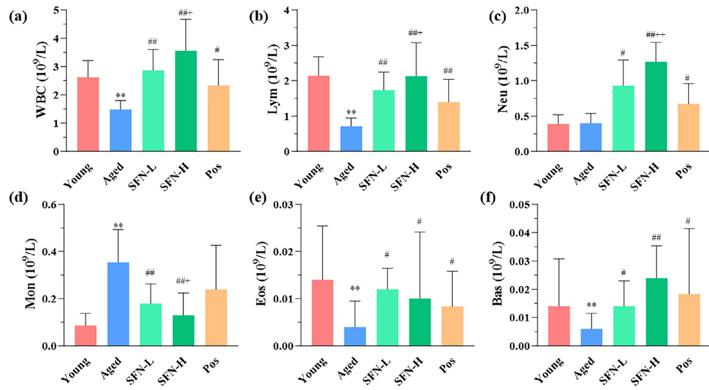
WBC count results indicated that mice from the Aged group were significantly immunosuppressed. T cells occupied the main lymphocyte lineages.

The present study illuminated the skin protective mechanism of SFN by network pharmacology and proteomics analyses in a natural aging mouse model. SFN therapy showed significant alterations in skin structure, redox balance, and composition of immune cell populations after an intervention duration of 2 months.”

https://onlinelibrary.wiley.com/doi/10.1002/mnfr.70281 “Integrative Network Pharmacology and Proteomics Decipher the Immunomodulatory Mechanism of Sulforaphane Against Intrinsic Skin Aging” (not freely available)


I rated this study Wasted resources for using sulforaphane doses not relevant to humans. I usually don’t curate such studies. Its lowest sulforaphane 50 mg/day dose is a ((50 mg * .081) * 70 kg) = 284 mg human equivalent.

I decided to curate it for its informative young controls vs. aged controls results in the above graphic. WBC counts are available on almost every standard human blood test.

This study’s young and aged groups per Grok: “A 2-month-old mouse aging for two more months (reaching 4 months) is approximately equivalent to a human aging from about 12–15 years to 17–21 years old. An 18-month-old mouse (human equivalent: ~45–50 years) aging for two months would be roughly equivalent to a human aging from ~45–50 years to ~51–58 years.”

Glucosinolate and isothiocyanate human interventions

A 2025 review covered human evidence from glucosinolate and isothiocyanate research through April 2025:

“Glucosinolates (GSLs) and their breakdown products, isothiocyanates (ITCs), are biogenesis compounds with anti-inflammatory, antioxidant, and anticancer properties, mediated through key pathways such as Nrf2, NF‐κB, and epigenetic regulation. However, their limited and variable bioavailability remains a key challenge. This review summarises the current clinical evidence on GSLs and ITCs, with a focus on their health effects and metabolic fate in humans.”

https://www.mdpi.com/2304-8158/14/16/2876 “Bioavailability, Human Metabolism, and Dietary Interventions of Glucosinolates and Isothiocyanates: Critical Insights and Future Perspectives”


In the above graphic, notice how the inactive myrosinase column has no small intestine participation, but the active myrosinase column does. This point wasn’t adequately emphasized, that for complete effects, an individual has to do whatever they can to thoroughly chew or otherwise activate myrosinase to hydrolyze glucosinolates before swallowing.

Researchers don’t rely on individuals taking responsibility for their own health, of course. Just swallow these pills, we’ll do it for you, as if humans are lab rats. This lack of emphasis is understandable, if not optimal.

This review provided longish coverage of studies, which is preferable to the usual treatment of citing a reference without much explanation. Compare, for example, my longish curation of the 2023 Eat broccoli sprouts for your high intensity interval training with its reference 68 summary below:

“Another study investigated the effects of consuming GSL-rich broccoli sprout (GRS) supplements on oxidative stress and physiological adaptations to intense exercise training. In a randomised, double-blind, crossover design, nine healthy participants consumed either a GRS supplement (75 g of sprouts) or a placebo twice daily over a 7-day high-intensity interval training period. The findings revealed that GRS supplementation significantly reduced markers of oxidative stress, including carbonylated proteins in skeletal muscle and plasma myeloperoxidase levels, compared to the placebo condition. Furthermore, GRS intake led to reduced lactate accumulation during submaximal exercise and enhanced exercise performance, as indicated by a longer time to exhaustion during maximal exercise tests. At the molecular level, supplementation with GRS was associated with elevated Nrf2 protein levels in muscle tissue, suggesting activation of endogenous antioxidant defence mechanisms. In addition, GRS intake mitigated nocturnal hypoglycaemic episodes and lowered average blood glucose levels, indicating improved glucose regulation during intense training. Collectively, these results suggest that GRS supplementation may enhance physiological adaptations to high-intensity exercise by reducing oxidative stress and supporting metabolic homeostasis.”


Oats sprouts treat gut inflammation

A 2025 rodent study investigated differing effects of regular oats and oat sprouts to treat induced colitis:

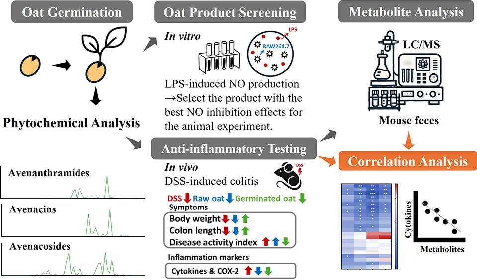
“This study aims to test our hypothesis that germinated oats exert stronger anti-inflammatory effects than raw oats due to their higher levels of bioactive phytochemicals. First, the nitric oxide (NO) production assay was used to screen [22] commercially available oat seed products and identify the product with the highest anti-inflammatory activity after germination [for five days]. The selected oat seed product was then produced in larger quantities and further evaluated in an in vivo study using the dextran sulfate sodium (DSS)-induced colitis mouse model to compare the anti-inflammatory effects of phytochemical extracts from germinated and raw oats.

The guideline states that for a healthy U.S.-style dietary pattern at a 2000 calorie level, a daily intake of 6 ounces of grains is recommended, with at least 3 ounces (84 g) coming from whole grains (WGs). For a 60 kg human, consuming 3 ounces of WGs per day translates to a 17.2 g/kg daily dose in mice. Given that the daily food intake of a 20 g mouse is approximately 2.5 g, the 17.2 g/kg daily dose corresponds to 14% of the total diet as WGs. Therefore, the 7 and 21% WG equivalent doses used in this study are relevant to human consumption.

Germination led to an overall increase in the content of all avenanthramides (AVAs) and avenacins (AVCs) as well as some avenacosides (AVEs):

  • For AVAs, the compounds 2c, 2p, 2f, 2cd, 2pd, and 2fd significantly increased by 10.0-, 6.3-, 9.6-, 20.7-, 10.6-, and 4.6-fold, respectively, which is consistent with previous reports.
  • This study is the first to report an increase in AVCs after germination, with AVC-A2, B2, A1, and B1 contents significantly increasing by 2.5-, 2.2-, 3.6-, and 4.2-fold, respectively.
  • Although germination resulted in a decrease in certain AVEs, it significantly increased the levels of AVE-C, Iso-AVE-A, AVE-E, and AVE-F by 1.8-, 3.3-, 3.3-, and 5.0-fold, respectively. Notably, AVE-E has been previously reported to have the strongest anti-inflammatory activity among all of the major AVEs.

In summary, germination enhances the anti-inflammatory properties of oats in both cells and DSS-induced colitis in mice by increasing levels of bioactive phytochemicals. Correlation analysis showed a significant inverse relationship between pro-inflammatory cytokines and phytochemical content in feces, especially AVAs and their microbial metabolites.

The observation of a stronger anti-inflammatory effect in the low-dose germinated oat group compared with the high-dose group is intriguing and warrants further investigation. One possible explanation is the phenomenon of hormesis, where low doses of bioactive compounds can exert beneficial effects, while higher doses may lead to diminished efficacy or even adverse effects. Further studies involving a broad range of doses would be valuable to define the effective intake range and provide insight into the underlying mechanisms.

It is possible that AVAs, AVEs, and AVCs act synergistically to enhance the overall anti-inflammatory efficacy, potentially by targeting different inflammatory pathways or modulating each other’s bioavailability and activity. Further investigation into the synergistic interactions among these compounds is warranted.”

https://pubs.acs.org/doi/10.1021/acs.jafc.5c02993 “Phytochemical-Rich Germinated Oats as a Novel Functional Food To Attenuate Gut Inflammation”


I’ve eaten 3-day-old Avena sativa oat sprouts (started from 20 grams of groats) every day for 4.5 years now, and haven’t had gut problems. Here’s what they looked like this morning:

Get a little stress into your life, Part 2

A 2025 reply to a letter to the editor cited 56 references to elaborate on Part 1 and related topics:

“A positive effect does not necessarily mean benefit, and positive effects on individual organisms may mean adverse effects on other coexisting organisms. However, a vast literature shows that hormetic stimulation can result in benefits depending on the context, for instance, clear growth, yield, and survival improvement.

There is some energetic cost to support hormetic stimulation, with a likely positive energy budget, which might also have negative consequences if there is insufficient energy substrate, especially under concurrent severe environmental challenges. Moreover, hormetic preconditioning could be particularly costly when there is a mismatch between the predicted environment and the actual environment the same individuals or their offspring might face in the future.

Hormesis should not be unilaterally linked to positive and beneficial effects without considering dose levels. For any research to answer the question of whether a stimulation represents hormesis and whether it is beneficial, robust dose–response evaluations are needed, which should be designed a priori for this purpose, meeting the requirements of the proper number, increment, and range of doses.

Both additivity and synergism are possible in the hormetic stimulatory zone, depending also on the duration of exposure and the relative ratio of different components. This might happen, for example, when a chemical primes stress pathways (e.g., heat shock proteins and antioxidants), thus enabling another chemical to trigger hormesis (defense cross-activation) and/or because combined low subtoxicity may modulate receptors (e.g., aryl hydrocarbon receptor and nuclear factor erythroid 2-related factor 2) differently than individual exposures (receptor binding synergy).

Moreover, even when stimulation occurs in the presence of individual components, stimulation may no longer be present when combined, and therefore, effects of mixtures cannot be accurately predicted based on the effects of individual components. There may be hormesis trade-offs; hormesis should be judged based on fitness-critical end points.

While often modeled mathematically, hormesis is fundamentally a dynamic biological process and should not be seen as a purely mathematical function, certainly not a linear one. Much remains to be learned about the role of hormesis in global environmental change, and an open mind is needed to not miss the forest for the trees.”

https://pubs.acs.org/doi/10.1021/acs.est.5c05892 “Correspondence on ‘Hormesis as a Hidden Hand in Global Environmental Change?’ A Reply”


Reference 38 was a 2024 paper cited for:

“Hormetic-based interventions, particularly priming (or preconditioning), do not weaken organisms but strengthen them, enhancing their performance and health under different environmental challenges, which are often more massive than the priming exposure.

The catabolic aspect of hormesis is primarily protective whereas the anabolic aspect promotes growth, and their integration could optimize performance and health. The concept of preconditioning has also gained widespread attention in biomedical sciences.”

https://www.sciencedirect.com/science/article/abs/pii/S1568163724004069 “The catabolic – anabolic cycling hormesis model of health and resilience” (not freely available)


Reference 40 was a 2021 review that characterized hormesis as a hallmark of health:

“Health is usually defined as the absence of pathology. Here, we endeavor to define health as a compendium of organizational and dynamic features that maintain physiology.

Biological causes or hallmarks of health include features of:

  • Spatial compartmentalization (integrity of barriers and containment of local perturbations),
  • Maintenance of homeostasis over time (recycling and turnover, integration of circuitries, and rhythmic oscillations), and
  • An array of adequate responses to stress (homeostatic resilience, hormetic regulation, and repair and regeneration).

Disruption of any of these interlocked features is broadly pathogenic, causing an acute or progressive derailment of the system.

A future ‘medicine of health’ might detect perilous trajectories to intercept them by targeted interventions well before the traditional ‘medicine of disease’ comes into action.”

https://www.sciencedirect.com/science/article/pii/S0092867420316068 “Hallmarks of Health”


Activate Nrf2 with far-infrared light

A 2025 rodent study investigated effects of far-infrared light on Alzheimer’s disease models. I’ll focus on its Nrf2 findings:

“Far-infrared radiation (FIR) is commonly utilized as a complementary treatment of a range of disease, for example, insomnia and rheumatoid arthritis. In this research, we explored how FIR light impacts cognitive functions of TgCRND8 AD mice and elucidated its underlying molecular mechanism.

Infrared radiation is a form of electromagnetic energy that has wavelengths between 750 nm and 1000 μm, which are longer than visible light. International Commission on Illumination categorizes infrared light as three sub-divisions according to the wavelength: (1) near-infrared radiation (0.7–1.4 μm), (2) middle infrared radiation (1.4–3.0 μm), and (3) far-infrared radiation (3.0–1000 μm).

Nrf-2/ HO-1 signaling, a key endogenous antioxidant system, helps mitigate oxidative stress and enhances expression of various endogenous genes. Activation of HO-1 during inflammatory conditions may serve as an adaptive response to reduce cytotoxicity through various mechanisms.

In this study, we applied EFFIT LITE® as the FIR spectrum transmitter which stably radiates an FIR spectrum with a wavelength of 4–20 μm, and the device was put within 1 cm directly above the head of the 3-month-old TgCRND8 mice for 30 min exposure once every day. FIR light notably enhanced cognitive function and spatial memory of TgCRND8 mice after 28-days consecutive treatment.

Underlying molecular mechanisms involve suppression of Aβ deposition, hyperphosphorylation of tau, and neuroinflammation through modulating Jak-2/Stat3 and Nrf-2/HO-1 pathways. Our current experimental findings amply indicate that FIR light is a potential non-pharmacological therapy for AD.”

https://link.springer.com/article/10.1007/s12017-025-08860-2“Far-Infrared Radiation Ameliorates the Cognitive Dysfunction in an Alzheimer’s Disease Transgenic Mouse via Modulating Jak-2/Stat3 and Nrf-2/HO-1 Pathways”


This study measured Nrf2 and its quickly-induced downstream enzyme HO-1 effects of daily far-infrared light exposure for 30 minutes. We’d have to see measurements of Nrf2’s more-slowly induced and longer-lasting downstream xenobiotic detoxifying enzyme NQO1 to compare far-infrared light Nrf2 activation effects with those of natural plant compounds.

Betaine as an exercise mimetic

A 2025 human study investigated effects of long-term exercise:

“Exercise has well-established health benefits, yet its molecular underpinnings remain incompletely understood. We conducted an integrated multi-omics analysis to compare effects of acute vs. long-term exercise in healthy males.

Acute exercise induced transient responses, whereas repeated exercise triggered adaptive changes, notably reducing cellular senescence and inflammation and enhancing betaine metabolism. Exercise-driven betaine enrichment, partly mediated by renal biosynthesis, exerts geroprotective effects and rescues age-related health decline in mice.

Betaine binds to and inhibits TANK-binding kinase 1 (TBK1), retarding the kinetics of aging.

Betaine effectively alleviated senescence phenotypes by reduced senescence-associated β-galactosidase (SA-β-Gal)-positive cells, decreased p21 expression, lowered DNA damage indicator γ-H2A.X, and elevated heterochromatin mark H3K9me3. Betaine treatment also enhanced cellular antioxidant capacity, as evidenced by increased NRF2 phosphorylation and reduced ROS accumulation.

These findings systematically elucidate the molecular benefits of exercise, and position betaine as an exercise mimetic for healthy aging.”

https://doi.org/10.1016/j.cell.2025.06.001 “Systematic profiling reveals betaine as an exercise mimetic for geroprotection” (not freely available) Thanks to Dr. Weimin Ci for providing a copy.


Taurine and mitochondrial health

A 2025 review subject was taurine’s beneficial effects on mitochondria:

“Taurine has multiple and complex functions in protecting mitochondria against oxidative-nitrosative stress. We introduce a novel potential role for taurine in protecting from deuterium (heavy hydrogen) toxicity. This can be of crucial impact to either normal or cancer cells that have highly different mitochondrial redox status.

Deuterium is an isotope of hydrogen with a neutron as well as a proton, making it about twice as heavy as hydrogen. We first explain the important role that the gut microbiome and gut sulfomucin barrier play in deuterium management. We describe synergistic effects of taurine in the gut to protect against deleterious accumulation of deuterium in mitochondria, which disrupts ATP synthesis by ATPase pumps.

Taurine’s derivatives, N-chlorotaurine (NCT) and N-bromotaurine (NBrT), produced through spontaneous reaction of taurine with hypochlorite and hypobromite, have fascinating regulatory roles to protect from oxidative stress and beyond. We describe how taurine could potentially alleviate deuterium stress, primarily through metabolic collaboration among various gut microflora to produce deuterium depleted nutrients and deuterium depleted water (DDW), and in this way protect against leaky gut barrier, inflammatory bowel disease, and colon cancer.

Taurine cannot be metabolized by human cells, but gut microbes are able to break it down to release sulfite, which then gets oxidized to sulfate anions that become available to support synthesis of sulfomucins. Taurine protects against many diseases linked to mitochondrial defects, such as aging, metabolic syndrome, cancer, cardiovascular diseases and neurological disorders.

We present a novel view that gut microbes play an essential role in providing deuterium depleted (deupleted) nutrients, especially, butyrate, to the host colonocytes forming the gut barrier. We propose that sulfomucins synthesized by goblet cells not only protect the barrier from pathogens, but also trap and sequester deuterium, thus reducing mitochondrial deuterium levels, resulting in improved mitochondrial health.

Due to taurine, redox buffer glutathione (GSH) further stabilizes the membrane potential. GSH not only reduces radical oxygen species (ROS) during oxidative stress, but it also assists in production of deupleted water in mitochondria.

Spontaneous oxidation of two GSH molecules to produce GSSG in the presence of hydrogen peroxide yields two molecules of DDW. Just as for glutathione, bilirubin can produce DDW indefinitely through chronic recycling between bilirubin and biliverdin, capturing a deupleted proton in NADPH to produce a DDW molecule in each cycle.

A novelty that arises from this investigation is introduction of the role that deuterium plays in mitochondrial disease, and ways in which taurine may facilitate maintenance of low deuterium in mitochondrial ATPase pumps. Excess deuterium causes a stutter in the pumps, which leads to inefficiencies in ATP production and an increase in ROS.”

https://pmc.ncbi.nlm.nih.gov/articles/PMC11717795/ “Taurine prevents mitochondrial dysfunction and protects mitochondria from reactive oxygen species and deuterium toxicity”


Stay away from NAC

A 2025 rodent study added several reasons to avoid non-emergency use of N-acetylcysteine:

“We previously showed that antioxidants induced an impairment of negative feedback of the hypothalamus-pituitary-adrenal (HPA) axis in rats, in parallel to a down-regulation of glucocorticoid receptor (GR) and nuclear factor erythroid 2-related factor 2 (Nrf2) expression in the pituitary gland. This study evaluated the role of the Nrf2-heme-oxygenase-1 (HO-1) pathway on impairment of negative feedback of the HPA axis induced by N-acetylcysteine (NAC).

Male Swiss-Webster mice were orally supplemented with NAC for 5 consecutive days. The Nrf2-HO-1 pathway activator cobalt protoporphyrin IX (CoPPIX) was injected intraperitoneally on days 2 and 5 after starting NAC supplementation.

NAC reduced expression of Nrf2 in the pituitary of mice. NAC induced adrenal enlargement and hypercorticoidism, along with a decrease in GRα expression and an increase of GRβ expression in the pituitary gland.

We observed that dietary supplementation with NAC ( Figure 4A ) significantly increased plasma corticosterone levels in mice 24h ( Figure 4B ) as well as 15 days ( Figure 4C ) after the last administration of the antioxidant with the same magnitude of response (3.5-fold and 3.4-fold, respectively).

Chronic activation of the HPA axis can have damaging effects on immune, cardiovascular, metabolic, and neural functions, increasing the risk of immune system dysfunction, mood disorders, and metabolic and cardiovascular diseases. To prevent these deleterious effects of chronic hypercortisolism, HPA axis function is controlled by a glucocorticoid-dependent negative feedback system that is essential for ending the stress response.

These findings showed that NAC reduced Nrf2-HO-1 pathway activation in the pituitary gland, in a mechanism probably related to a local downregulation of GRα and an up-regulation of GRβ, leading to a failure of negative feedback of the HPA axis and consequently to the hyperactivity of this neuroendocrine axis.”

https://pmc.ncbi.nlm.nih.gov/articles/PMC11827418/ “Activation of the Nrf2/HO-1 pathway restores N-acetylcysteine-induced impairment of the hypothalamus-pituitary-adrenal axis negative feedback by up-regulating GRα expression and down-regulating GRβ expression into pituitary glands”


A human equivalent to this study’s NAC dose is (150 mg x .081) x 70 kg = 851 mg. Human supplements are sold in 600 mg and 1000 mg doses.

Grok 3 replied to a question: Human equivalent time to 15 days in male Swiss-Webster mice aged between 4 and 6 weeks? by stating: “15 days in male Swiss-Webster mice aged 4 to 6 weeks corresponds to approximately 4.1 human years, advancing their equivalent human age from about 10–12 years to roughly 14–16 years.” Four+ years seems like a long time for NAC to steadily and continuously affect humans’ HPA axes per the above graphic. What do you think?

Previously mentioned reasons to avoid daily use of NAC were in the lower part of A good activity for bad weather days.