All about the betaine

A trio of papers on betaine, the first being a 2021 series of thorough rodent experiments relating betaine and gut microbiota, and cause and effect:

“Compared with lean individuals, adipose tissues in obese individuals secrete high levels of pro-inflammatory cytokines such as TNF-α, IL-1β, and IL-6, inducing:

  • Systemic inflammation;
  • Insulin resistance;
  • Large amounts of carcinogenic factors; and
  • Increasing risk of certain types of cancer such as melanoma, colon cancer, and liver cancer.

Prebiotics obtained from fruits and vegetables can regulate host lipid metabolism and glucose homeostasis by reversing gut dysbiosis in obese individuals.

kgmi_a_1862612_f0005_oc

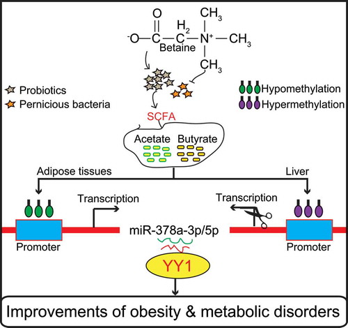
Results of this study show that dietary betaine alleviated gut microbiota imbalance in obese mice, and reduced development of obesity and obesity-related complications. Regulation of the miR-378a-YY1 regulatory axis by gut microbial acetate and butyrate was a critical mechanism for modulating:

  • White adipose tissue browning;
  • Classical brown adipose tissue activation; and
  • Lipid and glucose homeostasis

in obese mice after betaine supplementation.

These findings offer novel insights into underlying mechanisms by which gut microbiota affect host metabolism and host immune system, and demonstrate that the betaine-gut microbiota-derived signal axis is a potential therapeutic target in obesity and metabolic syndrome.”

https://www.tandfonline.com/doi/full/10.1080/19490976.2020.1862612 “Dietary betaine prevents obesity through gut microbiota-drived microRNA-378a family”


A second 2021 paper was a meta-analysis of effects on human cardiovascular biomarkers:

“Betaine supplementation had a significant effect on concentrations of:

  • Betaine;
  • Total cholesterol;
  • Low-density lipoprotein (LDL);
  • Homocysteine [negative effect]; and
  • Methionine.

Betaine supplementation did not affect serum concentrations of:

  • Triglycerides;
  • High-density lipoprotein (HDL);
  • Fasting blood glucose;
  • C-reactive protein;
  • Liver enzymes alanine aminotransferase (ALT), aspartate aminotransferase (AST), gamma-glutamyl transferase (GGT); and
  • Blood pressure.

Our meta-analysis supports the advantage of a lower dose of betaine supplementation (<4 g/d) on homocysteine concentrations without the lipid-augmenting effect observed with a higher dosage.”

https://www.tandfonline.com/doi/abs/10.1080/10408398.2021.1902938 “Effects of betaine supplementation on cardiovascular markers: A systematic review and Meta-analysis” (not freely available)


A third paper was a 2014 cereal analysis of betaine and its precursor choline that found a 224% increase in betaine from 62 to 139 μg/g and a 31% increase in choline from microwaving oats:

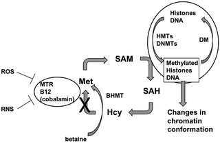
“Betaine and its precursor choline are important components of one-carbon metabolism, remethylating homocysteine into methionine and providing methyl groups for DNA methylation. Cereals are the main source of betaine in diet.

During cooking processes which did not involve removal of water (in this case oat porridge microwaved using instant oats) appeared to lead to creation of betaine. Explanations for this phenomenon could be that betaine is synthesised during the process, or that heating with water liberates betaine from cereal matrix, enhancing efficiency of extraction.”

https://www.sciencedirect.com/science/article/abs/pii/S0308814613012247 “Cereal foods are the major source of betaine in the Western diet – Analysis of betaine and free choline in cereal foods and updated assessments of betaine intake” (not freely available)


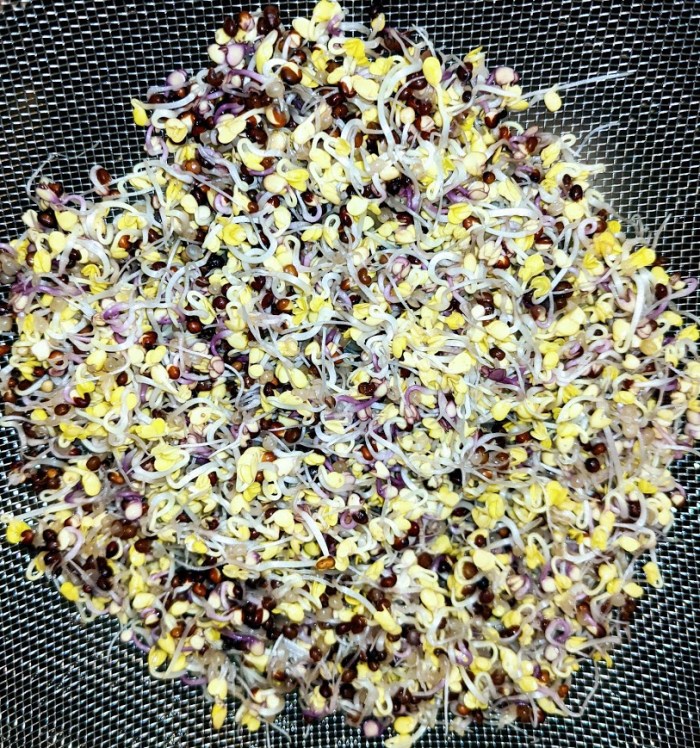
Another 2021 betaine (aka trimethyl glycine) study was curated in Ride the waves of gene expression with betaine for its role in preventing nerve disease. I take 1.5 grams of a betaine supplement every morning and evening when eating hulled Avena sativa 3-day-old oat sprouts.

I found the first two papers from their citing a 2016 human and rodent study Dietary Betaine Supplementation Increases Fgf21 Levels to Improve Glucose Homeostasis and Reduce Hepatic Lipid Accumulation in Mice, which was linked in a comment on this 2021 video:

Small intestine alkaline phosphatase

This 2021 rodent study used small intestine alkaline phosphatase (IAP)-overexpressed subjects on a high-fat, high-cholesterol diet to investigate effects:

“To examine direct effects of increased IAP expression on barrier function and development of metabolic diseases, we developed intestine-specific IAP transgenic mice (IAPTg) overexpressing human chimeric IAP. We evaluated effects of intestine-specific IAP overexpression on Western-type diet (WD)–induced atherosclerosis in Ldlr−/ mice.

Diets low in fiber deprive intestinal bacteria of essential nutrients. Luminal bacteria turn to alternate sources of energy, namely, the carbohydrate-rich mucosal layer. This enhances direct contact between gut bacteria and intestinal epithelial layer, and promotes inflammation and intestinal barrier dysfunction.

Increase in IAP improves intestinal barrier function by not only dephosphorylating LPS and limiting its translocation to systemic circulation, but also by improving mucosal layer. Furthermore, IAP overexpression results in attenuated WD-induced weight gain and significantly reduced absorption of dietary lipids, leading to attenuation of total plasma cholesterol and TG levels, as well as hepatic lipids. This improved metabolic profile results in significant reduction in WD-induced atherosclerosis in Ldlr−/−IAPTg mice.

overexpressed IAP

IAP overexpression results in attenuated WD-induced weight gain and significantly reduced absorption of dietary lipids, leading to attenuation of total plasma cholesterol and TG levels, as well as hepatic lipids. This improved metabolic profile results in significant reduction in WD-induced atherosclerosis in Ldlr−/−IAPTg mice.

Increases in IAP can significantly attenuate effects of WD feeding on intestinal barrier function. It is noteworthy that IAP is also shown to be involved in innate immunity, and its activity is positively correlated to intestinal levels of IgA in mice and fecal immunoglobulins in humans.

The list of nutrients and food components/supplements that increase IAP continues to grow (galactooligosaccharides, glucomannan, vitamin D3), providing a novel opportunity to develop simple strategies for modulation of diet/nutrition to target metabolic diseases, including diabetes, fatty liver disease, atherosclerosis, or heart disease.”

https://www.ahajournals.org/doi/10.1161/CIRCRESAHA.120.317144 “Over-Expression of Intestinal Alkaline Phosphatase Attenuates Atherosclerosis”


This study used ileal samples from the small intestine’s last section for its findings. It complemented Take FOS or inulin to increase your gut’s alkaline phosphate activity which used large intestine samples to demonstrate effects of  increased IAP activity.

Eat to make your gut microbiota happy, and expect reciprocity.

A biological age snapshot from a year ago

This May 3, 2020 two-hour video on Optimizing Biological Age was instructive:


Content was great! I recommend the longish Q & A, especially at 1:23 regarding inflammation.

It was a snapshot in that researchers on this conference call were interested in improving people’s health. Few recognized at the time a globally coordinated effort to herd humans into one tracking database using an injurious jab as a pretext.

optimizing biological age

The next phase of reversing aging and immunosenescent trends

Dr. Greg Fahy earlier this week provided an update on the November 2020 TRIIM-X follow-on to the September 2019 TRIIM curated in Reversal of aging and immunosenescent trends. Emphasis was on reproducibility:

23:45 Dr. Steve Horvath reanalyzed TRIIM for the plasma portion of Levine’s PhenoAge epigenetic clock. Results were congruent with four other epigenetic clocks showing a 2.5 year reduction of biological age.

39:20 TRIIM-X preliminary results started with C-Reactive protein.

43:05 No backsliding in epigenetic age deceleration between TRIIM and TRIIM-X!

continued epigenetic age deceleration

55:07 Q & A session starts with how TRIIM-X controls for supplements. Answers for resveratrol and calorie restriction, emphasizing that CR doesn’t reverse aging.

1:10 TRIIM-X took photos of subjects’ hair at baseline!


Great update! The last 20 minutes emphasized a need for capital in aging research. TRIIM-X has another 1.5 years to go, and other aging research projects needing funding were mentioned.

Don’t know what happened to the unmentioned 3000 IU vitamin D and 50 mg zinc recommendations of TRIIM. So I asked. Dr. Fahy replied:

“They are still there! Just not mentioned!”

Thought briefly about enrolling in TRIIM-X, but there’s no way anyone but me gets to experiment with my body.

Take FOS or inulin to increase your gut’s alkaline phosphatase activity

This 2020 rodent study ran a series of experiments relating gut health factors. I left some items out that Google Translate didn’t handle well.

“This study investigated effects of food factors on colonic microbiota, fermentation products, mucins, immunoglobulin A (IgA) and alkaline phosphatase (ALP) activity. Colonic ALP activity was positively correlated with colonic luminal variables such as fecal mucin level, ratio of Bifidobacterium spp., and level of n-butyrate, which are associated with a more favorable colonic environment. We propose that the increase of colonic ALP activity induced by fermentable non-digestible carbohydrates may be important for protection of gut epithelial homeostasis.

Since glucomannan was found to induce colon ALP activity, effects of other dietary fibers were also investigated. Ingestion of water-soluble dietary fibers pectin and inulin significantly increased colon tissue and fecal ALP activity of high-fat diet-fed rats.

Ingestion of chitosan, an insoluble dietary fiber, had no effect on colonic ALP activity. This indicates that colonic ALP activity may be induced by indigestible sugars such as fermentable water-soluble dietary fiber.

ALP activity of the large intestine tissue of rats fed a high-fat diet was significantly increased by ingestion of indigestible oligosaccharides fructooligosaccharide (FOS), galactooligosaccharide (GOS), raffinose (RAF) and lactulose (LAC). Mucin, n-butyric acid, and Bifidobacterium spp. significantly increased, and Clostridium coccoides was significantly reduced.

ALP activity

In the digestible isomaltooligosaccharide (IMOS)-added diet group, large intestine ALP activity, ALP gene expression, mucin, organic acid, and intestinal flora showed no effect.

In order to investigate nutritional conditions on inducing colon ALP activity by oligosaccharide intake, the difference in lipid quality ingested was examined under a high-fat diet.

  • When soybean oil and lard were used as dietary fats, the difference in quality of dietary lipids did not affect large intestine ALP activity and IAP-I expression in the FOS-free diet.
  • When FOS was added, ALP activity and IAP-I expression in the large intestine tissue were significantly increased under the condition of a high lard diet as compared with a high soybean oil diet.
  • Mucin content behaved similarly to colon tissue ALP activity and IAP-I expression.

mucin content

In this study, it was newly found that the effect of indigestible oligosaccharide intake on increasing colon ALP activity differs depending on nutritional conditions such as type of lipid. Furthermore, it was found that the increase in colon ALP activity under indigestible sugar intake has a positive correlation with factors involved in maintaining function of the intestinal environment, such as mucin.”

https://www.jstage.jst.go.jp/article/jsnfs/74/1/74_9/_article “Modulation of the Colonic Luminal Environment by Food Factors” (in Japanese)


I arrived at this study by it citing a 2011 study Vitamin K1 (Phylloquinone) or Vitamin K2 (Menaquinone-4) Induces Intestinal Alkaline Phosphatase Gene Expression. More on IAP in this 2020 video:

Your bones influence your brain

This 2020 review subject was brain-bone crosstalk:

“Multiple stress, mood and neurodegenerative brain disorders are associated with osteoporosis. Skeletal diseases display impaired brain development and function.

Along with brain and bone pathologies, trauma events highlight strong interaction of both organs. While brain-derived molecules affecting bone include central regulators – transmitters of the sympathetic, parasympathetic and sensory nervous system – bone-derived mediators altering brain function are released from bone cells and marrow.

ijms-21-04946-g001

Osteoblast-derived hormone osteocalcin (OCN) exerts neuroprotective effects. Studies revealed a bidirectional dependence of brain and bone through bone cell-derived modulators that directly affect behavioral and cognitive function.

The main bone-derived mediator affecting the brain is OCN, which is exclusively synthesized by osteoblasts. OCN was recently discovered to transverse the BBB to enter the CNS, where it promotes spatial learning and memory while preventing anxiety-like behavior or even depression.

Cognitive function and circulating levels of OCN are proposed to inversely correlate with age. Maternal osteocalcin regulates embryonic brain development by enhancing monoamine neurotransmitters and their synthesis.

Clinical observations provide key evidence for a bidirectional communication between brain and bone tissue, which is strongly supported by experimental studies that unraveled underlying mechanistic pathways and identified molecular mediators involved in this crosstalk.”

https://www.mdpi.com/1422-0067/21/14/4946/htm “Crosstalk of Brain and Bone-Clinical Observations and Their Molecular Bases”


The first paper of Vitamin K2 – What can it do? said:

Osteocalcin γ-carboxylation is the main mechanism of action through which Vitamin K2 improves bone health.”

This paper didn’t mention Matrix Gla Protein (MGP) carboxylation, and said a contrary:

“Undercarboxylated, bioactive OCN, initially considered as an inhibitor of bone mineralization, participates in systemic body regulation and homeostasis.”

The 2019 paper cited was Osteocalcin‑GPRC6A: An update of its clinical and biological multi‑organic interactions (Review):

“Osteocalcin is a small protein present in two forms: Carboxylated (cOC) and undercarboxylated (ucOC). Only ucOC can signal as a hormone while cOC cannot.”

It went on to downplay cOC, and also didn’t mention MGP carboxylation.

I think it’s a question of balance. cOC stays in your bones. Carboxylated MGP influences calcium to go into your bones instead of your blood vessel walls. Two good things.

An outstanding review of Vitamin K deficiency and disease

This 2019 review focused on one Vitamin K-deficiency biomarker. All parts I’ve quoted are outside the liver, so Vitamin K deficiency ≈ Vitamin K2 deficiency.

This is a hard read with many technical details, but sometimes that’s how researchers do it:

“Active MGP (matrix Gla protein), once released into extracellular space, acts as a local inhibitor of calcification. Widespread expression of MGP points to a role of MGP that by far exceeds its well-known function as local inhibitor of calcification.

Recent research confirmed this concept, usually by measuring plasma dp-ucMGP (desphospho-uncarboxylated MGP), a biomarker reflecting poor vitamin K status:

1160fig02

Vitamin K plays a pivotal role in maintaining bone health. Increasing evidence also implicates MGP in maintaining bone health.

In the Health, Aging and Body Composition study, 791 older community-dwelling adults underwent magnetic resonance imaging to measure bilateral knee structural features. The highest [25%] compared with the lowest fourth of the dp-ucMGP distribution had higher odds of having:

  • Meniscus damage;
  • Osteophytes;
  • Bone marrow lesions; and
  • Subarticular cysts.

Regarding Vitamin K supplementation:

  • Studies showed a dose-dependent decrease in circulating dp-ucMGP with an 86% decrease already observed after 4 weeks of substitution by 360 μg menaquinone-7 [in 50 hemodialysis patients];
  • In a randomized double-blind trial of 244 postmenopausal women followed up for 3 years, arterial stiffness as captured by aortic pulse wave velocity or stiffness index β, decreased in intervention compared with control group.

These results should be considered as hypothesis-generating in view of small sample size, and because there were no between-group differences in vitamin K–induced changes in elastic properties of the carotid artery.

Plasma dp-ucMGP levels ranging from 1.4 to 4.6 μg/L were optimal in terms of risk of mortality and macrovascular cardiovascular illness (4.6 μg/L threshold corresponding to the 65th percentile of dp-ucMGP distribution).

Vitamin K supplementation before irreversible organ damage sets in might find its application in prevention of a wide range of disabling diseases. Circulating dp-ucMGP levels might be measured over time to track risk of vascular complications.”

https://www.ahajournals.org/doi/10.1161/HYPERTENSIONAHA.119.12412 “Vitamin K–Dependent Matrix Gla Protein as Multifaceted Protector of Vascular and Tissue Integrity”


I usually don’t give 5+ stars to reviews. This one was different.

Yes, there could be factors other than this one Vitamin K deficiency biomarker involved in study findings. Sure, these coauthors cited their own studies. Its overall purpose, though, was to inform readers.

I’ll summarize this paper as providing evidence for a biomarker of Vitamin K2 deficiency being implicated in the development and progression of many diseases.

Vitamin K2 – What can it do?

A trio of papers on Vitamin K2, the first being a 2021 review that emphasized dual effects:

“Osteoporosis (OP) is the most common bone disease that affects elderly men and women. It is a metabolic skeletal disorder caused by an imbalance between bone formation and resorption, leading to a loss of bone mass and quality, skeletal structure deterioration, and an increased risk of fractures.

Vascular calcification is defined as ectopic deposition of mineral matrix in vessel wall. It occurs prevalently in aging and primary chronic conditions (hypertension, diabetes mellitus, and chronic kidney disease), representing an important risk factor for cardiovascular morbidity and mortality.

Studies have provided support for a close link between bone and vascular health. Findings suggest that bone loss in OP may promote and increase the risk of cardiovascular events and vascular atherosclerosis.

Vitamin K2 is involved in a phenomenon in which a low calcium deposition in bone tends to be associated with a parallel increase of calcium deposition in vessel wall as a consequence of impaired calcium metabolism. Most production of Vitamin K2 in humans takes place in intestines. However, the amount derived from intestinal bacteria is poorly absorbed, and is not able to reach concentrations required to exert physiological functions.

Vitamin K2‘s ability to reduce loss of bone mineral density and fracture risk, as well as to improve bone quality, has been described by several clinical studies, which have confirmed that osteocalcin (OC) γ-carboxylation is the main mechanism of action through which this natural compound is able to improve bone health. Clinical evidence suggests an analogous protective role of Vitamin K2 at the vascular level, emphasizing a strict association between:

  • Vitamin serum level;
  • Matrix gla protein (MGP) γ-carboxylation levels;
  • Reduction of vascular smooth muscle cells osteogenic trans-differentiation; and
  • Possible risk of cardiovascular events.”

https://www.mdpi.com/2072-6643/13/4/1222/htm “The Dual Role of Vitamin K2 in ‘Bone-Vascular Crosstalk’: Opposite Effects on Bone Loss and Vascular Calcification”


A second 2021 review emphasized aging:

“Vitamin K can:

  • Carboxylate OC (a protein capable of transporting and fixing calcium in bone);
  • Activate MGP (an inhibitor of vascular calcification and cardiovascular events); and
  • Carboxylate Gas6 protein (involved in brain physiology and a cognitive decline and neurodegenerative disease inhibitor).

By improving insulin sensitivity, Vitamin K lowers diabetes risk. It also exerts antiproliferative, proapoptotic, autophagic effects, and has been associated with a reduced risk of cancer.

The most common [Vitamin K2] subtypes in humans are the short-chain MK[menaquinone]-4, which is the only MK produced by systemic conversion of phylloquinone [Vitamin K1] to menaquinone, and MK-7 through MK-10, which are synthesized by bacteria. The main sources of Vitamin K2 are fermented foods, cheeses, eggs, and meats.”

https://www.mdpi.com/2076-3921/10/4/566/htm “The Role of Vitamin K in Humans: Implication in Aging and Age-Associated Diseases”


The third paper – somehow not cited by these two reviews – was a 2006 human study that performed four experiments:

“The synthetic short-chain vitamin K1 is commonly used in food supplements, but recently the natural long-chain MK-7 has also become available as an over-the-counter supplement. The purpose of this paper was to compare in healthy volunteers absorption and efficacy of K1 and MK-7.

Serum vitamin K species were used as a marker for absorption and OC carboxylation as a marker for activity. Both K1 and MK-7 were absorbed well, with peak serum concentrations at 4 hours after intake.

A major difference was:

  • Very long half-life time of MK-7, resulting in much more stable serum levels; and
  • Accumulation of MK-7 to higher levels (7- to 8-fold) during prolonged intake.

MK-7 induced more complete carboxylation of OC.

Vitamin K2 vs K1

Accumulation and efficacy of K vitamins during long-term daily administration. Participants received in a crossover design either K1 (○) or MK-7 (•) or placebo; in the latter case only K1 (▴) could be detected.

  • (A) Circulating levels of vitamin K; baseline levels for K1 were subtracted; no MK-7 could be detected at baseline.
  • (B) Ratio between circulating carboxylated and undercarboxylated osteocalcin (cOC/ucOC); at baseline the ratio was 1.74 for MK-7, 1.8 for K1, and 1.7 for the placebo group.

MK-7 accumulated during the first 2 weeks until it reached a plateau level of about 10 nM (6 μg/L), whereas K1 remained slightly above placebo values during the entire study period. Efficacy of both K vitamins for OC carboxylation was monitored using the ratio between circulating cOC and ucOC, and it turned out that within 3 days both vitamins had induced increased cOC.

But only by taking MK-7 did the effect continue to increase during the entire study period.

Taken together, these data demonstrate considerable differences between MK-7 and K1:

  • Higher and more stable serum levels are reached with MK-7; and
  • MK-7 has a higher efficacy in both hepatic and extrahepatic protein carboxylation.”

https://ashpublications.org/blood/article/109/8/3279/23729/Vitamin-K-containing-dietary-supplements “Vitamin K–containing dietary supplements: comparison of synthetic vitamin K1 and natto-derived menaquinone-7″


I’ve tried various things over the years to address hypertension. I stopped high blood pressure medications briefly to see if each intervention worked. They all haven’t, presumably because I didn’t address causes.

More recently, I broke my left big toe on furniture while walking around in the dark last month, and haven’t recovered. No pictures from walking on the beach at sunrise because it still isn’t possible. 😦

A link between these two health conditions could be Vitamin K2. I don’t eat fermented foods because of their high sodium, or dairy products, and haven’t supplemented Vitamin K2.

Next week I’ll start a 300 μg MK-7 daily dose. Current Vitamin D3 dose is 3800 IU, compared to the second paper of Part 2 of Vitamin K2 – What can it do? which is 400 μg MK-7 and 3200 Vitamin D3.

Eat broccoli sprouts instead of antibiotics

This 2020 cell study investigated antibiotic effects of broccoli sprout compounds:

“In this work, we asked whether isothiocyanates (ITCs) could act synergistically with each other to increase antibacterial effect. A set of aliphatic ITCs, such as iberin, iberverin, alyssin, erucin, sulforaphene, erysolin, and cheirolin was tested in combination with sulforaphane against E. coli.

All tested ITCs exhibit strong antimicrobial effect individually. Synergistic action observed for iberin, iberverin, and alyssin led to minimal inhibitory concentration necessary for antibacterial effect four- to eight-fold lower than for individual ITCs.

Effectiveness of antimicrobial effect is correlated with both type of ITC used and bacterial growth conditions. The combination of several fold lower concentration of ITCs gives a similar effect as much higher amounts of individual ITCs.

Antimicrobial action of sulforaphane analogs was impaired by specific amino acids. Antibacterial effect of ITC treatment is related to stringent response induction, which is triggered by amino acid starvation.

The use of ITCs as antibacterial agents can be advantageous, as there are very few examples of bacterial resistance to these compounds.”

https://www.frontiersin.org/articles/10.3389/fmicb.2020.591802/full “Induction of the Stringent Response Underlies the Antimicrobial Action of Aliphatic Isothiocyanates”


One of this study’s references was the 2016 Relationship between Chemical Structure and Antimicrobial Activities of Isothiocyanates from Cruciferous Vegetables against Oral Pathogens which found that broccoli and red cabbage compound indole-3-carbinol and mustard compound benzyl isothiocyanate were even more potent antibiotics than half of the aliphatic isothiocyanates in this study:

antibiotic isothiocyanates

Our ancestors evolved to deal with everyday bacteria, viruses, and other pathogens. Not sure about the current virus developed to herd humans into an agenda.

Train your immune system every day! disclosed that I was in Milan, Italy on the same February 22-23, 2020 weekend that ten towns were closed south of Milan. I’ve never experienced any symptoms.

  • One factor in immune response was that fifteen years previous, I’d taken daily steps with yeast cell wall β-glucan to guard against the phenotypical immune system collapse of old age.
  • Another factor was that I’d ridden the filthy Washington DC Metro twice a day to-and-from work for years, and had already been exposed to who knows what.

Treat your gut microbiota well. Give them what they want – including cruciferous sprouts – instead of prescription antibiotics, and expect reciprocity.

Eat oats and regain cognitive normalcy

This 2020 rodent study investigated effects of different diets:

“The present study aimed to evaluate effects of β-glucan on the microbiota gut-brain axis and cognitive function in an obese mouse model induced by a high-fat and fiber-deficient diet (HFFD). After long-term supplementation for 15 weeks, β-glucan prevented HFFD-induced cognitive impairment, assessed behaviorally by object location, novel object recognition, and nesting building tests:

  • Long-term β-glucan supplementation suppressed microglia activation and inflammation in hippocampus of HFFD-fed mice;
  • β-glucan attenuated deleterious engulfment of synapses by activation of microglia seen in HFFD mice;
  • β-glucan significantly prevented upregulation of TNF-α, IL-1β, and IL-6 mRNA expression in hippocampus; and
  • A broad-spectrum antibiotic intervention abrogated β-glucan-induced improvement in cognitive function, highlighting the essential role of gut microbiota to mediate cognitive function and behavior.

We found that short-term β-glucan supplementation did not change cognitive behavior in HFFD fed mice. HFFD feeding for 7 days dramatically changed gut microbial profile, with β-glucan-fed mice clustered apart from HFFD-fed mice sample, suggesting:

  • Quick changes in gut microbiota are induced by short-term β-glucan consumption and
  • Possible causality of gut microbiota profile on cognition.

7% β-glucan 7% nondigestible fiber

β-glucan supplementation increased place discrimination ratio in object location test compared with HFFD mice; however, there was no significant difference in total exploration time with objects during test phases between the two groups. Higher place discrimination index in β-glucan supplementation group was not due to better general performance, but increased recognition memory.

Results provide consistent evidence linking increased β-glucan intake to improved:

  • Gut microbiota profile;
  • Intestinal barrier function;
  • Reduced endotoxemia; and
  • Enhanced cognitive function via more optimized synaptic and signaling pathways in critical brain areas.

It is speculative that β-glucan improvement of gut microbiota composition, but not necessarily diversity per se, may be most critical for improved cognition. Enhanced consumption of β-glucan-rich foods is an easily implementable nutritional strategy to attenuate diet-induced cognitive decline.”

https://microbiomejournal.biomedcentral.com/articles/10.1186/s40168-020-00920-y “β-glucan attenuates cognitive impairment via the gut-brain axis in diet-induced obese mice”


This study did well by elaborating It’s the fiber, not the fat and Eat oats to prevent diabetes related findings. How many humans eat themselves into essentially the same situation as this HFFD group with no gut-microbiota-friendly dietary fiber?

Experiments were with β-glucan 1,3/1,4 found in oats. β-glucan 1,3/1,6 has separate effects, especially on innate immunity.

It’s a coin toss on whether observed cognitive improvement was due to 7% β-glucan soluble fiber, 7% indigestible fiber, or both since they were part of the same HFBG diet. I eat both fibers, beginning with Avena nuda oats for breakfast.

Ride the waves of gene expression with betaine

This 2021 cell study investigated a dietary supplement’s role in preventing nerve disease:

“A loss of epigenetic control has been implicated in development of neurodegenerative diseases. Previous studies have implicated aberrant DNA and histone methylation in multiple sclerosis (MS) disease pathogenesis.

We have previously reported that methyl donor betaine is depleted in MS and is linked to changes in histone H3 trimethylation (H3K4me3) in neurons. We have also shown that betaine increases histone methyltransferase activity by activating chromatin bound betaine homocysteine S-methyltransferase (BHMT).

A hallmark of MS is the death of oligodendrocytes, the cells responsible for wrapping axons in myelin in the central nervous system and maintaining a healthy sheath. In demyelinating diseases like MS, oligodendrocyte progenitor cells (OPCs) fail to differentiate and make more myelin, resulting in sclerotic lesions.

Promoting differentiation of OPCs and generation of myelin is of great interest as a novel MS therapy. Waves of gene regulation (repression and activation) need to occur to promote myelination.

This BHMT-betaine methylation pathway ensures availability of S-adenosylmethionine (SAM) for a variety of DNA and histone methylation processes. OPC survival and differentiation are dependent upon DNA and histone methylation, and both processes require SAM.

journal.pone.0250486.g001

BHMT uses betaine to remethylate homocysteine to methionine. Betaine can be taken in through the diet or synthesized through the oxidation of choline in mitochondria.

We demonstrated that oligodendrocyte gene expression can be modulated by betaine supplementation through the BHMT-betaine methylation pathway. Our study suggests that dietary betaine supplementation may prove to be a therapeutic agent for MS and other demyelinating disorders.”

https://journals.plos.org/plosone/article?id=10.1371/journal.pone.0250486 “The BHMT-betaine methylation pathway epigenetically modulates oligodendrocyte maturation”


I started taking betaine 16 years ago. Didn’t know of these effects until reading this study.

Treating psychopathological symptoms will somehow resolve causes? had more on betaine (aka trimethyl glycine). Current dose is 1.5 grams twice daily.

Are rodent models of human neurodegenerative diseases realistic?

This 2020 stem cell review argued against rodent models of human neurodegenerative diseases:

“Neuronal loss is not caused solely by intrinsic degenerative processes but rather via impaired interactions with surrounding glia and other brain cells. Dysfunctional astrocytes do not provide sufficient nutrients and antioxidants to neurons, while dysfunctional microglia cannot efficiently clear pathogens and cell debris from extracellular space, resulting in chronic inflammatory processes in the brain.

Human glia, especially astrocytes, differ significantly in morphology and function from their mouse counterparts. Recent advances in stem cell technology make it possible to reprogram human patients’ somatic cells to induced pluripotent stem cells (iPSC) and differentiate them further into patient‐specific glia and neurons, thus providing a source of human brain cells.

stem3309-fig-0002-m

Astrocytes do not efficiently utilize energy resources and cannot provide adequate metabolic support to neurons. A coculture of healthy human neurons with diseased astrocytes impaired neuronal calcium responses to glutamate and γ‐aminobutyric acid (GABA) as compared to coculture with healthy human astrocytes.

Treatment with sulforaphane:

  • Normalized basal level glycolysis;
  • Decreased basal level Aβ42 secretion; as well as
  • Ameliorated inflammatory response to pro‐inflammatory cytokines TNF-α and IL1-β in PSEN1 mutant iPSC astrocytes.

It is essential to make sure that what we see in the dish is the real patient‐specific phenotype. Transplantation of human brain organoids containing microglia into mice could provide a novel tool for drug screening in vivo.”

https://stemcellsjournals.onlinelibrary.wiley.com/doi/full/10.1002/stem.3309 “Metabolic and immune dysfunction of glia in neurodegenerative disorders: Focus on iPSC models”


This review’s thesis seems plausible. However, one problem with in vitro stem cell studies is that they often don’t have a control group.

Giving children allergies with pets

This 2021 human study investigated development and persistence of allergies:

“Allergic rhinitis (AR) is a common IgE-mediated disorder involving troublesome symptoms of nasal congestion, nasal itch, sneezing, and associated eye symptoms. Like many chronic health conditions, AR stems from complex gene–environment interactions.

130 subjects with AR were recruited. Control population included 154 healthy children who underwent a regular physical examination in the same ear, nose and throat clinic as AR patients. Individuals with history of asthma or atopic dermatitis were excluded.

AR analysis

Plenty of contradictory associations exist as whether furred pet exposure (cats and dogs) may be a risk or a protective factor for AR development. Discrepancies are likely due to the ubiquitous nature of pet allergens, while pet owners are more concerned about sanitation and many other hygiene-related reasons.

Interaction of early-life pet exposure with methylation level of ADAM33 increased the risk for AR onset 1.423 times more in children. This study provides evidence that:

  • Early-life pet exposure and low methylation level of ADAM33 increase AR risk in children; and
  • The interaction between pet exposure and methylation level of ADAM33 may play an important role in development of AR.”

https://aacijournal.biomedcentral.com/articles/10.1186/s13223-021-00526-5 “Interaction between early-life pet exposure and methylation pattern of ADAM33 on allergic rhinitis among children aged 3–6 years in China”


There’s nothing children can do about who their parents were. Exposing them to pet allergens, though, may be another example of early-life experiences causing lifelong effects.

Week 56 of Changing to a youthful phenotype with sprouts

1. Per Improving healthy compounds of broccoli sprouts and Broccoli sprouts’ immune effects, this week I added mustard sprouts and red cabbage sprouts to my twice-daily routine of eating 3-day-old microwaved broccoli sprouts.

At first, I started mustard and red cabbage seeds with the same 10.7 gram weight (one tablespoon) of seeds. They grew well such that after three days, mustard sprouts weighed an average 61.2 g, and red cabbage sprouts weighed 60.3 g average. Both of these were slightly less than broccoli sprouts’ 65.5 g average.

3-day-old mustard sprouts substantially mellowed out from mustard seeds’ effects. After microwaving mustard sprouts to ≤ 60°C (140°F) and letting them sit for five minutes, I still felt constant nose burn while eating them. 3-day-old red cabbage sprouts were milder than broccoli sprouts, so no difficulties.

The main problem with doing one tablespoon seed weights of all three Brassicaceae species consistently was that 61.2 + 60.3 + 65.5 = 187 g (6.6 ounces) twice a day was too much for me. I eat a lot of low-calorie fibrous food everyday to make my gut microbiota happy. An extra 4+ oz increase at the same time as twice-daily broccoli sprouts put my stomach over the top.

I changed to make equal contents (one teaspoon) of these three Brassicaceae species be the 10.7 g (one tablespoon) that I started sprouting twice a day.

2. I haven’t seen relevant mustard and red cabbage 3-day-old sprout studies, only 7+ day microgreen and mature plant studies. Evidence is limited in determining effects of cutting my estimated 52 mg of daily sulforaphane intake from broccoli sprouts by two-thirds starting this week.

A. I’ve eaten a clinically-relevant amount of sulforaphane every day for 4+ times longer than any clinical trial. I’ve experienced many positive effects described in studies, and look forward to further improvements.

Reducing sulforaphane intake from broccoli sprouts to 17 mg is still within boundaries of measurable effects. As an example, Upgrade your brain’s switchboard with broccoli sprouts found effects from a daily sulforaphane 17.3 mg (100 µmol) intake. Plus red cabbage’s main glucosinolate, like broccoli sprouts, is glucoraphanin, which may hydrolyze to sulforaphane.

B. Mustard’s main glucosinolate, sinigrin, hydrolyzes to allyl isothiocyanate, and is in the same aliphatic group as broccoli’s glucoraphanin, which hydrolyzes to sulforaphane. An example of their similar effects was in a citation of Eat broccoli sprouts for DIM:

“Isothiocyanates are both inducers and substrates for Phase II enzymes as glutathione-S-transferases, and polymorphisms of these enzymes have a significant impact.”

Mustard’s myrosinase enzyme activities over and above broccoli myrosinase were highlighted in cited studies of Does sulforaphane reach the colon? Don’t know whether mustard sprouts’ myrosinase ≤ 60°C boosts broccoli and red cabbage sprouts’ hydrolyzation of glucoraphanin into sulforaphane.

C. Here’s a graphic from a 2010 study RED CABBAGE, A VEGETABLE RICH IN HEALTH-RELATED GLUCOSINOLATES which compared red cabbage glucoraphanin content with white cabbage:

red cabbage glucoraphanin vs white cabbage

The seeds I received were an “Agnostic” variety. In clarification correspondence with my supplier, I received a response “It means in this use ‘Generic’ or Variety not stated. Meaning it is just whatever variety of Red cabbage we bought and we don’t know the exact specifics.” 🙄

Red cabbage anthocyanins are greater than broccoli anthocyanins, which was highlighted in Colorize your diet, Red cabbage pigments and the brain, and Measuring bioavailability.

Figure 5 of Lab analyses of broccoli sprout compounds had analysis of three red cabbage cultivars’ 9-day-old sprouts. Glucosinolates are on top, hydrolysis products on the bottom. Glucoraphanin is red 4MSOB in A, and sulforaphane is red 4MSOB-ITC in C:

red cabbage 9-day-old sprouts

D. In summary, I don’t think I’ve significantly reduced broccoli sprouts’ effects by substituting two-thirds weight with two other Brassicaceae species. I haven’t noticed that growth characteristics / compounds interfered with each other.

Still looking for mustard and red cabbage 3-day-old sprout studies. My current Brassicaceae species composite is tasty, and doesn’t cause mustard nose burn.

3. This Brassicaceae species composite isn’t photogenic:

PXL_20210502_214348538

Red cabbage sprouts by themselves are pretty.

PXL_20210504_212505224

4. I still eat 3-day-old oat sprouts twice a day per Sprouting hulled oats. I don’t eat them with Brassicaceae species, but with Avena nuda oats in the morning, and AGE-less chicken vegetable soup in the evening.