Eat broccoli sprouts for your HIIT, Part 2

A 2025 human study followed up Eat broccoli sprouts for your high intensity interval training using red kale sprouts:

“Glucosinolate-rich broccoli sprouts combined with intense exercise training for 7 days have been shown to reduce blood lactate concentrations during exercise, attenuate hypoglycemic events, improve physical performance, and reduce markers of oxidative stress. This study aimed to investigate the acute, dose-dependent effects of glucosinolate-rich red kale sprouts (GRS) on blood lactate and blood glucose following the ingestion of three different doses.

Fifteen healthy participants [11 females, 4 males] consumed 37.5 g or 75 g of GRS or an isocaloric placebo blended into a beverage on three separate occasions. The participants cycled on an ergometer at three submaximal work rates before and three hours after ingestion.

Intake of glucosinolate-rich sprouts acutely decreased blood lactate levels during submaximal cycling and increased blood glucose levels at rest. The largest reduction in blood lactate was observed at the 37.5 g dose compared to placebo.

To identify the dose of GRS that results in the lowest blood lactate concentration during submaximal exercise, we applied a quadratic modeling approach. The optimal dose for minimizing lactate accumulation was calculated as 44 g of GRS.

In our previous study, we found a tendency towards a lower respiratory exchange ratio after one week of supplementation. Moreover, studies have demonstrated that mitochondrial oxidation of long-chain and short-chain fatty acids is depressed in the absence of Nrf2, and accelerated when Nrf2 is constitutively active.

We observed a reduction in myeloperoxidase levels approximately three hours after GRS intake, suggesting a decrease in oxidative stress. This finding indicates that the adaptive compensatory system may act rapidly, likely within just a few hours of GRS consumption.

A limitation is that we did not assess whether the lower lactate levels translated into improved performance. Theoretically, if the reduction in lactate results from the activation of pyruvate dehydrogenase, it could enhance performance by channeling more pyruvate into mitochondria for efficient oxidation, reducing reliance on glycolysis, and thereby sparing muscle glycogen. Alternatively, if the lower lactate levels are due to increased activity of the hepatic Cori cycle, lactate could be more rapidly converted to glucose, possibly supporting glycogen resynthesis or maintaining blood glucose levels during exercise. Both mechanisms could potentially contribute to improved performance.

Acute intake of small doses of GRS followed by submaximal ergometer cycling results in changes in lactate and glucose metabolism that could be beneficial for exercise performance.”

https://www.biorxiv.org/content/10.1101/2025.04.15.648889v1.full “A glucosinolate-rich beverage lowers blood lactate concentrations during submaximal exercise”


These researchers chose red kale sprouts of undisclosed age over the predecessor study’s broccoli raab five-day old sprouts, and two other undisclosed cruciferous vegetable sprouts.

This study is in its preprint phase. Items that could be clarified before publishing in final form include:

  • In the Abstract section, reference findings to red kale sprouts rather than broccoli sprouts;
  • Characterize the lactate U-shaped dose-response curve as hormesis; and
  • Reference other hormesis findings for context.

Nrf2 activators and transcriptomic clocks

Two preprint studies looked at making transcriptional aging clocks using Nrf2 activators. Let’s start with a 2025 nematode study that used constant exposure to sulforaphane at different concentrations:

“To explore the potential of sulforaphane as a candidate natural compound for promoting longevity more generally, we tested the dose and age-specific effects of sulforaphane on C. elegans longevity, finding that it can extend lifespan by more than 50% at the most efficacious doses, but that treatment must be initiated early in life to be effective. We then created a novel, gene-specific, transcriptional aging clock, which demonstrated that sulforaphane-treated individuals exhibited a “transcriptional age” that was approximately four days younger than age-matched controls, representing a nearly 20% reduction in biological age.

The clearest transcriptional responses were detoxification pathways, which, together with the shape of the dose-response curve, indicates a likely hormetic response to sulforaphane. The hormetic, stress-pathway inducing properties of sulforaphane may indicate that many beneficial dietary supplements work in a fairly generic fashion as mild toxins rather than being driven by the biochemical properties of the compounds themselves (e.g., as antioxidants).

These results support the idea that robust longevity-extending interventions can act via global effects across the organism, as revealed by systems level changes in gene expression.”

https://www.biorxiv.org/content/10.1101/2025.05.11.653363v1 “The broccoli derivative sulforaphane extends lifespan by slowing the transcriptional aging clock”

There are difficulties in researchers translating nematode studies to mammals and humans. Nematodes lack a homolog to the Keap1 protein, which is sulforaphane’s main mammalian target to activate Nrf2.


A 2024 study developed various mammalian epigenetic clocks:

“A unified transcriptomic model of mortality that encompasses both aging and various models of lifespan-shortening and longevity interventions (i.e., mortality clocks) has been lacking. We conducted an RNA-seq analysis of mice subjected to 20 compound treatments in the Interventions Testing Program (ITP).

We sequenced the transcriptomes of a large cohort of ITP mice subjected to various neutral and longevity interventions, expanded the dataset with publicly available gene expression data representing organs of mice and rats across various strains and lifespan-regulating interventions, connected these models with survival data, and performed a meta-analysis of aggregated 4,539 rodent samples, which allowed us to identify multi-tissue transcriptomic signatures of aging, mortality rate, and maximum lifespan.

Aging and mortality were characterized by upregulation of genes involved in inflammation, complement cascade, apoptosis, and p53 pathway, while oxidative phosphorylation, fatty acid metabolism, and mitochondrial translation were negatively associated with mortality, both before and after adjustment for age.

Utilizing the aggregated dataset, we developed rodent multi-tissue transcriptomic clocks of chronological age, lifespan-adjusted age, and mortality. While the chronological clock could distinguish the effect of detrimental genetic and dietary models, it did not show a decrease in biological age in response to longevity interventions. In contrast, clocks of lifespan-adjusted age and mortality both captured aging-associated dynamics and correctly predicted the effect of lifespan-shortening and extending interventions.

Transcriptomic biomarkers developed in this study provide an opportunity to identify interventions promoting or counteracting molecular mechanisms of mortality, and characterize specific targets associated with their effects at the level of cell types, intracellular functional components, and individual genes. Our study underscores the complexity of aging and mortality mechanisms, the interplay between various processes involved, and the clear potential for developing therapies to extend healthspan and lifespan.”

https://www.biorxiv.org/content/10.1101/2024.07.04.601982v1.full “Transcriptomic Hallmarks of Mortality Reveal Universal and Specific Mechanisms of Aging, Chronic Disease, and Rejuvenation”


This second study’s references included an ITP study curated in Astaxanthin and aging, which stated:

“Despite the fact that the average diet contained 1840 ppm astaxanthin (only 46% of the target), median lifespans of male UM-HET3 mice were significantly improved. Amounts of dimethyl fumarate (DMF) in the diet averaged 35% of the target dose, which may explain the absence of lifespan effects.”

So screw-ups in making both astaxanthin and DMF mouse chows ended up with study data that didn’t measure the full lifespan impacts of activating transcription factor Nrf2. I’ll assert that such faulty data may have deviated this second study by downplaying Nrf2 activation’s impact on aging, chronic disease, and rejuvenation.

Sponsors may be less likely to be presented sulforaphane and other Nrf2 activator candidates for future aging and chronic disease studies as this first study suggests, thinking that these have already been studied in mammals. Well, maybe these compounds haven’t been accurately studied. There’s no effective way to fix a rodent study’s missing DMF Nrf2 data and faulty astaxanthin Nrf2 data to train an epigenetic clock in this second study.

I could be wrong about this second study using faulty astaxanthin Nrf2 data. It was cited as Reference 27 in the Introduction as an ITP study, but not specifically cited in the Method section. I don’t know how findings such as one of Nrf2’s target genes (“Remarkably, one of the top genes positively associated with maximum lifespan and negatively associated with chronological age and expected mortality was Gpx1, encoding the selenoprotein glutathione peroxidase 1″) and a Nrf2 specific pathway (Phase II) (“Pathways positively associated with lifespan and negatively with mortality, both before and after adjustment for age, included..xenobiotic metabolism..”) were made without Reference 27. Neither of the above studies has been peer reviewed yet.


TFEB and autophagy

Two 2024 papers that cited Precondition your defenses with broccoli sprouts, starting with an in vitro study of influences on auditory cell function:

“Although various studies have focused on the effect of oxidative stress on the inner ear as an inducer of age-related hearing loss (ARHL), there are no effective preventive approaches for ARHL.

We focused on the function of TFEB and the impact of intracellular ROS as a potential target for ARHL treatment in a NaAsO2-induced auditory premature senescence model. Our results suggested that short exposure to NaAsO2 leads to DNA damage, lysosomal damage and mitochondrial damage in auditory cells, triggering temporary signals for TFEB transport into the nucleus and, as a result, causing insufficient autophagic flux and declines in lysosomal function and biogenesis and mitochondrial quality.

41420_2024_2139_Fig6_HTML

This is the first report to indicate that the inactivation of TFEB directly causes oxidative stress (NaAsO2)-induced premature auditory senescence and SASP induction via decreases in autophagic flux and lysosomal dysfunction, with a lowered pH at the transcriptional level and, as a consequence, ROS production with decreasing mitochondrial quality in auditory cells. The activator of TFEB might have a pivotal antiaging effect in the inner ear.”

https://www.nature.com/articles/s41420-024-02139-4 “Premature senescence is regulated by crosstalk among TFEB, the autophagy lysosomal pathway and ROS derived from damaged mitochondria in NaAsO2-exposed auditory cells”


These researchers used exposure concentrations and durations that had no relevance to humans. Human irrelevance made it difficult to assess the above graphic that shows both TFEB activation and inactivation as stress-related. “No effective preventive approaches for ARHL” was asserted as a given, although “TFEB activation via transport into the nucleus contributes to anti-senescence activity in auditory cells and represents a new therapeutic target for ARHL” was also stated.

Just like the two papers in Eat broccoli sprouts for your hearing, preconditioning’s importance wasn’t investigated. So this study didn’t have findings about how mild TFEB activation or inactivation might precondition auditory cells for other stress that might damage hearing.


Next is a review of muscle regeneration and autophagy:

“Satellite cells, also known as muscle stem cells when activated, are essential for muscle repair. These adult stem cells typically remain in a dormant state. In response to tissue injury, these cells are rapidly activated and divided to generate new stem cells, which proliferate to form myoblasts, which further differentiate into myocytes to repair damaged muscle tissue. However, muscle regeneration can be significantly impaired under various conditions due to dysfunctional satellite cell activity.

mTORC1 activity is suppressed during amino acid starvation, leading to autophagy activation. Under these conditions, TFEB, TFE3, and MITF translocate to the nucleus, where they enhance the transcription of genes involved in autophagy and lysosomal function. When nutrients are abundant, mTORC1 suppresses autophagy. This inhibition ensures that resources are directed toward growth and proliferation rather than cellular recycling.

Chronic injuries are typically associated with sustained metabolic or oxidative stress, leading to prolonged or impaired autophagy. While autophagy serves a compensatory and beneficial role in acute injuries, its role in chronic muscle diseases is more complex. On the one hand, autophagy alleviates oxidative stress and mitigates aging. On the other hand, dysregulated autophagy may contribute to muscle fibrosis and loss of muscle mass.

The function of autophagy varies across different stages of satellite cell activity. Autophagy:

  1. Maintains cellular homeostasis by clearing damaged organelles.
  2. Preserves the number of satellite cells by antagonizing apoptosis.
  3. Sustains the quiescence of satellite cells by reducing reactive oxygen species (ROS).
  4. Promotes the activation of satellite cells by supplying energy.
  5. Facilitates the differentiation of satellite cells by mitochondrial remodeling.”

ijms-25-11901-g003-550

https://www.mdpi.com/1422-0067/25/22/11901 “Autophagy in Muscle Regeneration: Mechanisms, Targets, and Therapeutic Perspective”


I’ve curated a few other of the 110 papers that cited the 2020 “Sulforaphane activates a lysosome-dependent transcriptional program to mitigate oxidative stress” over the years, to include:

Sulforaphane’s effects on autism and liver disease;

Bridging Nrf2 and autophagy; and

Eat broccoli sprouts to maintain your cells.

Polyphenol Nrf2 activators

Two 2024 reviews by the same group that published Sulforaphane in the Goldilocks zone investigated dietary polyphenols’ effects as “hormetic nutrients”:

“Polyphenols display biphasic dose–response effects by activating at a low dose the Nrf2 pathway resulting in the upregulation of antioxidant vitagenes [see diagram]. We aimed to discuss hormetic nutrients, including polyphenols and/or probiotics, targeting the Nrf2 pathway and vitagenes for the development of promising neuroprotective and therapeutic strategies to suppress oxidative stress, inflammation and microbiota deregulation, and consequently improve cognitive performance and brain health.

antioxidants-13-00484-g001

Hormetic nutrition through polyphenols and/or probiotics targeting the antioxidant Nrf2 pathway and stress resilient vitagenes to inhibit oxidative stress and inflammatory pathways, as well as ferroptosis, could represent an effective therapy to manipulate alterations in the gut microbiome leading to brain dysfunction in order to prevent or slow the onset of major cognitive disorders. Notably, hormetic nutrients can stimulate the vagus nerve as a means of directly modulating microbiota-brain interactions for therapeutic purposes to mitigate or reverse the pathophysiological process, restoring gut and brain homeostasis, as reported by extensive preclinical and clinical studies.”

https://www.mdpi.com/2076-3921/13/4/484 “Hormetic Nutrition and Redox Regulation in Gut–Brain Axis Disorders”


I’m not onboard with this study’s probiotic assertions because most of the cited studies contained unacknowledged measurement errors. Measuring gut microbiota, Part 2 found:

“The fecal microbiome does not represent the overall composition of the gut microbiome. Despite significant roles of gut microbiome in various phenotypes and diseases of its host, causative microbes for such characteristics identified by one research fail to be reproduced in others.

Since fecal microbiome is a result of the gut microbiome rather than the representative microbiome of the GI tract of the host, there is a limitation in identifying causative intestinal microbes related to these phenotypes and diseases by studying fecal microbiome.”

These researchers also erroneously equated isothiocyanate sulforaphane’s Nrf2-activating mechanisms with polyphenols activating Nrf2.


This research group did better in clarifying polyphenols’ mechanisms in a review of hormetic dose-response effects of the polyphenol rosmarinic acid:

“This article evaluates whether rosmarinic acid may act as a hormetic agent, mediating its chemoprotective effects as has been shown for similar agents, such as caffeic acid, a derivative of rosmarinic acid.

Rosmarinic acid enhanced memory in institute of cancer research male mice in the Morris water maze (escape latency).

untitled

Of importance in the evaluation of rosmarinic acid are its bioavailability, metabolism, and tissue distribution (including the capacity to affect and/or cross the BBB and its distribution and half-life within the brain). In the case of polyphenols, including rosmarinic acid, they are typically delivered at low doses in the diet and, in most instances, they do not escape first-pass metabolism, with the prominent chemical forms being conjugates of glucuronides and sulfates, with or without methylation.

These conjugated metabolites are chemically distinct from the parent compound, showing considerable differences in size, polarity, and ionic form. Their biological actions are quite different from the parent compound.

Bioavailability studies reveal that maximum concentrations in plasma typically do not exceed 1 µM following consumption of 10–100 mg of a single phenolic compound, with the maximum concentration occurring typically less than 2 h after ingestion, then dropping quickly thereafter. In the case of the in vitro studies assessed herein, and with few exceptions, most of the studies employed concentrations >10 µM with some studies involving concentrations in the several hundred µM range, with the duration of exposure typically in the range of 24–72 h, far longer duration than the very short time interval of a few minutes to several hours in human in vivo situations.

We strongly recommend that all experiments using in vitro models to study biological responses to dietary polyphenols use only physiologically relevant flavonoids and their conjugates at appropriate concentrations, provide evidence to support their use, and justify any conclusions generated. When authors fail to do this, referees and editors must act to ensure that data obtained in vitro are relevant to what might occur in vivo.”

https://www.degruyter.com/document/doi/10.1515/med-2024-1065/html “The chemoprotective hormetic effects of rosmarinic acid”

Failed aging paradigms

A 2024 paper with 81 coauthors presented different views of aging:

“This article highlights the lack of consensus among aging researchers on fundamental questions such as the definition, causes, and onset of aging as well as the nature of rejuvenation. Our survey revealed broad disagreement and no majority opinion on these issues.

We obtained 103 responses (∼20% of which were submitted anonymously). The respondents included 29.8% professors, 25% postdoctoral fellows, 22.1% graduate students, 13.5% industry professionals, and 9.6% representing other categories (a total of eight additional groups).

When does aging begin? At 20 years (22%), gastrulation (18%), conception (16.5%), gametogenesis (13%), 25 years (11%), birth (8%), 13 years (5%), and 9 years (4%). Nobody chose the only remaining option (30 years).

m_pgae499f3

It is clear from responses that aging remains an unsolved problem in biology. While most scientists think they understand the nature of aging, apparently their understanding differs. Where some may stress the importance of targeting underlying mechanisms, others focus on ameliorating the phenotypes.”

https://academic.oup.com/pnasnexus/article/3/12/pgae499/7913315?login=false “Disagreement on foundational principles of biological aging”


I’ll assert that these researchers were unable to incorporate information outside of their chosen paradigm. This would explain why only 18% understood the embryonic stage of gastrulation as aging’s start, although the 2022 paper Epigenetic profiling and incidence of disrupted development point to gastrulation as aging ground zero in Xenopus laevis provided epigenetic clock evidence that:

“It is not birth, marriage, or death, but gastrulation which is truly the most important time in your life.”


I’ve cited Josh Mitteldorf’s work about aging a few times. His paradigm of aging is in his 2017 book Cracking the Aging Code: The New Science of Growing Old – And What It Means for Staying Young that:

“Aging has an evolutionary purpose: to stabilize populations and ecosystems.”

However, there isn’t evidence of such causal inheritance mechanisms that would begin an organism’s aging during embryogenesis, i.e., that an embryo’s development of aging elements at gastrulation is causally affected by population and ecosystem factors.


Dr. Goodenowe recently had a casual conversation Episode 8 – Perpetual Health, Exploring The Science Behind Immortality where he asserted items such as:

“What we’re all fighting is entropy. Entropy is the tendency of all things to reach a level of randomness. Aging is not a disease. It’s just apathy and entropy. The body just doesn’t care – people don’t pay attention.

This notion that we are programmed for death is wrong. We’re not programmed to die. We actually teach ourselves to die. The body learns how to die, so as your function decreases, it adjusts. It appears to be programmed because of the association with chronological age.”

I haven’t seen any of his papers that put these and his other assertions up for review. For example, I doubt the entropy-caused randomness assertion would survive peer review per Stochastic methylation clocks?:

“Entropic theories of aging have never been coherent, but they are nevertheless experiencing a resurgence in recent years, primarily because neo-Darwinist theories of aging are all failing. I find this ironic, because the neo-Darwinist theories arose precisely because scientists realized that the Second Law of Thermodynamics does not apply to living systems.”


The funny thing about failed aging paradigms is that quite a few of their treatments improve healthspan, but not lifespan. If they don’t “target aging underlying mechanisms” they “ameliorate aging phenotypes.” None so far have positively affected both human healthspan and lifespan.

PXL_20241129_174732711.MP~2

Fourteen cruciferous microgreens

A 2024 study investigated beneficial properties of cruciferous microgreens grown for 12 days:

“Fourteen microgreens were ranked morphologically, phytochemically, and sensorially: (A) watercress, (B) broccoli, (C) pak choi, (D) red cabbage, (E) tatsoi, (F) red mizuna, (G) green mizuna, (H) white mustard, (I) red mustard, (J) purple-top white globe turnip, (K) red globe radish, (L) cauliflower, (M) white cabbage, (N) rocket.

12 cruciferous microgreens

  • Watercress and pak choi microgreens had the highest levels of phenolic compounds.
  • Red mustard and red cabbage microgreens revealed the major content of glucosinolates.
  • Cauliflower microgreens had the highest ascorbic acid, TPC, and consumer acceptance.
  • Radish and cauliflower microgreens topped the quality indices ranking.”

https://www.sciencedirect.com/science/article/abs/pii/S0963996924008822 “Optimal Brassicaceae family microgreens from a phytochemical and sensory perspective” (not freely available) Thanks to Dr. Florencia Alloggia for providing a copy.


This study determined “optimal” in a very broad sense, which didn’t lend itself to specific recommendations. For example:

  • A twelve-day growing duration and individual cultivars were selected with no references to how they were chosen as optimal;
  • Measurements weren’t taken along the way to discover informative compositional changes from a phytochemical and sensory perspective; and
  • Measurements such as ascorbic acid and phenolics after twelve days also didn’t reflect several of these compounds’ reactivities and purposes in earlier plant growth phases.

PXL_20240729_101428384

Eat broccoli sprouts to reduce knee pain?

A 2024 preprint published results of feasibility trial NCT03878368:

“High glucosinolate broccoli soup is a novel approach to managing osteoarthritis (OA) that is widely accessible and can be used on a large scale. This study shows that it is an acceptable way of delivering dietary bioactives and has potential for therapeutic benefit.

Limitations of the study:

1. COVID-19 curtailed data collection and restricted sample size below that originally planned, however we remained able to derive meaningful interpretation and meet our original study aims.

2. The study had a short time scale (12 weeks). A longer study would be useful to understand how a long-term intervention might be accepted, important for chronic conditions such as OA.

3. The full sample size fell short of the number anticipated, therefore we were unable to use the data to provide a reliable estimate of sample size for a full trial.

4. Participants were excluded if they did not like broccoli to maximise compliance and retention, and so a food intervention should account for this in future developments. While most patients tolerated the soups well, two patients withdrew because they did not like the soup.”

https://www.medrxiv.org/content/10.1101/2024.06.20.24309233v1.full-text “The BRoccoli In Osteoarthritis (BRIO study) – A randomised controlled feasibility trial to examine the potential protective effect of broccoli bioactives, (specifically sulforaphane), on osteoarthritis”


The glucoraphanin dose used was the highest of three tested in 2017 via NCT02300324:

“This study seeks to quantify the exposure of human tissues to glucoraphanin and sulforaphane following consumption of broccoli with contrasting Myb28 genotypes. Myrosinases are intentionally denatured during soup manufacture. Threefold and fivefold higher levels of sulforaphane occur in the circulation following consumption of Myb28V/B and Myb28V/V broccoli soups, respectively.

6b

Myb28V/V and Myb28B/V broccoli soups contained 452 ± 10.6 μmoles glucoraphanin per 300 mL portion and 280 ± 8.8 μmoles glucoraphanin per 300 mL portion respectively, approximately five- and threefold greater glucoraphanin levels compared to Myb28B/B broccoli soup that contained 84 ± 2.8 μmoles glucoraphanin per 300 mL.

The percentage of sulforaphane excreted in 24 h relative to the amount of glucoraphanin consumed varies among volunteers from 2 to 15%, but does not depend on the broccoli genotype.”

https://onlinelibrary.wiley.com/doi/10.1002/mnfr.201700911 “Bioavailability of Glucoraphanin and Sulforaphane from High-Glucoraphanin Broccoli”


Unlike these two papers, I don’t depend primarily on my gut microbiota for results. Microwaving 3-day-old broccoli sprouts to 60°C to create 80% bioavailable sulforaphane then immediately eating it is way more efficient. If depending on an individual’s gut microbiota to convert glucoraphanin into sulforaphane, the best that can be expected is 15% bioavailability.

Don’t think an osteoarthritis clinical trial that depends on a person’s gut microbiota could have steady, predictable results when there could be more than 700% variability (2% to 15%) among subjects’ sulforaphane conversions. If a treatment subject doesn’t have relief from knee pain, there would have to be additional methods to detect that subject’s effective sulforaphane dose based on their gut microbiota conversion ability. Would these researchers suggest that subject change their gut microbiota? What study has reliable results for that?

PXL_20240628_095215629

Astaxanthin and aging

A 2023 rodent study investigated two NRF2-activating compounds for their effects in increasing median and maximum lifespan:

“In genetically heterogeneous (UM-HET3) mice, the Nrf2 activator astaxanthin (Asta) extended the median male lifespan by 12%. Astaxanthin (Asta) is a naturally occurring xanthophyll carotenoid that is an efficient Nrf2 activator, with potent antioxidant activity, broad health applications, and excellent safety.

Asta is distributed systemically and incorporated into cellular membranes, where it spans and stabilizes the lipid bilayer and reduces lipid peroxidation. Asta localizes in mitochondria and protects against mitochondrial dysfunction.

It has anti-inflammatory properties, showing equivalent efficacy to prednisolone in an animal model. Geroprotective mechanisms of Asta regulate FOXO3, Nrf2, Sirt1, and Klotho, and the influence of Asta on autophagy via modulation of AMPK (a direct upstream regulator of mTOR), PI3K/Akt, and MAPK (JNK and p38) signaling pathways.

The present Interventions Testing Program (ITP) study is the first evaluation of Asta in a mammalian lifespan model, so the target dose of 4000 ppm in the diet is based on chronic mammalian studies other than lifespan. Despite the fact that the average diet contained 1840 ppm Asta (only 46% of the target), median lifespans of male UM-HET3 mice were significantly improved.astaxanthin male survival

Asta and dimethyl fumarate (DMF) are both Nrf2 inducers; while both had low concentrations sometimes in the diet, we used about 30 times more Asta, which may explain why it increased the lifespan in males while DMF had no effect. Amounts of DMF in the diet averaged 35% of the target dose, which may explain the absence of lifespan effects.”

https://link.springer.com/article/10.1007/s11357-023-01011-0 “Astaxanthin and meclizine extend lifespan in UM‑HET3 male mice; fisetin, SG1002 (hydrogen sulfide donor), dimethyl fumarate, mycophenolic acid, and 4‑phenylbutyrate do not significantly affect lifespan in either sex at the doses and schedules used”


This study repeated an astaxanthin supplier’s claims without investigating its low bioavailability issues mentioned in Astaxanthin bioavailability. No explanations were forthcoming for unintentional low doses of astaxanthin and DMF in the treatment chows.

A human equivalent for the intended astaxanthin dose was 22 mg (4000 ppb x .081 x 70 kg), whereas the actual dose human equivalent was 10 mg (1840 ppb x .081 x 70 kg). Dose/response studies weren’t performed, so no conclusions could be drawn as to whether the target dose or other astaxanthin doses may be optimal for increasing lifespan.

A previous ITP study of another commercial NRF2 activator (PB125) found no lifespan benefits. Maybe one day, ITP or others will come around to testing sulforaphane that has 80% bioavailability (regardless of sex) and dose/response studies, which should end the uncertainty about NRF2’s anti-aging effects.

Eat broccoli sprouts for your bladder?

A 2024 clinical trial investigated effects of people adding a cup of cruciferous vegetables to their daily diet for six months:

“We developed a 6-month behavioral dietary intervention (Power to Redefine Your Health [POW-R Health]) designed to increase Cruciferae intake and isothiocyanate (ITC) levels in non–muscle-invasive bladder cancer (NMIBC) survivors, with the long-term goal of reducing bladder cancer recurrence, progression, and mortality.

Orally ingested ITCs are rapidly and almost exclusively delivered to the bladder and concentrated in the urine, showing 2 to 3 orders of magnitude higher concentration of ITCs and metabolites in the urine than in the plasma within 3 hours of dosing. Storage of urine in the bladder further enhances exposure of malignant cells to ITCs.

We estimated dietary ITC intake and measured urinary ITC levels. With the exception of urinary ITC levels in µM, the treatment arm had a significantly higher increase in levels in all variables compared with the control arm from baseline to 6-month follow-up.

full-jnccn230138t2

Our POW-R Health intervention increased Cruciferae intake by 0.94 cups/day and urinary ITC levels by 11.1 μmol/g creatinine compared with the control arm, with an average increase of urinary concentration of 10.4 μM, the desirable dose level of urinary ITCs needed to stop or kill at least 50% of bladder cancer cells in in vitro models. To our knowledge, this is the only study that showed an intervention’s success in significantly and meaningfully increasing both Cruciferae intake and urinary ITC levels among NMIBC survivors.

Our simple dietary intervention only requires NMIBC survivors to consume 1 cup of Cruciferae a day, is of low cost to participants (cost of Cruciferae), and is easily accessible (available at grocery stores). If a future RCT demonstrates that the intervention significantly reduces bladder cancer recurrence and progression, it would be an easily scalable strategy to prevent NMIBC recurrence and progression, which occurs within 5 years in most NMIBC survivors.”

https://jnccn.org/view/journals/jnccn/aop/article-10.6004-jnccn.2023.7086/article-10.6004-jnccn.2023.7086.xml “Outcomes of a Dietary Intervention to Reduce Bladder Cancer Recurrence and Progression in Survivors of Non–Muscle-Invasive Bladder Cancer”


These researchers took a result of “an average increase of urinary concentration of 10.4 μM” and touted it as evidence of the intervention’s success. The first paragraph of clinical trial NCT04548193’s primary objective was:

“Develop an evidence-based behavioral intervention to increase cruciferous vegetable intake, with the goal of attaining desirable urinary isothiocyanates (ITC) levels effective for anti-cancer activities.”

No mention in this study’s Discussion section of why 10.4 ± 22.5 µM was so wildly variable. After all, nobody’s measurements of excreted ITCs (aka bioavailability) were below zero as the standard deviation implies (10.4 – 22.5 µM ?).

How much did “estimated dietary ITC intake” contribute to variability in this trial’s measurement goal? Reference 26 “Total isothiocyanate yield from raw cruciferous vegetables commonly consumed in the United States” shared a few coauthors with this study, and had items such as:

“We observed up to 345-fold difference in isothiocyanate yield among nine samples of mustard green (ranging from 0.4 to 137.9 μmol/100g wet weight).”

Reference 25 “Effects of cooking methods on total isothiocyanate yield from cruciferous vegetables” also shared a few coauthors with this study, and had items such as:

“The efficiency and amount of ITCs generated by gastrointestinal microflora could be relatively low and vary substantially by individuals.”

But differences in cooking methods and raw cruciferous vegetables weren’t the only explanations for this primary outcome’s wild variability. These researchers knew or should have known about the 2016 https://onlinelibrary.wiley.com/doi/abs/10.1002/mnfr.201600766 “Stabilized Sulforaphane for Clinical Use: Phytochemical Delivery Efficiency” (not freely available).

Per that study of 10 healthy people’s metabolisms after ingesting a 200 μmol isothiocyanate sulforaphane amount (35 mg), urinary % of dose amount excreted ranged from 19.5% to 86.9%. Statistics wouldn’t add any plausible explanations of why there were >four times individual differences in ITC bioavailability.

So we’ll have to wait for follow-on studies.


Kingfisher

PXL_20240215_182326756

Eat broccoli sprouts for your liver, Part 2

A 2023 review cited Part 1 and caught up other relevant research on sulforaphane effects through early 2023:

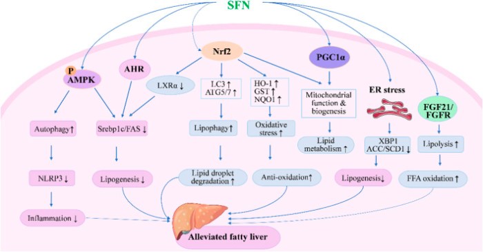
“A growing number of studies have reported that sulforaphane (SFN) could significantly ameliorate hepatic steatosis and prevent development of fatty liver, improve insulin sensitivity, attenuate oxidative damage and liver injury, induce apoptosis, and inhibit proliferation of hepatoma cells through multiple signaling pathways.

SFN inhibits lipogenesis and oxidative stress while enhancing lipid droplet degradation through modulating expression of genes involved in lipid synthesis, metabolism, and oxidation. SFN modulates autophagy, lipolysis, mitochondrial function, and ER stress to alleviate fatty liver through AMPK-, AHR-, PGC1α-, and FGF21-mediated pathways.

fphar-14-1256029-g001

There is still a gap between basic research and clinical application of SFN. More efficient delivery systems and precise dose schedules of SFN are expected to be developed in future studies, which would improve its solubility, stability, and bioavailability, and reduce inter-individual variations in humans.”

https://www.frontiersin.org/articles/10.3389/fphar.2023.1256029/full “Therapeutic potential of sulforaphane in liver diseases: a review”


These reviewers did alright gathering papers. That’s only part of what needed to be done, with the other part being reading, understanding, and interpreting these papers.

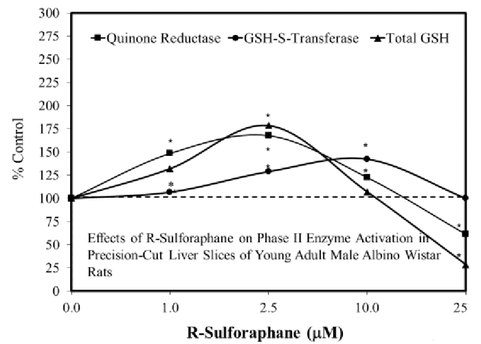
First example: Sulforaphane in the Goldilocks zone was cited [reference 12], but applicability to this review with its main point “The stimulatory zone for in vitro studies proved to be consistently in the 1-10 μM range” as in Figure 10 “Effects of R-sulforaphane on phase II enzyme activation in precision-cut liver slices of young adult male Albino Wistar rats” wasn’t understood:

figure 10

These reviewers complained:

“Few dose-response studies on SFN have been reported, and the range of its effective doses is unclear. Doses used in most animal studies have exceeded the highest dose of SFN used in humans.”

So it might have taken a little bit more effort, but these reviewers could have highlighted studies where sulforaphane liver treatments were in the 1-10 μM potentially therapeutic range.

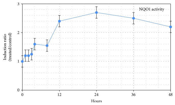
Another example: these reviewers said “The half-life of SFN is very short due to its rapid metabolism in the human body.” They missed a point that the second paper in How much sulforaphane is suitable for healthy people? [reference 46] made in section 6.4. “NQO1 Pharmacokinetics following SFN Ingestion:”

“Maximal induction of NQO1 occurred at around 24 hours, declining thereafter (Figure 8). This peak represents an approximate 2.8-fold induction over baseline.

These findings are useful when considering the effect of SFN as an intervention material in acute compared with chronic conditions. A significant increase in NQO1 occurred between 6 and 12 hours, a timeframe that may not be sufficiently responsive for management of an acute state, leaving one to conclude that NQO1 induction is best suited to chronic conditions where a rapid response may not be necessary.”

OMCL2019-2716870.008

Sulforaphane’s effects of inducing NQO1 for its cytoprotective, antioxidant, and other functions lasts for days, regardless of when sulforaphane leaves the bloodstream.

Oat sprout stressors

Two 2023 Avena sativa oat sprout studies, starting with one that found different effects during germination from varying temperature and relative humidity:

“This study evaluated effects of temperature (20, 25, and 30°C) and relative humidity (RH, 55, 60, and 65%) as abiotic stressors during oat germination. We identified eighty polyphenols, nine avenanthramides, twelve lignans, and five phytosterols.

  • 100% germination was achieved at 25°C/60% RH from day 3, yielding the longest radicle size.
  • The highest content of most phenolic acids, avenanthramides, and lignans occurred at 30°C/65% RH, where 100% germination was attained by day 5, but with a shorter radicle size.
  • The best flavonoid and phytosterol profile was obtained at 20°C/55% RH, achieving only a 67% germination rate by day 5.

sprouted oat temp rh

By considering germination conditions, end-users can harness the versatility of oat sprouting to meet their specific needs and maximize potential benefits of this promising cereal crop. For instance, manufacturers of functional foods and beverages could consider using sprouts from conditions that yield high polyphenol content for products targeting antioxidant benefits, whereas nutraceutical manufacturers could focus on sprouting conditions that result in elevated levels of avenanthramides, well-known for their health-promoting properties.”

https://www.sciencedirect.com/science/article/abs/pii/S0308814623027917Impact of temperature and humidity conditions as abiotic stressors on the phytochemical fingerprint of oat (Avena sativa L.) sprouts” (not freely available) Thanks to Dr. Iza F. Pérez-Ramírez for providing a copy.


Another study compared and contrasted eight sprouted grains to their ungerminated grains and to each other. I’ll highlight oat sprout results:

“The method used was germination for up to 72 h at temperatures ranging from 19–23°C. Oat germination rate was 80%.

Linoleic acid (omega-6) was the predominant fatty acid in oat grain powder, followed by similar amounts of oleic and palmitic acids and smaller amounts of stearic and linolenic (omega-3) acids. Since omega-6 content remained unchanged and omega-3 quantity increased slightly in sprouted oats, the omega-6/omega-3 ratio decreased.”

https://www.mdpi.com/2304-8158/12/17/3306 “Effect of Germination on Fatty Acid Composition in Cereal Grains”


My kitchen cupboard’s oat sprouting conditions are closer to this second study’s temperature, where relative humidity wasn’t specified. I doubt that kitchen winter-time relative humidity ever rises to the 55% lower threshold of the first study for more than a few minutes.

At this time of year in Sprouting hulled oats, I got a 97% germination rate over three days with an estimated 21°C (70°F) and a relative humidity closer to 30% than 55%. Couldn’t tell you why the first study’s germination rate with 20°C/55% RH was only 67% at day 5, or why the second study’s germination rate was only 80% at day 3 with 19–23°C.


PXL_20231223_175744134

Suboccipital release

Here’s a 2023 paper on the subject, but let’s first see what it is, from Michigan State University:

https://hal.bim.msu.edu/CMEonLine/Autonomic/Sympathetic/Treatment/SuboccipitalRelease/start.html

and University of Wisconsin:

https://www.fammed.wisc.edu/suboccipital-release-technique/


An instruction paper for osteopaths:

“This technique treats migraines, headaches, and neck pain. It can also be used as an adjunct for treating conditions with autonomic dysfunction.

Before performing a suboccipital release, the patient should have a neurologic and musculoskeletal exam of the neck and upper extremities. Ensure that the patient does not have contraindications such as: Acute cervical fractures; concern for neurovascular compromise; focal neurologic deficit.”

https://www.ncbi.nlm.nih.gov/books/NBK582126/ “Osteopathic Manipulative Treatment: Suboccipital Release”


I read a half-dozen other 2023 papers comparing this technique with other suboccipital techniques. I’m not posting links because these papers didn’t completely specify their technique, what was its history, and what was its safety. It’s extremely easy to traumatize this area of our bodies.

I similarly won’t post links to other 2023 papers that claimed this technique improved other body problems, such as ankle range of motion, hamstring tightness, and lumbar pain. Mechanistic explanations are required, not just “they are connected by one neural system,” hand-waving, and woo.


This search for recent research was disappointing. I’ve performed suboccipital release on myself at least twice a day for years the same way I saw physical therapists do it to their patients while I was in physical therapy 13 years ago. Maybe there’s a video about self-suboccipital release that didn’t involve gadgets, but I couldn’t find it.

The point of this technique is to evoke a relaxation response. It usually relaxes areas from the neck to my feet. Sometimes it works quickly, other times it takes a while.

In any event – you and I are different, and I’m definitely not recommending suboccipital release for you. It isn’t a cure-all for neck pain, sitting most of the day, bad posture, poor ergonomic setup, being stuck in traffic, being glued to your phone, etc.

What you expect may not be what you find

I’m halfway through a 90-day trial of plasmalogens coincident with improving peroxisomal function via resistance exercise and time-restricted eating. I haven’t curated related 2023 papers I’ve read concerning plasmalogens, peroxisomes, sphingolipids, ceramides, and mitochondrial interactions with these, mainly because I haven’t seen human-pertinent aspects similar to Dr. Goodenowe’s efforts.

The 2023 papers I’ve read have more to do with researcher incentives rather than actual human benefits. I’d guess that researchers care about these related subjects to the extent that they want to be the first to publish arcane details about them, like peroxisomes in the parotid salivary gland.

One area I expected to see a difference at the regimen’s beginning was in my peripheral nervous system Schwann cells. Instead, I had taste and smell improvements in my primary olfactory nervous system olfactory ensheathing cells, which are highly similar to Schwann cells. I was also happy to experience an immediate halt to my ulnar nerve elbow pain after what I interpret as ProdromeNeuro effects and perhaps coincident ProdromeGlia effects on items upstream of Schwann cells.

Here are three papers on Schwann cells that I haven’t yet seen as applicable to my current regimen, starting with a 2022 review:

“We summarise contributions of neurotransmitter receptors in regulation of morphogenetic events of glial cells, with particular attention paid to the role of acetylcholine receptors in Schwann cell physiology. This redundant and complex integrated regulation system could be explained as a mechanism of preserving glial cell physiology. In case of a single receptor signalling dysfunction, other neurotransmitters can overcome the deficit, preserving functions of glia and health of the nervous system.

Increased knowledge in medicinal chemistry and in bioinformatics accompanied by drug delivery studies might open a fascinating therapeutic perspective for cholinergic mimetics for treatment of several nervous system pathologies, and in reducing neuroinflammation both in the central and peripheral nervous systems.”

https://www.mdpi.com/2227-9059/11/1/41 “Emerging Roles of Cholinergic Receptors in Schwann Cell Development and Plasticity”


A 2023 study investigated the vagus nerve’s Schwann cells’ impact with gut function:

“The vagus nerve is the longest extrinsic cranial nerve in the body. It regulates gut physiology through the intrinsic nervous system (myenteric and submucosal plexus) and enteric glial cells interactions, which participate in controlling intestinal absorption, secretion, immune homeostasis, and motility.

Normal intestinal motility is critical for nutrition assimilation and several biological functions. The loss of normal gut function aggravates inflammation, oxidative stress, and other cellular stressors.”

https://bmcbiotechnol.biomedcentral.com/articles/10.1186/s12896-023-00781-x “A critical role for erythropoietin on vagus nerve Schwann cells in intestinal motility”


I haven’t curated a Buck Institute for Research on Aging sponsored study for a while, since their 2015 A study of how “age” itself wasn’t a causal factor for wound-healing differences detracted from science and their 2020 Linear thinking about biological age clocks wasted resources.

This 2023 rodent study couldn’t investigate anything outside of Buck’s limited paradigm’s echo chamber. This sponsor would rather break their arms patting themselves on their backs pretending they’re advancing science than fund relevant human research successes that do advance science:

“Following peripheral nerve injury, successful axonal growth and functional recovery require Schwann cell (SC) reprogramming into a reparative phenotype. This work provides the first characterization of senescent SCs and their influence on axonal regeneration in aging and chronic denervation.”

https://www.embopress.org/doi/full/10.15252/emmm.202317907 “Senescent Schwann cells induced by aging and chronic denervation impair axonal regeneration following peripheral nerve injury”


PXL_20231207_185309349

An ulnar nerve anecdote

Two 2023 papers demonstrated the weak-sauce treatments currently offered to resolve elbow ulnar nerve pain:

“This case report investigated the use of ultrasound-guided nerve hydrodissection and platelet releasate injection for treating ulnar neuritis at the elbow.

  • The patient’s symptoms were first managed with home exercise and ulnar nerve hydrodissection at the elbow, which decreased but did not resolve her pain.
  • Platelet releasate injection of the ulnar nerve at the elbow was subsequently performed. Six weeks post-procedure, the patient reported additional pain improvement.

Despite these results, the patient was not completely symptom-free. Persistent symptoms were attributed to her concomitant neurogenic thoracic outlet syndrome.”

https://www.cureus.com/articles/133241-platelet-releasate-injection-as-a-novel-treatment-for-ulnar-neuritis-at-the-elbow-a-case-report/ “Platelet Releasate Injection as a Novel Treatment for Ulnar Neuritis at the Elbow: A Case Report”

When a diagnosis concludes with the word ‘syndrome’, we can be assured that medical professionals don’t know any specific cause. Expect physical therapy and/or drugs and/or surgery to be recommended, which will only address symptoms, not causes.

These practitioners proposed two experimental treatments, and somehow, the patient agreed to be a lab rat for both. If they were repeatedly questioned as to whether those two treatments would address causes, I’d expect responses similar to “That’s all we can do for you.”

In line with this decade’s revelations about the medical profession, the patient was also gaslighted. These practitioners asserted “changes to the patient’s lifestyle” as a reason neither treatment worked, although no such lifestyle changes were indicated.

Medical professionals are people whose early life experiences impel them to control other people with a license, among other driving factors. They won’t discuss items outside their ideas and beliefs, because these are defenses against their and their patients’ realities.


Next is a study of 111 elbow neuropathy patients (average age 55, median follow-up period of 880 days), one third of whom had various surgeries:

“There are three main potential mechanisms of recovery after nerve lesion: (1) resolution of conduction block, (2) collateral reinnervation, and (3) nerve regeneration.

  • Nerve function in chronic focal compression/entrapment neuropathies seems to improve mainly due to resolution of the conduction block and collateral reinnervation.
  • Contribution of nerve regeneration seems to be minor.

The majority of axons lost in chronic focal neuropathies probably never recover. Further studies using quantitative methods are needed to validate present findings.”

https://www.mdpi.com/2077-0383/12/12/3906 “No Major Nerve Regeneration Seems to Occur during Recovery of Ulnar Neuropathy at the Elbow”


Another interesting thing may have unexpectedly started with my 90-day trial of Prodrome Glia and Neuro products. Here’s an abbreviated look at what I’m tracking that omits intermittent fasting data:

day 7-25

Left ulnar nerve pain stayed the same or decreased two hours after a ProdromeGlia loading dose from Days 11-21. After adding a ProdromeNeuro loading dose at Day 22, my left ulnar nerve pain has unexpectedly stopped.

Any resistance exercise I’ve done during the past month would have aggravated my left ulnar nerve prior to the current regimen. Yesterday I clumped together reverse curls, regular bicep curls, bench presses, and triceps extensions, in that order, two sets each. I used lower weights than in the past, squeezed at the top of concentric motion, and returned slowly with eccentric motion for each rep.

Today on Day 25 the exercised muscles burn as expected, especially due to eccentric motion. But my left ulnar nerve is fine.

At the beginning, I thought that ProdromeGlia might eventually have an effect on left ulnar nerve pain, but not ProdromeNeuro. The first paper noted “The ulnar nerve begins in the axilla as a continuation of the medial cord of the brachial plexus, originally arising from the C8 and T1 nerve roots of the spinal cord.” I’ll guess that something upstream of my left ulnar nerve may also be involved in recent results.

Don’t agree with the second paper’s unevidenced assertion that “The majority of axons lost in chronic focal neuropathies probably never recover.” I’ve had intermittent left ulnar nerve numbness and pain for over five years, which is a lot longer than the 880-day median follow-up period of that paper.

Dr. Goodenowe presented his combined daily plasmalogen precursor dose as ~100 mg/kg. My analogous combined daily plasmalogen precursor loading doses are 7200 mg, appropriate for a person who weighs 72 kg. I weigh 155 lbs. / 70 kg.

More testing is warranted, of course. Maybe I’m just in-between an intermittent occurrence of left ulnar pain. So far, the way my current regimen is playing out, every day has something to make it Thanksgiving Day.

A smell and taste anecdote

Two 2023 papers, starting with a study of smell and taste disorders:

“This study investigates the impact of etiology on the epidemiologic profile, disease severity, type of treatment, and therapy outcome in smell and taste disorders.

Hyposmia has a prevalence of about 15%, while approximately 5% of the population suffers from anosmia. Multiple innervation of the taste mucosa with fibers from the seventh, ninth, and tenth cranial nerves assures robustness of the gustatory system compared to smell.

Conservative therapy employs corticosteroids, antibiotics, vitamins and and minerals as well as functional rehabilitation by olfactory training. Data regarding outcome of therapy were only available for 71 (26.3%) of patients. Only the sinunasal etiology was significantly more likely to show improvement after therapy (27.4% show improvement vs. 9.6% show no improvement).”

https://link.springer.com/article/10.1007/s00405-023-07967-1 “Characteristics of smell and taste disorders depending on etiology: a retrospective study”

This study was a little light on describing effective treatments for smell and taste problems. For example, olfactory training was said to have good therapeutic response. Looking it up, though, it seems to be whatever each practitioner feels like doing.


A review introduced the subject of olfactory ensheathing cells:

“Olfactory ensheathing cells (OECs) are glial cells of the primary olfactory nervous system, which are composed of the olfactory nerve and outer nerve fiber layer of the olfactory bulb. The primary olfactory nervous system is unique in that it can constantly regenerate.

It is now possible to remove olfactory bulb tissue and olfactory mucosa (outermost layer and lamina propria, which belong to the central nervous system and peripheral nervous system, respectively), which also suggests the potential value of OECs therapy in central nervous system and peripheral nervous system diseases. OECs can survive and renew in the central nervous system, and have been widely used in nerve regeneration and tissue repair.

Schwann cells (SCs) form the myelin sheath of the peripheral nerve, protect and nourish neurons, and play an irreplaceable role in the repair of peripheral nerve injury. There is no transcriptional difference between OECs and SCs. OECs are highly similar to SCs, and express the biomarkers of SCs.

fimmu-14-1280186-g002

Functional mechanisms of OECs in the treatment of neurological diseases include neuroprotection, immune regulation, axon regeneration, improvement of nerve injury microenvironment and myelin regeneration, which also includes secreted bioactive factors. Results obtained in clinical trials are not very satisfactory, and the effectiveness of these cell-based therapies remains to be proved.”

https://www.frontiersin.org/articles/10.3389/fimmu.2023.1280186/full “Potential therapeutic effect of olfactory ensheathing cells in neurological diseases: neurodegenerative diseases and peripheral nerve injuries”


Something interesting may have unexpectedly started with my 90-day trial of Prodrome Glia and Neuro products. Here’s an abbreviated look that omits my intermittent fasting and resistance exercise data:

day 7-15

Both product labels have a loading dose suggestion of 4-8 softgels (2 to 4 times the standard two-softgel dose) for 1-3 months. Two days after I started a Glia loading dose, my sense of smell, then sense of taste, were noticeably better.

I’ll guess that my primary olfactory nervous system glial cells are responding to these changes. At the beginning I thought that my peripheral nervous system Schwann cells might be affected regarding my left ulnar nerve. Since olfactory ensheathing cells are highly similar to Schwann cells, it doesn’t seem to be that much of a stretch to think that they could also be affected by my current regimen.

More testing is warranted, of course. I’ve had diminished smell and taste for decades, though. If the gardenias, roses, magnolias, honeysuckles, and other scents in past summers that had fainter scents than I remembered come across stronger, so much the better.

IMG_20200425_154336