Ancient DNA fragments enable adult neurogenesis

A 2025 rodent study investigated mechanisms by which erythropoietin (EPO) enables adult neurogenesis and cognitive function:

“We mapped epigenomic and transcriptional landscapes of adult mouse hippocampus under recombinant human EPO (rhEPO) treatment. We discovered significant lineage-specific remodelling of chromatin accessibility predominantly in newly formed pyramidal neurons, highlighting a robust EPO-driven neurogenic response. Notably, transposable elements (TEs), particularly ancient LINEs and SINEs, emerged as critical cis-regulatory elements (cCREs).

EPO is known to be upregulated in the brain under hypoxic or injury conditions, and it has been considered a natural neuroprotective agent. We demonstrated that EPO, a traditionally hematopoietic hormone, can profoundly reprogram the adult neural epigenome to drive neurogenesis.

EPO may activate a specific subclass of dormant regulatory elements to drive nearby genes. Such a mechanism would represent a previously unappreciated mode of gene regulation: the de novo recruitment of ancient genomic elements to drive a contemporary cellular response.

Our data support the model that EPO drives differentiation of progenitors rather than inducing widespread cell division. The net effect is an enrichment of pyramidal neurons at the cost of interneurons. Pyramidal neurons integrate in the hippocampal circuitry, leading to potential implications for mood, memory, cognitive enhancement, and recovery from brain injury.

We propose a conserved evolutionary mechanism at play: ancient TEs embedded in the genome have been repurposed as cCREs in neural cells, and during an EPO-induced neurogenic stimulus, the brain taps into this reservoir of regulatory elements to rapidly reshape gene expression. In evolutionary terms, this represents an efficient strategy.”

https://www.biorxiv.org/content/10.1101/2025.10.13.682070v1.full “Transposable Element-Mediated Epigenomic Remodeling Drives Erythropoietin-Induced Neurogenesis in the Adult Hippocampus”


Activating Nrf2 pathways with sunlight

A 2025 review subject was non-electrophilic Nrf2 activators:

“NRF2 can be induced via:

  1. Non-specific electrophile/ROS generation,
  2. Disruption of the NRF2–KEAP1 protein–protein interaction,
  3. Autophagy-mediated KEAP1 degradation,
  4. Direct modulation of NRF2 protein stability, and
  5. Post-transcriptional/post-translational modifications.

Except for a single intervention, therapeutic hypothermia, every non-pharmacological strategy with defined mechanisms employs more than one of these routes, most frequently pairing post-translational modification with either protein-stability regulation or limited electrophile production. This combinatorial activation elevates both NRF2 abundance and transcriptional competence while minimizing the liabilities of purely electrophilic agents and circumventing the efficacy limitations.

Classical electrophilic NRF2 activators, despite potent activation potential, exhibit paradoxically reduced therapeutic efficacy relative to single antioxidants, attributable to concurrent oxidative stress generation, glutathione depletion, mitochondrial impairment, and systemic toxicity. Although emerging non-electrophilic pharmacological activators offer therapeutic potential, their utility remains limited by bioavailability and suboptimal potency.”

https://www.mdpi.com/2076-3921/14/9/1047 “Non-Electrophilic Activation of NRF2 in Neurological Disorders: Therapeutic Promise of Non-Pharmacological Strategies”


These researchers exaggerated problems of electrophilic Nrf2 activators such as “mitochondrial impairment, and systemic toxicity” so they could have something to write about. Just like every intervention, the dose determines the response. I can’t imagine not eating broccoli sprouts in favor of brain zapping with electroconvulsive therapy or transcranial magnetic stimulation just to avoid sulforaphane’s temporary mild oxidative stress that activates Nrf2 for 15-20 minutes.

But there are limitations to how an unwell person can benefit from Nrf2 activation. For example, I haven’t curated many cancer papers because healthy body functioning can’t be assumed.

I walk the beach at sunrise, weather permitting, because it makes me feel good, and I’m always happy afterwards that I made the effort to get outside. That doing so combines two of the above non-electrophilic Nrf2 activators, physical exercise and photobiomodulation, hasn’t been a consideration.

These reviewers didn’t include human studies of sunlight’s effects. Nevermind that hospitals used to have sundecks for patients, and John Ott published relevant human and animal studies over fifty years ago.


Many studies have an undisclosed limitation in that they were performed without controlling for light. For example, knowing that mitochondria are light-activated, I don’t trust those studies’ in vivo, ex vivo, or in vitro results.

None of the 100 most recent 2025 photobiomodulation papers examined natural sunlight. Maybe it wouldn’t sell red light, green light, and blue light lasers and other products to show that people could produce the same effects themselves with sunlight at different times of the day? Would researchers damage their reputations to study a freely-available intervention, one where they don’t “do something”?

Sulforaphane and skin aging

A 2025 rodent study investigated sulforaphane effects on natural skin aging:

“Aging is a multifactorial process that progressively impairs skin integrity by diminishing dermal fibroblast function, which is macroscopically manifested by wrinkling, laxity, and pigmentary abnormalities. The potential mechanism by which sulforaphane (SFN) delays intrinsic skin aging was explored through skin proteomic sequencing and immune cell infiltration analysis. Associations between SFN administration and phenotypic changes in skin aging, immune cell populations, and key signaling pathway targets were further examined.

WBC count results indicated that mice from the Aged group were significantly immunosuppressed. T cells occupied the main lymphocyte lineages.

The present study illuminated the skin protective mechanism of SFN by network pharmacology and proteomics analyses in a natural aging mouse model. SFN therapy showed significant alterations in skin structure, redox balance, and composition of immune cell populations after an intervention duration of 2 months.”

https://onlinelibrary.wiley.com/doi/10.1002/mnfr.70281 “Integrative Network Pharmacology and Proteomics Decipher the Immunomodulatory Mechanism of Sulforaphane Against Intrinsic Skin Aging” (not freely available)


I rated this study Wasted resources for using sulforaphane doses not relevant to humans. I usually don’t curate such studies. Its lowest sulforaphane 50 mg/day dose is a ((50 mg * .081) * 70 kg) = 284 mg human equivalent.

I decided to curate it for its informative young controls vs. aged controls results in the above graphic. WBC counts are available on almost every standard human blood test.

This study’s young and aged groups per Grok: “A 2-month-old mouse aging for two more months (reaching 4 months) is approximately equivalent to a human aging from about 12–15 years to 17–21 years old. An 18-month-old mouse (human equivalent: ~45–50 years) aging for two months would be roughly equivalent to a human aging from ~45–50 years to ~51–58 years.”

Glucosinolate and isothiocyanate human interventions

A 2025 review covered human evidence from glucosinolate and isothiocyanate research through April 2025:

“Glucosinolates (GSLs) and their breakdown products, isothiocyanates (ITCs), are biogenesis compounds with anti-inflammatory, antioxidant, and anticancer properties, mediated through key pathways such as Nrf2, NF‐κB, and epigenetic regulation. However, their limited and variable bioavailability remains a key challenge. This review summarises the current clinical evidence on GSLs and ITCs, with a focus on their health effects and metabolic fate in humans.”

https://www.mdpi.com/2304-8158/14/16/2876 “Bioavailability, Human Metabolism, and Dietary Interventions of Glucosinolates and Isothiocyanates: Critical Insights and Future Perspectives”


In the above graphic, notice how the inactive myrosinase column has no small intestine participation, but the active myrosinase column does. This point wasn’t adequately emphasized, that for complete effects, an individual has to do whatever they can to thoroughly chew or otherwise activate myrosinase to hydrolyze glucosinolates before swallowing.

Researchers don’t rely on individuals taking responsibility for their own health, of course. Just swallow these pills, we’ll do it for you, as if humans are lab rats. This lack of emphasis is understandable, if not optimal.

This review provided longish coverage of studies, which is preferable to the usual treatment of citing a reference without much explanation. Compare, for example, my longish curation of the 2023 Eat broccoli sprouts for your high intensity interval training with its reference 68 summary below:

“Another study investigated the effects of consuming GSL-rich broccoli sprout (GRS) supplements on oxidative stress and physiological adaptations to intense exercise training. In a randomised, double-blind, crossover design, nine healthy participants consumed either a GRS supplement (75 g of sprouts) or a placebo twice daily over a 7-day high-intensity interval training period. The findings revealed that GRS supplementation significantly reduced markers of oxidative stress, including carbonylated proteins in skeletal muscle and plasma myeloperoxidase levels, compared to the placebo condition. Furthermore, GRS intake led to reduced lactate accumulation during submaximal exercise and enhanced exercise performance, as indicated by a longer time to exhaustion during maximal exercise tests. At the molecular level, supplementation with GRS was associated with elevated Nrf2 protein levels in muscle tissue, suggesting activation of endogenous antioxidant defence mechanisms. In addition, GRS intake mitigated nocturnal hypoglycaemic episodes and lowered average blood glucose levels, indicating improved glucose regulation during intense training. Collectively, these results suggest that GRS supplementation may enhance physiological adaptations to high-intensity exercise by reducing oxidative stress and supporting metabolic homeostasis.”


Get a little stress into your life, Part 2

A 2025 reply to a letter to the editor cited 56 references to elaborate on Part 1 and related topics:

“A positive effect does not necessarily mean benefit, and positive effects on individual organisms may mean adverse effects on other coexisting organisms. However, a vast literature shows that hormetic stimulation can result in benefits depending on the context, for instance, clear growth, yield, and survival improvement.

There is some energetic cost to support hormetic stimulation, with a likely positive energy budget, which might also have negative consequences if there is insufficient energy substrate, especially under concurrent severe environmental challenges. Moreover, hormetic preconditioning could be particularly costly when there is a mismatch between the predicted environment and the actual environment the same individuals or their offspring might face in the future.

Hormesis should not be unilaterally linked to positive and beneficial effects without considering dose levels. For any research to answer the question of whether a stimulation represents hormesis and whether it is beneficial, robust dose–response evaluations are needed, which should be designed a priori for this purpose, meeting the requirements of the proper number, increment, and range of doses.

Both additivity and synergism are possible in the hormetic stimulatory zone, depending also on the duration of exposure and the relative ratio of different components. This might happen, for example, when a chemical primes stress pathways (e.g., heat shock proteins and antioxidants), thus enabling another chemical to trigger hormesis (defense cross-activation) and/or because combined low subtoxicity may modulate receptors (e.g., aryl hydrocarbon receptor and nuclear factor erythroid 2-related factor 2) differently than individual exposures (receptor binding synergy).

Moreover, even when stimulation occurs in the presence of individual components, stimulation may no longer be present when combined, and therefore, effects of mixtures cannot be accurately predicted based on the effects of individual components. There may be hormesis trade-offs; hormesis should be judged based on fitness-critical end points.

While often modeled mathematically, hormesis is fundamentally a dynamic biological process and should not be seen as a purely mathematical function, certainly not a linear one. Much remains to be learned about the role of hormesis in global environmental change, and an open mind is needed to not miss the forest for the trees.”

https://pubs.acs.org/doi/10.1021/acs.est.5c05892 “Correspondence on ‘Hormesis as a Hidden Hand in Global Environmental Change?’ A Reply”


Reference 38 was a 2024 paper cited for:

“Hormetic-based interventions, particularly priming (or preconditioning), do not weaken organisms but strengthen them, enhancing their performance and health under different environmental challenges, which are often more massive than the priming exposure.

The catabolic aspect of hormesis is primarily protective whereas the anabolic aspect promotes growth, and their integration could optimize performance and health. The concept of preconditioning has also gained widespread attention in biomedical sciences.”

https://www.sciencedirect.com/science/article/abs/pii/S1568163724004069 “The catabolic – anabolic cycling hormesis model of health and resilience” (not freely available)


Reference 40 was a 2021 review that characterized hormesis as a hallmark of health:

“Health is usually defined as the absence of pathology. Here, we endeavor to define health as a compendium of organizational and dynamic features that maintain physiology.

Biological causes or hallmarks of health include features of:

  • Spatial compartmentalization (integrity of barriers and containment of local perturbations),
  • Maintenance of homeostasis over time (recycling and turnover, integration of circuitries, and rhythmic oscillations), and
  • An array of adequate responses to stress (homeostatic resilience, hormetic regulation, and repair and regeneration).

Disruption of any of these interlocked features is broadly pathogenic, causing an acute or progressive derailment of the system.

A future ‘medicine of health’ might detect perilous trajectories to intercept them by targeted interventions well before the traditional ‘medicine of disease’ comes into action.”

https://www.sciencedirect.com/science/article/pii/S0092867420316068 “Hallmarks of Health”


Treating a stomach infection with sulforaphane

A 2025 rodent study integrated metabolomics and lipidomics analyses to investigate how sulforaphane treats a Helicobacter pylori infection:

“Helicobacter pylori (H. pylori) is a microaerobic Gram-negative bacterium that colonizes the gastric mucosa. Approximately half of the global population is infected with this bacterium, and it is classified as a group 1 carcinogen.

However, H. pylori infection does not typically present with obvious symptoms in the early stages, making it difficult to detect. Daily dietary interventions may be a relatively effective method for its prevention and treatment.

This study established an H. pylori-infected mouse model, to which sulforaphane was orally administered. H. pylori-low-dose and H. pylori-high-dose represent 4 weeks of gavage with 5 mg/kg/d and 20 mg/kg/d of sulforaphane after H. pylori colonization.

Metabolomics and lipidomics analysis of the effects of sulforaphane treatment on mouse serum. Stacked bar chart of the metabolites regulated by (A) low-dose and (B) high-dose sulforaphane treatment compared to the differential metabolites between the control group and H. pylori group.

Results showed that H. pylori infection significantly altered host amino acid and lipid levels, specifically manifested as abnormal serum glycerophospholipids and metabolic imbalances of amino acids, bile acids, glycerophospholipids, ceramides, and peptides in the liver. Sulforaphane treatment reversed these metabolic abnormalities, with high-dose sulforaphane exhibiting more prominent regulatory effects.

High-dose sulforaphane effectively restored hepatic metabolic disorders of amino acids, bile acids, and lipids, and ameliorated abnormal serum glycerophospholipid profiles. Regulation of key pathways such as glycine metabolism and glutathione metabolism constitutes an important basis for sulforaphane’s anti-H. pylori infection effects.

This study provides a comprehensive metabolic basis for understanding the role of sulforaphane as a dietary intervention in preventing and managing H. pylori-associated gastric diseases and lays a foundation for subsequent clinical translational research.”

https://www.mdpi.com/1422-0067/26/16/7791 “Therapeutic Effects of Sulforaphane on Helicobacter pylori-Infected Mice: Insights from High-Coverage Metabolomics and Lipidomics Analyses of Serum and Liver”


A human equivalent to this study’s low sulforaphane dose is (5 mg x .081) x 70 kg = 28 mg, which is achievable by eating broccoli sprouts every day. Quadrupling 28 mg to a human equivalent of the study’s high sulforaphane dose would involve additional supplementation.

Another way to support this study’s glycine metabolism findings without high-dose sulforaphane is to supplement betaine (trimethylglycine) so that the body requires less choline-to-glycine synthesis. A synergistic effect can be achieved with taurine supplementation that enhances cysteine availability for the tripeptide (glutamate, cysteine, and glycine) glutathione synthesis by requiring less cysteine-to-taurine synthesis.

Inulin vs. FOS

A 2025 clinical trial compared inulin glycemic effects with FOS effects. I won’t curate its gut microbiota results as these have unresolved measurement problems:

“In this study, we conducted a randomized, double-blind investigation to examine the impact of inulin and fructooligosaccharides (FOS) on glycemic metabolism in overweight/obese and healthy adults.

Inulin and FOS are both fructans composed of fructose units, but they differ in their degree of polymerization (DP) and chain length, which lead to differences in their physicochemical properties and physiological effects. Inulin typically has a longer chain length, with a DP ≥ 10, resulting in lower solubility and slower fermentation in the distal colon. FOS consists of shorter chains, with a DP 2 to 9, presenting higher solubility and undergoing rapid fermentation in the proximal colon. These differences affect their impact on short-chain fatty acid (SCFA) production, gut microbiota modulation, and subsequently results in different effects on host metabolism.

131 participants were recruited and randomized into three groups: inulin (N = 44), FOS (N = 43), and control (N = 44). Each group was conducted with a daily supplement of 15 g FOS, inulin, and maltodextrin as placebo and lasted for 4 weeks. Dosage was determined based on our previous clinical trials in the healthy young population, which reported using 16 g/day has no risk of adverse effects. Subjects were still recommended to take a half dose in the first 2 days to promote adaptation to the prebiotics and minimize gastrointestinal symptoms. Products were suggested to add to drinks such as coffee, tea, or milk.

Inulin significantly reduced glucose levels at 1 h and 2 h during oral glucose tolerance test (OGTT), increased fasting insulin, and lowered homocysteine (HCY) levels in overweight/obese individuals. These effects were not observed in healthy individuals.

In contrast, although FOS significantly decreased HCY, it did not improve glycemic metrics in either group.”

https://bmcmedicine.biomedcentral.com/articles/10.1186/s12916-025-04189-6 “Differential effects of inulin and fructooligosaccharides on gut microbiota composition and glycemic metabolism in overweight/obese and healthy individuals: a randomized, double-blind clinical trial”


Prior research found:

“A 2010 gastrointestinal tolerance of chicory inulin products study indicated that 10 g/day of native inulin were well-tolerated in healthy, young adults. Over this dose would induce mild gastrointestinal symptoms.”

So a lead-in half-dose probably wouldn’t be needed for people to start a 10 gram inulin dose.

Activate Nrf2 with far-infrared light

A 2025 rodent study investigated effects of far-infrared light on Alzheimer’s disease models. I’ll focus on its Nrf2 findings:

“Far-infrared radiation (FIR) is commonly utilized as a complementary treatment of a range of disease, for example, insomnia and rheumatoid arthritis. In this research, we explored how FIR light impacts cognitive functions of TgCRND8 AD mice and elucidated its underlying molecular mechanism.

Infrared radiation is a form of electromagnetic energy that has wavelengths between 750 nm and 1000 μm, which are longer than visible light. International Commission on Illumination categorizes infrared light as three sub-divisions according to the wavelength: (1) near-infrared radiation (0.7–1.4 μm), (2) middle infrared radiation (1.4–3.0 μm), and (3) far-infrared radiation (3.0–1000 μm).

Nrf-2/ HO-1 signaling, a key endogenous antioxidant system, helps mitigate oxidative stress and enhances expression of various endogenous genes. Activation of HO-1 during inflammatory conditions may serve as an adaptive response to reduce cytotoxicity through various mechanisms.

In this study, we applied EFFIT LITE® as the FIR spectrum transmitter which stably radiates an FIR spectrum with a wavelength of 4–20 μm, and the device was put within 1 cm directly above the head of the 3-month-old TgCRND8 mice for 30 min exposure once every day. FIR light notably enhanced cognitive function and spatial memory of TgCRND8 mice after 28-days consecutive treatment.

Underlying molecular mechanisms involve suppression of Aβ deposition, hyperphosphorylation of tau, and neuroinflammation through modulating Jak-2/Stat3 and Nrf-2/HO-1 pathways. Our current experimental findings amply indicate that FIR light is a potential non-pharmacological therapy for AD.”

https://link.springer.com/article/10.1007/s12017-025-08860-2“Far-Infrared Radiation Ameliorates the Cognitive Dysfunction in an Alzheimer’s Disease Transgenic Mouse via Modulating Jak-2/Stat3 and Nrf-2/HO-1 Pathways”


This study measured Nrf2 and its quickly-induced downstream enzyme HO-1 effects of daily far-infrared light exposure for 30 minutes. We’d have to see measurements of Nrf2’s more-slowly induced and longer-lasting downstream xenobiotic detoxifying enzyme NQO1 to compare far-infrared light Nrf2 activation effects with those of natural plant compounds.

Plasmapheresis doesn’t reduce biological age

A 2025 clinical trial investigated effects of plasmapheresis as measured with epigenetic clocks:

“This study aimed to assess whether plasmapheresis without volume replacement with young plasma or albumin affects epigenetic age and other biomarkers in healthy adults. No significant epigenetic rejuvenation was observed based on epigenetic clock measurements. Instead, plasmapheresis was associated with increases in DNAmGrimAge, the Hannum clock, and the Dunedin Pace of Aging.

  1. The relatively small sample size of 34 finishing participants comprising of first-time plasma donors limits the statistical power and generalizability of our findings.
  2. Our cohort was restricted to individuals aged 40 to 60 years in accordance with Czech regulatory guidelines, which, although intentional to focus on an older population where rejuvenating effects might be most apparent, constrains evaluation of age-related differences across a broader demographic.
  3. The 18-week duration of the study, while sufficient to detect rapid alterations in key biomarkers under an intensive plasmapheresis protocol, may not fully capture the long-term implications of these changes.
  4. Due to our trial taking place during spring and summer months, we cannot fully separate the effects of increased sunlight exposure, outdoor physical activity, and dietary changes from the observed rises in Vitamin D and concurrent shifts in DNAm-based aging metrics. We did not collect objective measures of activity or diet, so these factors remain potential confounders.

The protocol of donating plasma every two weeks, although deemed safe by many countries around the world, is not yet well researched and cannot therefore be marked as benefiting to the donor right now. Further refinement to balance clearance of pro-aging factors with maintenance of systemic homeostasis is needed.”

https://www.nature.com/articles/s41598-025-05396-0 “Human clinical trial of plasmapheresis effects on biomarkers of aging (efficacy and safety trial)”


Betaine as an exercise mimetic

A 2025 human study investigated effects of long-term exercise:

“Exercise has well-established health benefits, yet its molecular underpinnings remain incompletely understood. We conducted an integrated multi-omics analysis to compare effects of acute vs. long-term exercise in healthy males.

Acute exercise induced transient responses, whereas repeated exercise triggered adaptive changes, notably reducing cellular senescence and inflammation and enhancing betaine metabolism. Exercise-driven betaine enrichment, partly mediated by renal biosynthesis, exerts geroprotective effects and rescues age-related health decline in mice.

Betaine binds to and inhibits TANK-binding kinase 1 (TBK1), retarding the kinetics of aging.

Betaine effectively alleviated senescence phenotypes by reduced senescence-associated β-galactosidase (SA-β-Gal)-positive cells, decreased p21 expression, lowered DNA damage indicator γ-H2A.X, and elevated heterochromatin mark H3K9me3. Betaine treatment also enhanced cellular antioxidant capacity, as evidenced by increased NRF2 phosphorylation and reduced ROS accumulation.

These findings systematically elucidate the molecular benefits of exercise, and position betaine as an exercise mimetic for healthy aging.”

https://doi.org/10.1016/j.cell.2025.06.001 “Systematic profiling reveals betaine as an exercise mimetic for geroprotection” (not freely available) Thanks to Dr. Weimin Ci for providing a copy.


Eat broccoli sprouts for your HIIT, Part 2

A 2025 human study followed up Eat broccoli sprouts for your high intensity interval training using red kale sprouts:

“Glucosinolate-rich broccoli sprouts combined with intense exercise training for 7 days have been shown to reduce blood lactate concentrations during exercise, attenuate hypoglycemic events, improve physical performance, and reduce markers of oxidative stress. This study aimed to investigate the acute, dose-dependent effects of glucosinolate-rich red kale sprouts (GRS) on blood lactate and blood glucose following the ingestion of three different doses.

Fifteen healthy participants [11 females, 4 males] consumed 37.5 g or 75 g of GRS or an isocaloric placebo blended into a beverage on three separate occasions. The participants cycled on an ergometer at three submaximal work rates before and three hours after ingestion.

Intake of glucosinolate-rich sprouts acutely decreased blood lactate levels during submaximal cycling and increased blood glucose levels at rest. The largest reduction in blood lactate was observed at the 37.5 g dose compared to placebo.

To identify the dose of GRS that results in the lowest blood lactate concentration during submaximal exercise, we applied a quadratic modeling approach. The optimal dose for minimizing lactate accumulation was calculated as 44 g of GRS.

In our previous study, we found a tendency towards a lower respiratory exchange ratio after one week of supplementation. Moreover, studies have demonstrated that mitochondrial oxidation of long-chain and short-chain fatty acids is depressed in the absence of Nrf2, and accelerated when Nrf2 is constitutively active.

We observed a reduction in myeloperoxidase levels approximately three hours after GRS intake, suggesting a decrease in oxidative stress. This finding indicates that the adaptive compensatory system may act rapidly, likely within just a few hours of GRS consumption.

A limitation is that we did not assess whether the lower lactate levels translated into improved performance. Theoretically, if the reduction in lactate results from the activation of pyruvate dehydrogenase, it could enhance performance by channeling more pyruvate into mitochondria for efficient oxidation, reducing reliance on glycolysis, and thereby sparing muscle glycogen. Alternatively, if the lower lactate levels are due to increased activity of the hepatic Cori cycle, lactate could be more rapidly converted to glucose, possibly supporting glycogen resynthesis or maintaining blood glucose levels during exercise. Both mechanisms could potentially contribute to improved performance.

Acute intake of small doses of GRS followed by submaximal ergometer cycling results in changes in lactate and glucose metabolism that could be beneficial for exercise performance.”

https://www.biorxiv.org/content/10.1101/2025.04.15.648889v1.full “A glucosinolate-rich beverage lowers blood lactate concentrations during submaximal exercise”


These researchers chose red kale sprouts of undisclosed age over the predecessor study’s broccoli raab five-day old sprouts, and two other undisclosed cruciferous vegetable sprouts.

This study is in its preprint phase. Items that could be clarified before publishing in final form include:

  • In the Abstract section, reference findings to red kale sprouts rather than broccoli sprouts;
  • Characterize the lactate U-shaped dose-response curve as hormesis; and
  • Reference other hormesis findings for context.

Sulforaphane as a senotherapy

A 2025 rodent study investigated cardioprotective effects of dasatinib / quercetin (a senolytic combination) and sulforaphane (senomorphic):

“Senolytics are molecules that selectively eliminate senescent cells (SCs). Senomorphics are drugs that suppress or mitigate the pro-inflammatory effects of the senescence-associated secretory phenotype (SASP) without killing SCs. Senomorphics decrease the number of SCs by diminishing the paracrine effect of senescence induction in neighboring cells, and by enhancing elimination of SCs by the immune system, which reduces their harmful effects.

We used middle-aged female rats fed a hypercaloric diet (HD) from 21 days to 15 months of age. Under our experimental conditions, rats exhibited cardiac hypertrophy and fibrosis, accumulation of senescent cells, changes in mitochondrial morphology, and oxidative stress. Rats were treated for 2 months with senolytic (dasatinib + quercetin, DQ) or senomorphic (sulforaphane, SFN) agents.

A novel aspect of recent research has been the crosstalk between organelles, particularly between the mitochondria and endoplasmic reticulum (ER), through specialized contact sites (MERCSs). However, there is still no consensus on the optimal distance between MERCSs and their positive or negative effects on disease progression.

HD rats showed cardiac improvement after both treatments. Although both strategies improved cardiomyocyte size and cardiac fibrosis, only DQ decreased LDH levels, whereas SFN positively affected cardiac damage proteins.

In general, no changes in structure or damage-associated enzymes were observed in control rats treated with DQ or SFN, indicating that senotherapies do not promote adverse effects on the heart, reinforcing the concept that they are safe for application in the clinical field. Data suggest a possible link mechanism between Nrf2 activation and MERCSs preservation, activated by SFN rather than by the DQ combination, which allowed cardiac structure maintenance in HD rats decreasing harmful effects of senescent cells.”

https://www.sciencedirect.com/science/article/abs/pii/S0955286325000865 “Cardioprotective effect of senotherapy in chronically obese middle-aged female rats may be mediated by a MERCSs/Nrf2 interaction” (not freely available) Thanks to Dr. Alejandro Silva for providing a copy.


Eat broccoli sprouts for your eyes, Part 3

Two 2025 papers cited Precondition your defenses with broccoli sprouts, starting with a review of age-related macular degeneration:

“AMD progression from intermediate to late AMD leads to a point of irreversible retinal pigmented epithelium (RPE) degeneration where treatment becomes worthless. Treating patients at the early/intermediate stages presents a better therapeutic window opportunity for AMD as the disease could potentially be prevented or slowed down.

Strong evidence points to RPE dysfunction at these stages, mainly through redox imbalance and lysosomal dysfunction in RPE oxidative injury. Restoring oxidative balance and lysosomal function may act as preventive and therapeutic measures against RPE dysfunction and degeneration.

Due to interaction with KEAP1, NRF2 is a ubiquitously expressed protein with a high turnover and half-life of about 20 minutes. Because the turnover of NRF2 is faster than KEAP1, newly synthesized NRF2 does not have free KEAP1 to bind and is translocated into the nucleus. Once in the nucleus, NRF2 dimerizes with sMAF and the complex binds to antioxidant response element (ARE) sequences, promoting the expression of ARE genes.

There is NRF2 involvement in most of the hallmarks of aging. Key transcriptional regulatory factors of related pathways, such as transcription factor EB (TFEB) and NRF2, may be targeted to restore homeostasis and/or prevent further RPE degeneration.”

https://www.mdpi.com/2076-3921/14/5/596 “Targeting Lysosomal Dysfunction and Oxidative Stress in Age-Related Macular Degeneration”

There were other informative tidbits throughout this review, such as:

  • “Anti-inflammatory effects of most electrophilic NRF2 activators are thought to be at least partly NRF2-independent, suggesting that these compounds lacking specificity may be advantageous for multitargeted pathologies.
  • TFEB can activate NRF2 under conditions devoid of oxidative stress.”

This paper also cited Bridging Nrf2 and autophagy when discussing the above graphic.


In this human cell and rodent study, several coauthors of the original 2020 study tested sulforaphane and TFEB interactions for ameliorating effects of a rare disease:

“Mutations in genes encoding lysosomal proteins could result in more than approximately 70 different lysosomal storage disorders. Niemann–Pick disease type C (NPC) is a rare lysosomal storage disorder caused by mutation in either NPC1 or NPC2 gene. Deficiency in NPC1 or NPC2 protein results in late endosomal/lysosomal accumulation of unesterified cholesterol.

Clinical symptoms of NPC include hepatosplenomegaly, progressive neurodegeneration, and central nervous system dysfunction, that is, seizure, motor impairment, and decline of intellectual function. So far there is no FDA-approved specific therapy for NPC.

Under stress conditions, that is, starvation or oxidative stress, TFEB is dephosphorylated and actively translocates into the nucleus, promoting expression of genes associated with lysosome and autophagy. TFEB overexpression or activation results in increased number of lysosomes, autophagy flux, and exocytosis.

Pharmacological activation of TFEB by sulforaphane (SFN), a previously identified TFEB agonist, significantly promoted cholesterol clearance in human and mouse NPC cells, while genetic inhibition (KO) of TFEB blocked SFN-induced cholesterol clearance. This clearance effect exerted by SFN was associated with upregulated lysosomal exocytosis and biogenesis. SFN treatment has no effect on the liver and spleen enlargement of Npc1 mice.

SFN is reportedly BBB-permeable, assuring a good candidate for efficient delivery to the brain, which is essential for targeting neurodegenerative phenotypes in neurological diseases including NPC. This is the first time that SFN was shown to directly activate TFEB in the brain.

Collectively, our results demonstrated that pharmacological activation of TFEB by a small-molecule agonist can mitigate NPC neuropathological symptoms in vivo. TFEB may be a putative target for NPC treatment, and manipulating lysosomal function via small-molecule TFEB agonists may have broad therapeutic potential for NPC.”

https://elifesciences.org/articles/103137 “Small-molecule activation of TFEB alleviates Niemann–Pick disease type C via promoting lysosomal exocytosis and biogenesis”


Eat broccoli sprouts to alleviate diabetic heart disease

A 2025 rodent study investigated sulforaphane’s effects on diabetic cardiomyopathy:

“The protective effect of cruciferae-derived sulforaphane (SFN) on diabetic cardiomyopathy (DCM) has garnered increasing attention. However, no studies have specifically explored its mechanistic involvement in cardiac substrate metabolism and mitochondrial function.

To address this gap, Type 2 diabetes mellitus (T2DM) db/db mice were orally gavaged with vehicle or 10 mg/kg body weight SFN every other day for 16 weeks, with vehicle-treated wild-type mice as controls. SFN intervention (SFN-I) alleviated hyperglycemia, dyslipidemia, HOMA-IR, serum MDA levels, and liver inflammation. SFN-I improved the lipotoxicity-related phenotype of T2DM cardiomyopathy, manifested as attenuation of diastolic dysfunction, cardiac injury, fibrosis, lipid accumulation and peroxidation, ROS generation, and decreased mitochondrial complex I and II activities and ATP content.

Although not fully understood, multiple systemic and cardiac-local mechanisms contribute to DCM, encompassing hyperglycemia, dyslipidemia, insulin resistance (IR), disturbances in cardiac substrate metabolism, lipotoxicity, glucotoxicity, mitochondrial dysfunction, oxidative stress, inflammation, and endoplasmic reticulum (ER) stress. Nrf2 and its downstream metallothionein also mediated the preventive effect of SFN on DCM, and may underlie the synergistic effect of SFN and zinc in DCM.

These results suggest that chronic oral SFN-I protects against DCM by mitigating overall metabolic dysregulation and inhibiting cardiolipotoxicity. The latter might involve controlling cardiac fatty acid metabolism and improving mitochondrial function, rather than promoting glucose metabolism.”

https://www.mdpi.com/2076-3921/14/5/603 “Oral Sulforaphane Intervention Protects Against Diabetic Cardiomyopathy in db/db Mice: Focus on Cardiac Lipotoxicity and Substrate Metabolism”


This study had numerous charts like the above showing it was better to not have a deviation from health (Ctrl) rather than incur injury (DCM) then try to fix it with sulforaphane (DCM + SFN). But the control group was wild-type mice, not mice genetically inclined to diabetes like the treatment groups.

The subjects’ starting points were at nine-weeks-old (equivalent to 18-25 year-old humans), and duration was 16 weeks. Grok 3 said: “A 25-week-old db/db mouse is roughly equivalent to a human aged 30–35 years chronologically, though its metabolic condition may mimic older human physiological states in diabetes and obesity research.”

A human equivalent of a 10 mg/kg sulforaphane dose is (.081 x 10 mg) = 56 mg orally administered every other day. That’s about how much total sulforaphane I estimated I took every day (52 mg) from Week 6 through Week 56 by eating microwaved broccoli sprouts twice daily.

No rationale was provided for the sulforaphane dose or the every-other-day dosing regimen. Since I took ~52 mg every day for almost a year, I’ll guess that this study may have had more definitive results with daily dosing. Or maybe add zinc per Zinc and broccoli sprouts – a winning combination.

Nrf2 activators and transcriptomic clocks

Two preprint studies looked at making transcriptional aging clocks using Nrf2 activators. Let’s start with a 2025 nematode study that used constant exposure to sulforaphane at different concentrations:

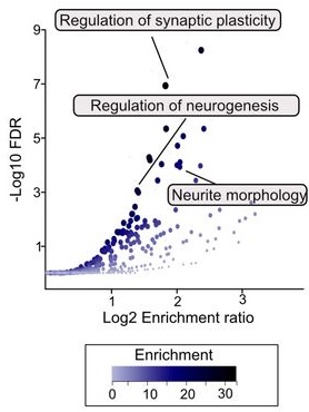
“To explore the potential of sulforaphane as a candidate natural compound for promoting longevity more generally, we tested the dose and age-specific effects of sulforaphane on C. elegans longevity, finding that it can extend lifespan by more than 50% at the most efficacious doses, but that treatment must be initiated early in life to be effective. We then created a novel, gene-specific, transcriptional aging clock, which demonstrated that sulforaphane-treated individuals exhibited a “transcriptional age” that was approximately four days younger than age-matched controls, representing a nearly 20% reduction in biological age.

The clearest transcriptional responses were detoxification pathways, which, together with the shape of the dose-response curve, indicates a likely hormetic response to sulforaphane. The hormetic, stress-pathway inducing properties of sulforaphane may indicate that many beneficial dietary supplements work in a fairly generic fashion as mild toxins rather than being driven by the biochemical properties of the compounds themselves (e.g., as antioxidants).

These results support the idea that robust longevity-extending interventions can act via global effects across the organism, as revealed by systems level changes in gene expression.”

https://www.biorxiv.org/content/10.1101/2025.05.11.653363v1 “The broccoli derivative sulforaphane extends lifespan by slowing the transcriptional aging clock”

There are difficulties in researchers translating nematode studies to mammals and humans. Nematodes lack a homolog to the Keap1 protein, which is sulforaphane’s main mammalian target to activate Nrf2.


A 2024 study developed various mammalian epigenetic clocks:

“A unified transcriptomic model of mortality that encompasses both aging and various models of lifespan-shortening and longevity interventions (i.e., mortality clocks) has been lacking. We conducted an RNA-seq analysis of mice subjected to 20 compound treatments in the Interventions Testing Program (ITP).

We sequenced the transcriptomes of a large cohort of ITP mice subjected to various neutral and longevity interventions, expanded the dataset with publicly available gene expression data representing organs of mice and rats across various strains and lifespan-regulating interventions, connected these models with survival data, and performed a meta-analysis of aggregated 4,539 rodent samples, which allowed us to identify multi-tissue transcriptomic signatures of aging, mortality rate, and maximum lifespan.

Aging and mortality were characterized by upregulation of genes involved in inflammation, complement cascade, apoptosis, and p53 pathway, while oxidative phosphorylation, fatty acid metabolism, and mitochondrial translation were negatively associated with mortality, both before and after adjustment for age.

Utilizing the aggregated dataset, we developed rodent multi-tissue transcriptomic clocks of chronological age, lifespan-adjusted age, and mortality. While the chronological clock could distinguish the effect of detrimental genetic and dietary models, it did not show a decrease in biological age in response to longevity interventions. In contrast, clocks of lifespan-adjusted age and mortality both captured aging-associated dynamics and correctly predicted the effect of lifespan-shortening and extending interventions.

Transcriptomic biomarkers developed in this study provide an opportunity to identify interventions promoting or counteracting molecular mechanisms of mortality, and characterize specific targets associated with their effects at the level of cell types, intracellular functional components, and individual genes. Our study underscores the complexity of aging and mortality mechanisms, the interplay between various processes involved, and the clear potential for developing therapies to extend healthspan and lifespan.”

https://www.biorxiv.org/content/10.1101/2024.07.04.601982v1.full “Transcriptomic Hallmarks of Mortality Reveal Universal and Specific Mechanisms of Aging, Chronic Disease, and Rejuvenation”


This second study’s references included an ITP study curated in Astaxanthin and aging, which stated:

“Despite the fact that the average diet contained 1840 ppm astaxanthin (only 46% of the target), median lifespans of male UM-HET3 mice were significantly improved. Amounts of dimethyl fumarate (DMF) in the diet averaged 35% of the target dose, which may explain the absence of lifespan effects.”

So screw-ups in making both astaxanthin and DMF mouse chows ended up with study data that didn’t measure the full lifespan impacts of activating transcription factor Nrf2. I’ll assert that such faulty data may have deviated this second study by downplaying Nrf2 activation’s impact on aging, chronic disease, and rejuvenation.

Sponsors may be less likely to be presented sulforaphane and other Nrf2 activator candidates for future aging and chronic disease studies as this first study suggests, thinking that these have already been studied in mammals. Well, maybe these compounds haven’t been accurately studied. There’s no effective way to fix a rodent study’s missing DMF Nrf2 data and faulty astaxanthin Nrf2 data to train an epigenetic clock in this second study.

I could be wrong about this second study using faulty astaxanthin Nrf2 data. It was cited as Reference 27 in the Introduction as an ITP study, but not specifically cited in the Method section. I don’t know how findings such as one of Nrf2’s target genes (“Remarkably, one of the top genes positively associated with maximum lifespan and negatively associated with chronological age and expected mortality was Gpx1, encoding the selenoprotein glutathione peroxidase 1″) and a Nrf2 specific pathway (Phase II) (“Pathways positively associated with lifespan and negatively with mortality, both before and after adjustment for age, included..xenobiotic metabolism..”) were made without Reference 27. Neither of the above studies has been peer reviewed yet.