Two 2025 papers cited Precondition your defenses with broccoli sprouts, starting with a review of age-related macular degeneration:
“AMD progression from intermediate to late AMD leads to a point of irreversible retinal pigmented epithelium (RPE) degeneration where treatment becomes worthless. Treating patients at the early/intermediate stages presents a better therapeutic window opportunity for AMD as the disease could potentially be prevented or slowed down.
Strong evidence points to RPE dysfunction at these stages, mainly through redox imbalance and lysosomal dysfunction in RPE oxidative injury. Restoring oxidative balance and lysosomal function may act as preventive and therapeutic measures against RPE dysfunction and degeneration.

Due to interaction with KEAP1, NRF2 is a ubiquitously expressed protein with a high turnover and half-life of about 20 minutes. Because the turnover of NRF2 is faster than KEAP1, newly synthesized NRF2 does not have free KEAP1 to bind and is translocated into the nucleus. Once in the nucleus, NRF2 dimerizes with sMAF and the complex binds to antioxidant response element (ARE) sequences, promoting the expression of ARE genes.
There is NRF2 involvement in most of the hallmarks of aging. Key transcriptional regulatory factors of related pathways, such as transcription factor EB (TFEB) and NRF2, may be targeted to restore homeostasis and/or prevent further RPE degeneration.”
https://www.mdpi.com/2076-3921/14/5/596 “Targeting Lysosomal Dysfunction and Oxidative Stress in Age-Related Macular Degeneration”
There were other informative tidbits throughout this review, such as:
- “Anti-inflammatory effects of most electrophilic NRF2 activators are thought to be at least partly NRF2-independent, suggesting that these compounds lacking specificity may be advantageous for multitargeted pathologies.
- TFEB can activate NRF2 under conditions devoid of oxidative stress.”
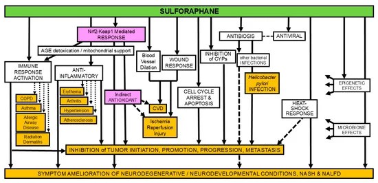
This paper also cited Bridging Nrf2 and autophagy when discussing the above graphic.
In this human cell and rodent study, several coauthors of the original 2020 study tested sulforaphane and TFEB interactions for ameliorating effects of a rare disease:
“Mutations in genes encoding lysosomal proteins could result in more than approximately 70 different lysosomal storage disorders. Niemann–Pick disease type C (NPC) is a rare lysosomal storage disorder caused by mutation in either NPC1 or NPC2 gene. Deficiency in NPC1 or NPC2 protein results in late endosomal/lysosomal accumulation of unesterified cholesterol.
Clinical symptoms of NPC include hepatosplenomegaly, progressive neurodegeneration, and central nervous system dysfunction, that is, seizure, motor impairment, and decline of intellectual function. So far there is no FDA-approved specific therapy for NPC.
Under stress conditions, that is, starvation or oxidative stress, TFEB is dephosphorylated and actively translocates into the nucleus, promoting expression of genes associated with lysosome and autophagy. TFEB overexpression or activation results in increased number of lysosomes, autophagy flux, and exocytosis.
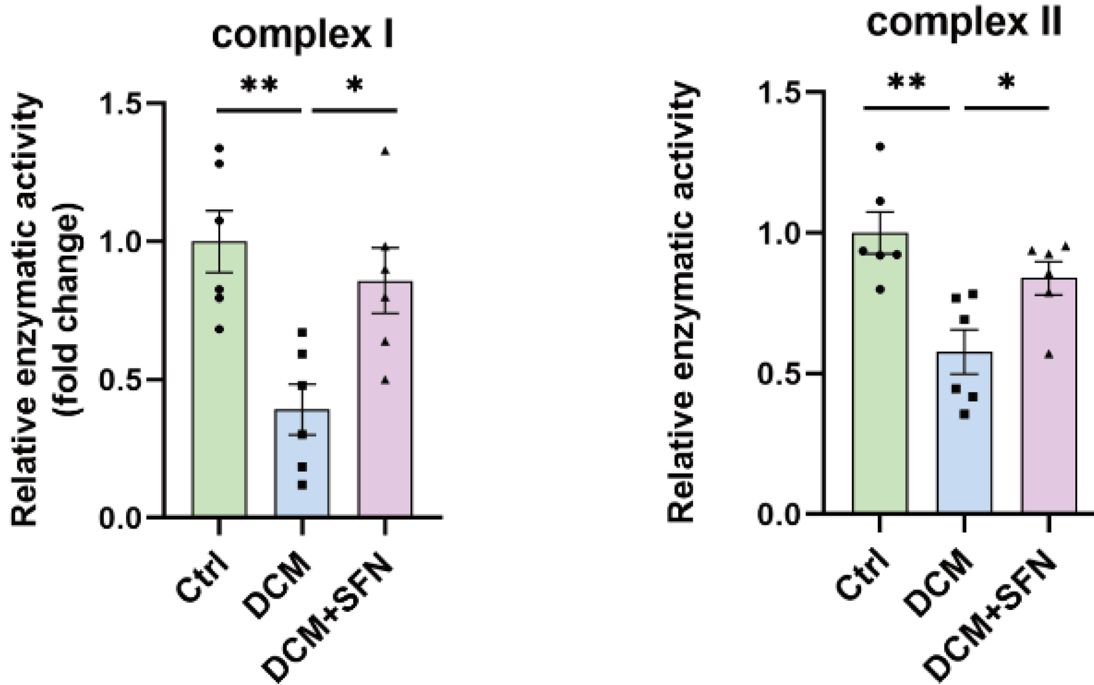
Pharmacological activation of TFEB by sulforaphane (SFN), a previously identified TFEB agonist, significantly promoted cholesterol clearance in human and mouse NPC cells, while genetic inhibition (KO) of TFEB blocked SFN-induced cholesterol clearance. This clearance effect exerted by SFN was associated with upregulated lysosomal exocytosis and biogenesis. SFN treatment has no effect on the liver and spleen enlargement of Npc1 mice.

SFN is reportedly BBB-permeable, assuring a good candidate for efficient delivery to the brain, which is essential for targeting neurodegenerative phenotypes in neurological diseases including NPC. This is the first time that SFN was shown to directly activate TFEB in the brain.
Collectively, our results demonstrated that pharmacological activation of TFEB by a small-molecule agonist can mitigate NPC neuropathological symptoms in vivo. TFEB may be a putative target for NPC treatment, and manipulating lysosomal function via small-molecule TFEB agonists may have broad therapeutic potential for NPC.”
https://elifesciences.org/articles/103137 “Small-molecule activation of TFEB alleviates Niemann–Pick disease type C via promoting lysosomal exocytosis and biogenesis”