Oats sprouts treat gut inflammation

A 2025 rodent study investigated differing effects of regular oats and oat sprouts to treat induced colitis:

“This study aims to test our hypothesis that germinated oats exert stronger anti-inflammatory effects than raw oats due to their higher levels of bioactive phytochemicals. First, the nitric oxide (NO) production assay was used to screen [22] commercially available oat seed products and identify the product with the highest anti-inflammatory activity after germination [for five days]. The selected oat seed product was then produced in larger quantities and further evaluated in an in vivo study using the dextran sulfate sodium (DSS)-induced colitis mouse model to compare the anti-inflammatory effects of phytochemical extracts from germinated and raw oats.

The guideline states that for a healthy U.S.-style dietary pattern at a 2000 calorie level, a daily intake of 6 ounces of grains is recommended, with at least 3 ounces (84 g) coming from whole grains (WGs). For a 60 kg human, consuming 3 ounces of WGs per day translates to a 17.2 g/kg daily dose in mice. Given that the daily food intake of a 20 g mouse is approximately 2.5 g, the 17.2 g/kg daily dose corresponds to 14% of the total diet as WGs. Therefore, the 7 and 21% WG equivalent doses used in this study are relevant to human consumption.

Germination led to an overall increase in the content of all avenanthramides (AVAs) and avenacins (AVCs) as well as some avenacosides (AVEs):

  • For AVAs, the compounds 2c, 2p, 2f, 2cd, 2pd, and 2fd significantly increased by 10.0-, 6.3-, 9.6-, 20.7-, 10.6-, and 4.6-fold, respectively, which is consistent with previous reports.
  • This study is the first to report an increase in AVCs after germination, with AVC-A2, B2, A1, and B1 contents significantly increasing by 2.5-, 2.2-, 3.6-, and 4.2-fold, respectively.
  • Although germination resulted in a decrease in certain AVEs, it significantly increased the levels of AVE-C, Iso-AVE-A, AVE-E, and AVE-F by 1.8-, 3.3-, 3.3-, and 5.0-fold, respectively. Notably, AVE-E has been previously reported to have the strongest anti-inflammatory activity among all of the major AVEs.

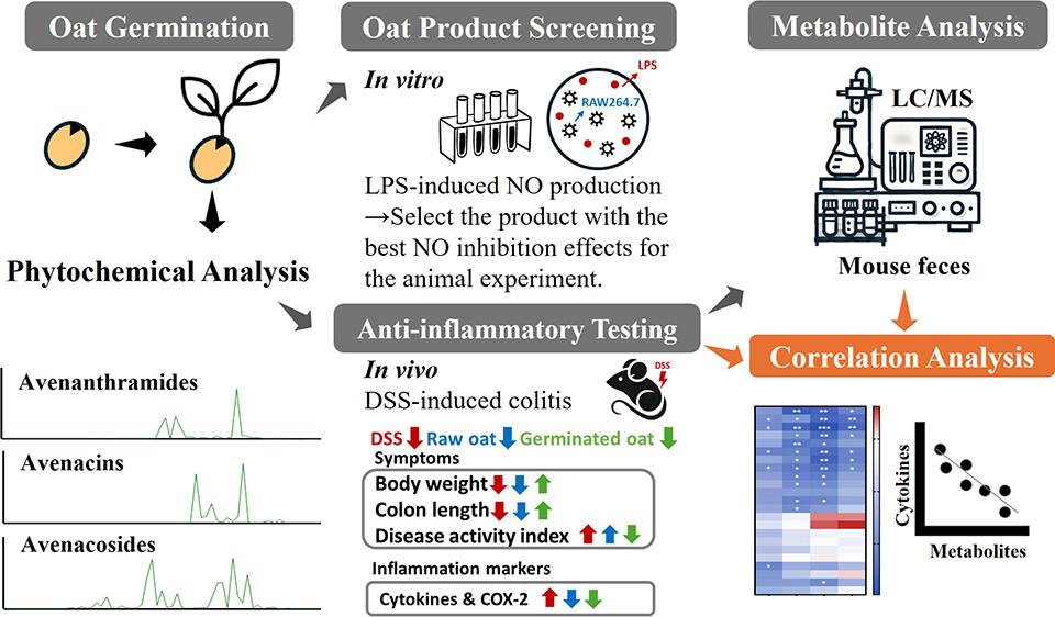
In summary, germination enhances the anti-inflammatory properties of oats in both cells and DSS-induced colitis in mice by increasing levels of bioactive phytochemicals. Correlation analysis showed a significant inverse relationship between pro-inflammatory cytokines and phytochemical content in feces, especially AVAs and their microbial metabolites.

The observation of a stronger anti-inflammatory effect in the low-dose germinated oat group compared with the high-dose group is intriguing and warrants further investigation. One possible explanation is the phenomenon of hormesis, where low doses of bioactive compounds can exert beneficial effects, while higher doses may lead to diminished efficacy or even adverse effects. Further studies involving a broad range of doses would be valuable to define the effective intake range and provide insight into the underlying mechanisms.

It is possible that AVAs, AVEs, and AVCs act synergistically to enhance the overall anti-inflammatory efficacy, potentially by targeting different inflammatory pathways or modulating each other’s bioavailability and activity. Further investigation into the synergistic interactions among these compounds is warranted.”

https://pubs.acs.org/doi/10.1021/acs.jafc.5c02993 “Phytochemical-Rich Germinated Oats as a Novel Functional Food To Attenuate Gut Inflammation”


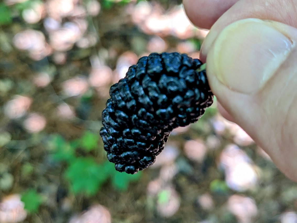
I’ve eaten 3-day-old Avena sativa oat sprouts (started from 20 grams of groats) every day for 4.5 years now, and haven’t had gut problems. Here’s what they looked like this morning:

Sulforaphane as a senotherapy, Part 2

A 2025 rodent study by the same group as Part 1 investigated similar subjects from a different experimental angle of senotherapy effects on brain and behavior rather than cardioprotective effects of dasatinib / quercetin (a senolytic combination) and sulforaphane (senomorphic):

“This is the first study to analyze the effect of senotherapy in the brain of a model of chronic obesity in middle-aged female rats. D + Q reduced the pro-inflammatory cytokines evaluated in the obesity model. It did not improve memory and learning nor the expression of molecules associated with the maintenance of synapses.

In contrast, sulforaphane (SFN), which without eliminating senescent cells, decreased pro-inflammatory factors, increased IL-10, as well as brain-derived neurotrophic factor BDNF, synaptophysin (SYP), and postsynaptic density protein 95 (PSD-95), which, in turn, were associated with an improvement in behavioral tests in obese rats. This suggests that modulating the senescence-associated secretory phenotype (SASP), rather than eliminating senescent cells, might have better effects.”

https://www.sciencedirect.com/science/article/pii/S0014488625001955 “Senotherapy as a multitarget intervention in chronic obesity: Modulation of senescence, neuroinflammation, dysbiosis, and synaptic integrity in middle-aged female Wistar rats”


Treating a stomach infection with sulforaphane

A 2025 rodent study integrated metabolomics and lipidomics analyses to investigate how sulforaphane treats a Helicobacter pylori infection:

“Helicobacter pylori (H. pylori) is a microaerobic Gram-negative bacterium that colonizes the gastric mucosa. Approximately half of the global population is infected with this bacterium, and it is classified as a group 1 carcinogen.

However, H. pylori infection does not typically present with obvious symptoms in the early stages, making it difficult to detect. Daily dietary interventions may be a relatively effective method for its prevention and treatment.

This study established an H. pylori-infected mouse model, to which sulforaphane was orally administered. H. pylori-low-dose and H. pylori-high-dose represent 4 weeks of gavage with 5 mg/kg/d and 20 mg/kg/d of sulforaphane after H. pylori colonization.

Metabolomics and lipidomics analysis of the effects of sulforaphane treatment on mouse serum. Stacked bar chart of the metabolites regulated by (A) low-dose and (B) high-dose sulforaphane treatment compared to the differential metabolites between the control group and H. pylori group.

Results showed that H. pylori infection significantly altered host amino acid and lipid levels, specifically manifested as abnormal serum glycerophospholipids and metabolic imbalances of amino acids, bile acids, glycerophospholipids, ceramides, and peptides in the liver. Sulforaphane treatment reversed these metabolic abnormalities, with high-dose sulforaphane exhibiting more prominent regulatory effects.

High-dose sulforaphane effectively restored hepatic metabolic disorders of amino acids, bile acids, and lipids, and ameliorated abnormal serum glycerophospholipid profiles. Regulation of key pathways such as glycine metabolism and glutathione metabolism constitutes an important basis for sulforaphane’s anti-H. pylori infection effects.

This study provides a comprehensive metabolic basis for understanding the role of sulforaphane as a dietary intervention in preventing and managing H. pylori-associated gastric diseases and lays a foundation for subsequent clinical translational research.”

https://www.mdpi.com/1422-0067/26/16/7791 “Therapeutic Effects of Sulforaphane on Helicobacter pylori-Infected Mice: Insights from High-Coverage Metabolomics and Lipidomics Analyses of Serum and Liver”


A human equivalent to this study’s low sulforaphane dose is (5 mg x .081) x 70 kg = 28 mg, which is achievable by eating broccoli sprouts every day. Quadrupling 28 mg to a human equivalent of the study’s high sulforaphane dose would involve additional supplementation.

Another way to support this study’s glycine metabolism findings without high-dose sulforaphane is to supplement betaine (trimethylglycine) so that the body requires less choline-to-glycine synthesis. A synergistic effect can be achieved with taurine supplementation that enhances cysteine availability for the tripeptide (glutamate, cysteine, and glycine) glutathione synthesis by requiring less cysteine-to-taurine synthesis.

Activate Nrf2 with far-infrared light

A 2025 rodent study investigated effects of far-infrared light on Alzheimer’s disease models. I’ll focus on its Nrf2 findings:

“Far-infrared radiation (FIR) is commonly utilized as a complementary treatment of a range of disease, for example, insomnia and rheumatoid arthritis. In this research, we explored how FIR light impacts cognitive functions of TgCRND8 AD mice and elucidated its underlying molecular mechanism.

Infrared radiation is a form of electromagnetic energy that has wavelengths between 750 nm and 1000 μm, which are longer than visible light. International Commission on Illumination categorizes infrared light as three sub-divisions according to the wavelength: (1) near-infrared radiation (0.7–1.4 μm), (2) middle infrared radiation (1.4–3.0 μm), and (3) far-infrared radiation (3.0–1000 μm).

Nrf-2/ HO-1 signaling, a key endogenous antioxidant system, helps mitigate oxidative stress and enhances expression of various endogenous genes. Activation of HO-1 during inflammatory conditions may serve as an adaptive response to reduce cytotoxicity through various mechanisms.

In this study, we applied EFFIT LITE® as the FIR spectrum transmitter which stably radiates an FIR spectrum with a wavelength of 4–20 μm, and the device was put within 1 cm directly above the head of the 3-month-old TgCRND8 mice for 30 min exposure once every day. FIR light notably enhanced cognitive function and spatial memory of TgCRND8 mice after 28-days consecutive treatment.

Underlying molecular mechanisms involve suppression of Aβ deposition, hyperphosphorylation of tau, and neuroinflammation through modulating Jak-2/Stat3 and Nrf-2/HO-1 pathways. Our current experimental findings amply indicate that FIR light is a potential non-pharmacological therapy for AD.”

https://link.springer.com/article/10.1007/s12017-025-08860-2“Far-Infrared Radiation Ameliorates the Cognitive Dysfunction in an Alzheimer’s Disease Transgenic Mouse via Modulating Jak-2/Stat3 and Nrf-2/HO-1 Pathways”


This study measured Nrf2 and its quickly-induced downstream enzyme HO-1 effects of daily far-infrared light exposure for 30 minutes. We’d have to see measurements of Nrf2’s more-slowly induced and longer-lasting downstream xenobiotic detoxifying enzyme NQO1 to compare far-infrared light Nrf2 activation effects with those of natural plant compounds.

Plasmapheresis doesn’t reduce biological age

A 2025 clinical trial investigated effects of plasmapheresis as measured with epigenetic clocks:

“This study aimed to assess whether plasmapheresis without volume replacement with young plasma or albumin affects epigenetic age and other biomarkers in healthy adults. No significant epigenetic rejuvenation was observed based on epigenetic clock measurements. Instead, plasmapheresis was associated with increases in DNAmGrimAge, the Hannum clock, and the Dunedin Pace of Aging.

  1. The relatively small sample size of 34 finishing participants comprising of first-time plasma donors limits the statistical power and generalizability of our findings.
  2. Our cohort was restricted to individuals aged 40 to 60 years in accordance with Czech regulatory guidelines, which, although intentional to focus on an older population where rejuvenating effects might be most apparent, constrains evaluation of age-related differences across a broader demographic.
  3. The 18-week duration of the study, while sufficient to detect rapid alterations in key biomarkers under an intensive plasmapheresis protocol, may not fully capture the long-term implications of these changes.
  4. Due to our trial taking place during spring and summer months, we cannot fully separate the effects of increased sunlight exposure, outdoor physical activity, and dietary changes from the observed rises in Vitamin D and concurrent shifts in DNAm-based aging metrics. We did not collect objective measures of activity or diet, so these factors remain potential confounders.

The protocol of donating plasma every two weeks, although deemed safe by many countries around the world, is not yet well researched and cannot therefore be marked as benefiting to the donor right now. Further refinement to balance clearance of pro-aging factors with maintenance of systemic homeostasis is needed.”

https://www.nature.com/articles/s41598-025-05396-0 “Human clinical trial of plasmapheresis effects on biomarkers of aging (efficacy and safety trial)”


Taurine and mitochondrial health

A 2025 review subject was taurine’s beneficial effects on mitochondria:

“Taurine has multiple and complex functions in protecting mitochondria against oxidative-nitrosative stress. We introduce a novel potential role for taurine in protecting from deuterium (heavy hydrogen) toxicity. This can be of crucial impact to either normal or cancer cells that have highly different mitochondrial redox status.

Deuterium is an isotope of hydrogen with a neutron as well as a proton, making it about twice as heavy as hydrogen. We first explain the important role that the gut microbiome and gut sulfomucin barrier play in deuterium management. We describe synergistic effects of taurine in the gut to protect against deleterious accumulation of deuterium in mitochondria, which disrupts ATP synthesis by ATPase pumps.

Taurine’s derivatives, N-chlorotaurine (NCT) and N-bromotaurine (NBrT), produced through spontaneous reaction of taurine with hypochlorite and hypobromite, have fascinating regulatory roles to protect from oxidative stress and beyond. We describe how taurine could potentially alleviate deuterium stress, primarily through metabolic collaboration among various gut microflora to produce deuterium depleted nutrients and deuterium depleted water (DDW), and in this way protect against leaky gut barrier, inflammatory bowel disease, and colon cancer.

Taurine cannot be metabolized by human cells, but gut microbes are able to break it down to release sulfite, which then gets oxidized to sulfate anions that become available to support synthesis of sulfomucins. Taurine protects against many diseases linked to mitochondrial defects, such as aging, metabolic syndrome, cancer, cardiovascular diseases and neurological disorders.

We present a novel view that gut microbes play an essential role in providing deuterium depleted (deupleted) nutrients, especially, butyrate, to the host colonocytes forming the gut barrier. We propose that sulfomucins synthesized by goblet cells not only protect the barrier from pathogens, but also trap and sequester deuterium, thus reducing mitochondrial deuterium levels, resulting in improved mitochondrial health.

Due to taurine, redox buffer glutathione (GSH) further stabilizes the membrane potential. GSH not only reduces radical oxygen species (ROS) during oxidative stress, but it also assists in production of deupleted water in mitochondria.

Spontaneous oxidation of two GSH molecules to produce GSSG in the presence of hydrogen peroxide yields two molecules of DDW. Just as for glutathione, bilirubin can produce DDW indefinitely through chronic recycling between bilirubin and biliverdin, capturing a deupleted proton in NADPH to produce a DDW molecule in each cycle.

A novelty that arises from this investigation is introduction of the role that deuterium plays in mitochondrial disease, and ways in which taurine may facilitate maintenance of low deuterium in mitochondrial ATPase pumps. Excess deuterium causes a stutter in the pumps, which leads to inefficiencies in ATP production and an increase in ROS.”

https://pmc.ncbi.nlm.nih.gov/articles/PMC11717795/ “Taurine prevents mitochondrial dysfunction and protects mitochondria from reactive oxygen species and deuterium toxicity”


Stay away from NAC

A 2025 rodent study added several reasons to avoid non-emergency use of N-acetylcysteine:

“We previously showed that antioxidants induced an impairment of negative feedback of the hypothalamus-pituitary-adrenal (HPA) axis in rats, in parallel to a down-regulation of glucocorticoid receptor (GR) and nuclear factor erythroid 2-related factor 2 (Nrf2) expression in the pituitary gland. This study evaluated the role of the Nrf2-heme-oxygenase-1 (HO-1) pathway on impairment of negative feedback of the HPA axis induced by N-acetylcysteine (NAC).

Male Swiss-Webster mice were orally supplemented with NAC for 5 consecutive days. The Nrf2-HO-1 pathway activator cobalt protoporphyrin IX (CoPPIX) was injected intraperitoneally on days 2 and 5 after starting NAC supplementation.

NAC reduced expression of Nrf2 in the pituitary of mice. NAC induced adrenal enlargement and hypercorticoidism, along with a decrease in GRα expression and an increase of GRβ expression in the pituitary gland.

We observed that dietary supplementation with NAC ( Figure 4A ) significantly increased plasma corticosterone levels in mice 24h ( Figure 4B ) as well as 15 days ( Figure 4C ) after the last administration of the antioxidant with the same magnitude of response (3.5-fold and 3.4-fold, respectively).

Chronic activation of the HPA axis can have damaging effects on immune, cardiovascular, metabolic, and neural functions, increasing the risk of immune system dysfunction, mood disorders, and metabolic and cardiovascular diseases. To prevent these deleterious effects of chronic hypercortisolism, HPA axis function is controlled by a glucocorticoid-dependent negative feedback system that is essential for ending the stress response.

These findings showed that NAC reduced Nrf2-HO-1 pathway activation in the pituitary gland, in a mechanism probably related to a local downregulation of GRα and an up-regulation of GRβ, leading to a failure of negative feedback of the HPA axis and consequently to the hyperactivity of this neuroendocrine axis.”

https://pmc.ncbi.nlm.nih.gov/articles/PMC11827418/ “Activation of the Nrf2/HO-1 pathway restores N-acetylcysteine-induced impairment of the hypothalamus-pituitary-adrenal axis negative feedback by up-regulating GRα expression and down-regulating GRβ expression into pituitary glands”


A human equivalent to this study’s NAC dose is (150 mg x .081) x 70 kg = 851 mg. Human supplements are sold in 600 mg and 1000 mg doses.

Grok 3 replied to a question: Human equivalent time to 15 days in male Swiss-Webster mice aged between 4 and 6 weeks? by stating: “15 days in male Swiss-Webster mice aged 4 to 6 weeks corresponds to approximately 4.1 human years, advancing their equivalent human age from about 10–12 years to roughly 14–16 years.” Four+ years seems like a long time for NAC to steadily and continuously affect humans’ HPA axes per the above graphic. What do you think?

Previously mentioned reasons to avoid daily use of NAC were in the lower part of A good activity for bad weather days.

Sulforaphane as a senotherapy

A 2025 rodent study investigated cardioprotective effects of dasatinib / quercetin (a senolytic combination) and sulforaphane (senomorphic):

“Senolytics are molecules that selectively eliminate senescent cells (SCs). Senomorphics are drugs that suppress or mitigate the pro-inflammatory effects of the senescence-associated secretory phenotype (SASP) without killing SCs. Senomorphics decrease the number of SCs by diminishing the paracrine effect of senescence induction in neighboring cells, and by enhancing elimination of SCs by the immune system, which reduces their harmful effects.

We used middle-aged female rats fed a hypercaloric diet (HD) from 21 days to 15 months of age. Under our experimental conditions, rats exhibited cardiac hypertrophy and fibrosis, accumulation of senescent cells, changes in mitochondrial morphology, and oxidative stress. Rats were treated for 2 months with senolytic (dasatinib + quercetin, DQ) or senomorphic (sulforaphane, SFN) agents.

A novel aspect of recent research has been the crosstalk between organelles, particularly between the mitochondria and endoplasmic reticulum (ER), through specialized contact sites (MERCSs). However, there is still no consensus on the optimal distance between MERCSs and their positive or negative effects on disease progression.

HD rats showed cardiac improvement after both treatments. Although both strategies improved cardiomyocyte size and cardiac fibrosis, only DQ decreased LDH levels, whereas SFN positively affected cardiac damage proteins.

In general, no changes in structure or damage-associated enzymes were observed in control rats treated with DQ or SFN, indicating that senotherapies do not promote adverse effects on the heart, reinforcing the concept that they are safe for application in the clinical field. Data suggest a possible link mechanism between Nrf2 activation and MERCSs preservation, activated by SFN rather than by the DQ combination, which allowed cardiac structure maintenance in HD rats decreasing harmful effects of senescent cells.”

https://www.sciencedirect.com/science/article/abs/pii/S0955286325000865 “Cardioprotective effect of senotherapy in chronically obese middle-aged female rats may be mediated by a MERCSs/Nrf2 interaction” (not freely available) Thanks to Dr. Alejandro Silva for providing a copy.


Eat broccoli sprouts for your eyes, Part 3

Two 2025 papers cited Precondition your defenses with broccoli sprouts, starting with a review of age-related macular degeneration:

“AMD progression from intermediate to late AMD leads to a point of irreversible retinal pigmented epithelium (RPE) degeneration where treatment becomes worthless. Treating patients at the early/intermediate stages presents a better therapeutic window opportunity for AMD as the disease could potentially be prevented or slowed down.

Strong evidence points to RPE dysfunction at these stages, mainly through redox imbalance and lysosomal dysfunction in RPE oxidative injury. Restoring oxidative balance and lysosomal function may act as preventive and therapeutic measures against RPE dysfunction and degeneration.

Due to interaction with KEAP1, NRF2 is a ubiquitously expressed protein with a high turnover and half-life of about 20 minutes. Because the turnover of NRF2 is faster than KEAP1, newly synthesized NRF2 does not have free KEAP1 to bind and is translocated into the nucleus. Once in the nucleus, NRF2 dimerizes with sMAF and the complex binds to antioxidant response element (ARE) sequences, promoting the expression of ARE genes.

There is NRF2 involvement in most of the hallmarks of aging. Key transcriptional regulatory factors of related pathways, such as transcription factor EB (TFEB) and NRF2, may be targeted to restore homeostasis and/or prevent further RPE degeneration.”

https://www.mdpi.com/2076-3921/14/5/596 “Targeting Lysosomal Dysfunction and Oxidative Stress in Age-Related Macular Degeneration”

There were other informative tidbits throughout this review, such as:

  • “Anti-inflammatory effects of most electrophilic NRF2 activators are thought to be at least partly NRF2-independent, suggesting that these compounds lacking specificity may be advantageous for multitargeted pathologies.
  • TFEB can activate NRF2 under conditions devoid of oxidative stress.”

This paper also cited Bridging Nrf2 and autophagy when discussing the above graphic.


In this human cell and rodent study, several coauthors of the original 2020 study tested sulforaphane and TFEB interactions for ameliorating effects of a rare disease:

“Mutations in genes encoding lysosomal proteins could result in more than approximately 70 different lysosomal storage disorders. Niemann–Pick disease type C (NPC) is a rare lysosomal storage disorder caused by mutation in either NPC1 or NPC2 gene. Deficiency in NPC1 or NPC2 protein results in late endosomal/lysosomal accumulation of unesterified cholesterol.

Clinical symptoms of NPC include hepatosplenomegaly, progressive neurodegeneration, and central nervous system dysfunction, that is, seizure, motor impairment, and decline of intellectual function. So far there is no FDA-approved specific therapy for NPC.

Under stress conditions, that is, starvation or oxidative stress, TFEB is dephosphorylated and actively translocates into the nucleus, promoting expression of genes associated with lysosome and autophagy. TFEB overexpression or activation results in increased number of lysosomes, autophagy flux, and exocytosis.

Pharmacological activation of TFEB by sulforaphane (SFN), a previously identified TFEB agonist, significantly promoted cholesterol clearance in human and mouse NPC cells, while genetic inhibition (KO) of TFEB blocked SFN-induced cholesterol clearance. This clearance effect exerted by SFN was associated with upregulated lysosomal exocytosis and biogenesis. SFN treatment has no effect on the liver and spleen enlargement of Npc1 mice.

SFN is reportedly BBB-permeable, assuring a good candidate for efficient delivery to the brain, which is essential for targeting neurodegenerative phenotypes in neurological diseases including NPC. This is the first time that SFN was shown to directly activate TFEB in the brain.

Collectively, our results demonstrated that pharmacological activation of TFEB by a small-molecule agonist can mitigate NPC neuropathological symptoms in vivo. TFEB may be a putative target for NPC treatment, and manipulating lysosomal function via small-molecule TFEB agonists may have broad therapeutic potential for NPC.”

https://elifesciences.org/articles/103137 “Small-molecule activation of TFEB alleviates Niemann–Pick disease type C via promoting lysosomal exocytosis and biogenesis”


Interpreting results of a Vitamin K2 clinical trial

This 2025 paper analyzed clinical trial subjects’ protocol adherence:

“The VitaK-CAC (vitamin K-coronary artery calcification) trial is a double-blind, randomized, placebo-controlled trial in patients with pre-existent CAC who were treated for two years with either placebo or the vitamin K2-analogue menaquinone-7 (MK-7) [360 mcg daily]. The present analysis of the VitaK-CAC trial assesses degree of adherence to supplementation with MK-7 during the implementation and persistence phases.

We estimated adherence in three different ways: 1) by pill counts, 2) by measuring plasma levels of MK-7, and 3) by measuring plasma levels of dephosphorylated, uncarboxylated matrix Gla-protein (dp-ucMGP), a marker of functional bioactivity of vitamin K.

  1. We estimated tablet intake by counting the number of pills participants returned to the laboratory at each follow-up visit. When 20% or less of the prescribed pills were returned, this was considered adequate adherence. Due to inconsistent bottle returns by many participants, reliable pill counts could be obtained for only half of the patients. No difference in evolution of the CAC score was found between patients with an adherence below or above 80%, as determined by pill counts.
  2. In patients receiving MK-7, an increase in plasma levels above the upper interquartile range (IQR) observed in the placebo group was taken as evidence of supplementation adherence. When this occurred both at 12 months and at 24 months, this was taken as a robust indicator of good persistence. When patients with persistence throughout the study, as based on the MK-7 levels, were compared to those who were not, the former group showed an attenuated progression of their CAC score.
  3. We considered a fall in dp-ucMGP by more than 10% as indicative of a positive effect of MK-7 supplementation and, hence, as a proxy for good adherence. Levels of dp-ucMGP rose significantly in both groups, although to a lesser extent in the group with active treatment. If the reduced rise in dp-ucMGP is considered a proxy for adherence, this suggests approximately 60% adherence in the MK-7 group. In 21 of the 75 patients (28%), the rise in MK-7 was sufficiently great for levels of dp-ucMGP to decrease.

There was no difference in the response of MK-7 levels and in the primary outcome (CAC progression) between patients with or without sufficient adherence. We cannot exclude the possibility that variations in absorption or metabolism have contributed to, or may even be entirely responsible for, the observed swings in MK-7 plasma levels.

None of the three methods that we applied in our study is absolutely reliable to estimate adherence to supplementation with MK-7. Measurement of MK-7 levels provides the best information.”

https://www.explorationpub.com/Journals/em/Article/1001321 “Adherence to dietary supplementation with menaquinone-7, a vitamin K2 analogue”

A paper described the clinical trial itself:

https://journals.lww.com/jhypertension/fulltext/2025/05001/menaquinone_7_slows_down_progression_of_coronary.39.aspx “MENAQUINONE-7 SLOWS DOWN PROGRESSION OF CORONARY ARTERY CALCIFICATION. A RANDOMIZED, PLACEBO-CONTROLLED TRIAL” (not freely available, NCT01002157)


Always more questions:

1. The registration summary included:

In animal studies, active supplementation of Vitamin K2 caused regression of existing arterial calcification. The aim of this randomized, double-blind, placebo-controlled clinical trial is to investigate whether daily supplementation of Vitamin K2 (Menaquinone-7) to patients with established CAC will lead to a decreased progression-rate of CAC after 24 months of follow-up in comparison to placebo.

Is this trial’s stated goal to decrease CAC progression to the point of an unstated goal of reversing existing arterial calcification?

2. What is the degree of success of this clinical trial? I don’t have access to the clinical trial paper, although I’ve requested a copy of it. Did any of the treatment or placebo group subjects reverse their CAC score over 12 or 24 months?

3. Which if any of the items discussed in Vitamin K2 and your brain were suggested to achieve a goal of reversing existing arterial calcification?


I’ve taken a higher MK-7 daily dose (600 mcg) over the past few years. I take it with a fat source, and without vitamins A, D3, and E to eliminate the possibility of competition from other fat-soluble vitamins.

I haven’t ever had CAC measured because I’m not particularly concerned about existing arterial calcification. I’ll update this post if needed after one of the trial’s researchers provides a copy of their paper.

Eat broccoli sprouts to alleviate diabetic heart disease

A 2025 rodent study investigated sulforaphane’s effects on diabetic cardiomyopathy:

“The protective effect of cruciferae-derived sulforaphane (SFN) on diabetic cardiomyopathy (DCM) has garnered increasing attention. However, no studies have specifically explored its mechanistic involvement in cardiac substrate metabolism and mitochondrial function.

To address this gap, Type 2 diabetes mellitus (T2DM) db/db mice were orally gavaged with vehicle or 10 mg/kg body weight SFN every other day for 16 weeks, with vehicle-treated wild-type mice as controls. SFN intervention (SFN-I) alleviated hyperglycemia, dyslipidemia, HOMA-IR, serum MDA levels, and liver inflammation. SFN-I improved the lipotoxicity-related phenotype of T2DM cardiomyopathy, manifested as attenuation of diastolic dysfunction, cardiac injury, fibrosis, lipid accumulation and peroxidation, ROS generation, and decreased mitochondrial complex I and II activities and ATP content.

Although not fully understood, multiple systemic and cardiac-local mechanisms contribute to DCM, encompassing hyperglycemia, dyslipidemia, insulin resistance (IR), disturbances in cardiac substrate metabolism, lipotoxicity, glucotoxicity, mitochondrial dysfunction, oxidative stress, inflammation, and endoplasmic reticulum (ER) stress. Nrf2 and its downstream metallothionein also mediated the preventive effect of SFN on DCM, and may underlie the synergistic effect of SFN and zinc in DCM.

These results suggest that chronic oral SFN-I protects against DCM by mitigating overall metabolic dysregulation and inhibiting cardiolipotoxicity. The latter might involve controlling cardiac fatty acid metabolism and improving mitochondrial function, rather than promoting glucose metabolism.”

https://www.mdpi.com/2076-3921/14/5/603 “Oral Sulforaphane Intervention Protects Against Diabetic Cardiomyopathy in db/db Mice: Focus on Cardiac Lipotoxicity and Substrate Metabolism”


This study had numerous charts like the above showing it was better to not have a deviation from health (Ctrl) rather than incur injury (DCM) then try to fix it with sulforaphane (DCM + SFN). But the control group was wild-type mice, not mice genetically inclined to diabetes like the treatment groups.

The subjects’ starting points were at nine-weeks-old (equivalent to 18-25 year-old humans), and duration was 16 weeks. Grok 3 said: “A 25-week-old db/db mouse is roughly equivalent to a human aged 30–35 years chronologically, though its metabolic condition may mimic older human physiological states in diabetes and obesity research.”

A human equivalent of a 10 mg/kg sulforaphane dose is (.081 x 10 mg) = 56 mg orally administered every other day. That’s about how much total sulforaphane I estimated I took every day (52 mg) from Week 6 through Week 56 by eating microwaved broccoli sprouts twice daily.

No rationale was provided for the sulforaphane dose or the every-other-day dosing regimen. Since I took ~52 mg every day for almost a year, I’ll guess that this study may have had more definitive results with daily dosing. Or maybe add zinc per Zinc and broccoli sprouts – a winning combination.

Year Five of Changing to a youthful phenotype with sprouts

1. I’ve continued daily practices from Year Four to experience another year without being sick! I’ll get a set of Labcorp tests in a week to see if anything is sneaking up on me.

Really think that Brassica clinical trials should last years, not weeks. Once people get over the fact that broccoli, red cabbage, and mustard sprouts will never taste good because their compounds are plants’ defenses against predators, they’ll overlook that in favor of health benefits. Avena sativa oat sprouts don’t have a palatability problem.

2. Daily supplements have changed a little:

  • Started taking a quercetin supplement suggested in a comment to Year One as helpful for seasonal allergies (it doesn’t do that for me). Repeatedly rinsing and soaking the salt out of capers for quercetin content became too much of a nuisance, and the results didn’t always taste right;
  • Stopped taking Prodrome supplements because of unsustainable high costs;
  • Started taking Ovega 3 algae oil DHA 420 mg/EPA 140 mg twice a day in their place;
  • Substituted flax oil 1400 mg once a day for Balance oil;
  • Started taking 2 g magnesium L-threonate;
  • Upped taurine intake from 5 to 6 grams;
  • Upped D3 by 25 mcg to a daily 4400 IU;
  • Reduced chondroitin sulfate by 1.8 g since my joints are doing fine;
  • Stopped soy lecithin in favor of eating three raw eggs.

3. I injured my left shoulder in May 2024 by overdoing upper body exercises, and stopped seven months to recover. Gained thirty pounds during that layoff, and have worked off ten pounds with new routines since then.

I’m no longer dogmatic about aerobic exercise / beach walks. I’ll go over to the beach before sunrise when it isn’t raining or windy, or wait until the afternoon for weather to improve, rather than walk 30 minutes a day irregardless.

The largest cause of coincidences, Part 2

Part 1 was informative, but this 2.5 hour interview with Dr. Suzanne Humphries provided a dozen times more information on health care and other subjects. Probably couldn’t have been on YouTube six months ago for more than a few minutes until it would have been censored.

I came across this interview by reading Why Is What We Feed Infants So Unhealthy?

“As I was finishing this article, Joe Rogan made the remarkable decision earlier this week to bring Suzanne Humphries onto his show to discuss the centuries of lies we’ve been told about vaccines (which amongst other things inspired this Substack) where she not only did that but also focused on the importance of breast feeding and the increased susceptibility formula fed infants have to vaccine injuries.”

I don’t specifically search for breast feeding topics, but my electronic services and devices seem to find them for me. Such as this suggestion:

2025 α-ketoglutarate research

I haven’t mentioned α-ketoglutarate for a while, although I’ve taken it twice a day for several years. Here are four 2025 papers on α-ketoglutarate, starting with a review of its role in bone health:

“α-Ketoglutarate (α-KG) serves as a pivotal intermediate in various metabolic pathways in mammals, significantly contributing to cellular energy metabolism, amino acid metabolism, and other physiological processes. α-KG may be a therapeutic target for a variety of bone-related diseases, such as osteoporosis, osteoarthritis, and rheumatoid arthritis, because of its role in maintaining metabolic balance of bone.

α-KG, as a rate-determining mitochondrial intermediate, is crucial in cell energy metabolism because it connects intracellular carbon and nitrogen metabolism between isocitrate and succinyl coenzyme A. Additionally, α-KG is closely involved in the amino acid cycle. As a precursor of amino acids such as glutamine and glutamic acid, α-KG plays a direct role in energy production and a wide range of cellular chemical reactions. α-KG provides an energy source, stimulating protein synthesis, inhibiting protein degradation in muscle, and serving as a significant metabolic fuel for gastrointestinal cells.

α-KG promotes osteogenic differentiation of stem cells, increases activity of osteoblasts to promote osteogenesis, and inhibits bone resorption activity of osteoclasts. α-KG in articular cartilage promotes differentiation and maturation of chondrocytes and formation of a cartilage matrix. The protective effect of α-KG on bone has practical value in treatment of abnormal bone loss symptoms in various bone tissue diseases.”

https://www.sciengine.com/ABBS/doi/10.3724/abbs.2025020 “Essential role of the metabolite α-ketoglutarate in bone tissue and bone-related diseases”


A rodent study explored adding α-KG to osteoarthritis treatment:

“Mesenchymal stem cell (MSC) therapy represents a promising treatment strategy for osteoarthritis (OA). Nevertheless, the therapeutic efficacy of MSCs may be attenuated under conditions of cellular senescence or when the available clinical quantity is insufficient. α-Ketoglutarate (AKG) exerts beneficial effects on skeletal tissues and activity of stem cells. The present study was designed to explore the potential of AKG in augmenting viability of MSCs and the potential of their combined utilization in treatment of OA.

AKG plays a crucial role in multiple biological processes. It is involved in regulating stem cell differentiation, exerts anti-apoptotic effects, modulates the body’s immune and inflammatory responses, contributes to muscle and bone development, and is essential for maintaining stability of the cartilage matrix.

Platelet-rich plasma (PRP) has been demonstrated to have protective effects on chondrocytes and can effectively repair damaged cartilage in OA. However, PRP has intractable problems in terms of product quality control and allogeneic application, and its long-term therapeutic effect gradually weakens.

Combining AKG’s regulation of cellular metabolism with the multi-directional differentiation and immunomodulatory functions of MSCs is likely to generate a synergistic effect. This combined treatment modality targets the complex pathological processes of OA, including cartilage damage, inflammatory responses, and extracellular matrix imbalance, in a more comprehensive manner than a single therapy.”

https://www.sciencedirect.com/science/article/pii/S2707368825000032 “The repair effect of α-ketoglutarate combined with mesenchymal stem cells on osteoarthritis via the hedgehog protein pathway”


A rodent study investigated whether α-KG has a role in determining frailty:

“Frailty is an age-related geriatric syndrome, for which the mechanisms remain largely unknown. We performed a longitudinal study of aging female (n = 40) and male (n = 47) C57BL/6NIA mice, measured frailty index, and derived metabolomics data from plasma samples.

We find that frailty related metabolites are enriched for amino acid metabolism and metabolism of cofactors and vitamins, include ergothioneine, tryptophan, and alpha-ketoglutarate, and present sex dimorphism. We identify B vitamin metabolism related flavin adenine dinucleotide and pyridoxate as female-specific frailty biomarkers, and lipid metabolism related sphingomyelins, glycerophosphoethanolamine and glycerophosphocholine as male-specific frailty biomarkers.

We were interested to observe whether metabolite abundance at any specific timepoint was associated with frailty at a future timepoint. Unfortunately, we didn’t observe any metabolites that showed an overall significant association with future FI (FIf) or future devFI (devFIf). When focusing only on the abundance of metabolites at the baseline time point (∼400 days), we found a single metabolite, alpha-ketoglutarate, was negatively associated with both FIf and devFIf.”

https://www.biorxiv.org/content/10.1101/2025.01.22.634160v1.full “Metabolomics biomarkers of frailty: a longitudinal study of aging female and male mice”


Wrapping up with a rodent study adding α-KG to exercise for its effects on depression and learning:

“aKG acts as a prophylactic and antidepressant to effectively counteract social avoidance behaviors by modulating BDNF levels in the hippocampus and nucleus accumbens. Exercise increases aKG levels in the circulation.

In mice, aKG supplementation prolongs lifespan and reduces aging-associated frailty. aKG supplementation also reverses aging in humans as measured by DNA methylation patterns.

aKG functions as a co-factor for epigenetic enzymes. Changes in the intracellular αKG/succinate ratio regulates chromatin modifications, including H3K27me3 and ten-eleven translocation (Tet)-dependent DNA demethylation. The ability of aKG to influence epigenetic status of cells may explain both its prophylactic and anti-depressant effects since transcriptional dysregulation and aberrant epigenetic regulation are unifying themes in psychiatric disorders. This may also explain its ability to differentially regulate BDNF expression in the hippocampus and NAc.

If exercise mediates its effects through aKG, aKG may be a pivotal component of an exercise pill along with lactate and BHB that can serve as both a prophylactic and antidepressant treatment for depression.”

https://www.sciencedirect.com/science/article/pii/S266717432500031X “α-ketoglutarate (aKG) is a circulatory exercise factor that promotes learning and memory recall and has antidepressant properties


Epigenetic clock analysis of a clinical trial

A 2025 paper performed post-hoc epigenetic clock analyses of a supplement and exercise clinical trial completed earlier this decade:

“We report results of a post hoc analysis among 777 participants of the DO-HEALTH trial on the effect of vitamin D (2,000 IU per day) and/or omega-3 (1 g (330 mg EPA plus 660 mg DHA from marine algae) per day) and/or a home exercise program (a strength-training exercise program performed for 30 min three times per week) on four next-generation DNA methylation (DNAm) measures of biological aging (PhenoAge, GrimAge, GrimAge2 and DunedinPACE) over 3 years. Omega-3 alone slowed the DNAm clocks PhenoAge, GrimAge2 and DunedinPACE, and all three treatments had additive benefits on PhenoAge.

Inclusion criteria were age 70 years and older, living at home, having no major health events (no cancer or myocardial infarction) in the 5 years before enrollment, having sufficient mobility to visit the study centers without help and having good cognitive function with a Mini-Mental State Examination score of at least 24. 777 provided consent for these analyses and had samples available after the application of the exclusion criteria. This group of individuals formed our analysis sample, which had the following characteristics: 59% were women; the mean age at baseline was 75 years; 30% had 25-hydroxyvitamin D (25(OH)D) levels of <20 ng ml−1; 53% were healthy agers as defined in the Nurses’ Health Study (free of major chronic diseases, disabilities, cognitive impairments and mental health limitations); and 88% were physically active (29% were active one to three times per week, and 59% were active more than three times per week). The Swiss participant subgroup represents a healthier and more active subgroup within the total DO-HEALTH population.

Overall, from baseline to year 3, standardized effects ranged from 0.16 to 0.32 units (2.9–3.8 months). In summary, our trial indicates a small protective effect of omega-3 treatment on slowing biological aging over 3 years across several clocks, with an additive protective effect of omega-3, vitamin D, and exercise based on PhenoAge.”

https://www.nature.com/articles/s43587-024-00793-y “Individual and additive effects of vitamin D, omega-3 and exercise on DNA methylation clocks of biological aging in older adults from the DO-HEALTH trial”

These epigenetic clock measurements of a subset of trial subjects was interesting, although I didn’t find it particularly relevant to what I do. I take twice as much Vitamin D and omega-3s everyday, do resistance exercises once or twice a week whenever I’ve recovered from the previous session, walk a few miles on the beach if the weather is nice, and other things.

I don’t bother with epigenetic clock measurements anymore because the free one (PhenoAge) is too variable to be personally accurate. For other clocks, it would be meaningless if all I got was a 2-3 month improvement over a three year period like this trial. Studies usually find that the most deficient subjects at the beginning are the ones that show the greatest improvements with effective treatments. Unhealthiness on any epigenetic clock parameter probably wouldn’t be my starting point, so I may not show even a one-month improvement over three years.


Dr. Goodenowe offered his opinion on the paper:

“DHA is a polyunsaturated fatty acid that is essential for maintaining youthful fluidity of the body’s membranes. While our bodies can make DHA from the essential omega-3 dietary fatty acid, as we get older, our ability to make DHA decreases and oxidative stress on our bodies increases. These two factors contribute to our membranes becoming stiffer and less pliable as we age, in other words, ‘older.’

Because getting older and losing function appear to go hand in hand, we equate aging with a loss of function. As such, we think that aging causes this loss of function, like a disease. Instead, the opposite is true, and it’s the loss of function that causes aging. To slow aging you need to focus on maintaining function.”

https://www.prevention.com/health/a63850396/vitamin-exercise-boost-longeivty-study/ “Scientists Find Taking This Vitamin Boosts Longevity, Add Years to Your Life”

Prevention magazine’s editors need to better proof their writers’ work before it gets published. Unlike the headline, the trial had nothing to do with adding years to human lifespan.