Dopamine may account for differences in cognitive performance

This 2015 German human study found:

“Dopamine may account for adult age differences in brain signal variability.”

The researchers administered amphetamine to the subjects to boost their dopamine levels, and measured their cognitive performance on several working memory tests under fMRI:

“Older adults expressed lower brain signal variability at placebo, but met or exceeded young adult..”

brain signal variability levels when on speed.

The order of the tests also influenced the results. Older adults who received amphetamine during the initial series of tests performed better on placebo during the second series of tests.


As is often done, the researchers focused on effects and not causes. I didn’t see questionnaires or investigation into possible historical or biological factors for reduced dopamine levels, leaving the researchers with age as the only correlated-but-not-causative explanation.

http://www.pnas.org/content/112/24/7593.full “Amphetamine modulates brain signal variability and working memory in younger and older adults”

How brains mature during critical periods

This 2015 German rodent study found:

“Once silent synapses are consolidated in any neural circuit, initial experience-dependent functional optimization and critical periods end.

Silent synapses are thought to be immature, still-developing excitatory synapses.”

The number of silent synapses related to visual processing was measured at ~50% at eye opening. Visual experience reduced this to 5% or less by adulthood in the study’s control group. Removing a protein in the subjects’ hippocampus silenced the synapses back up to ~50%, even in adults.

Critical periods are:

“Characterized by the absolute requirement for experience in a restricted time window for neural network optimization.

Although some functions can be substantially ameliorated after the CP [critical period], they are rarely optimally restored.”

Two human studies were cited on critical periods in second-language and musical skills development, Sensitive periods in human development: Evidence from musical training (not freely available).

The researchers generalized their findings as:

“Experience-dependent unsilencing of silent synapses constitutes an important general maturational process during CPs of cortical development of different functional domains and suggest an interplay with inhibitory circuits in regulating plasticity.”

http://www.pnas.org/content/112/24/E3131.full “Progressive maturation of silent synapses governs the duration of a critical period”

The effects of inescapable, uncontrollable, repeated stress on the hippocampus

This 2015 MIT rodent study found:

Behavioral stress impairs cognitive function via activation of a specific direct neural circuit from the basolateral amygdala to the dorsal hippocampus. Moreover, we delineate a molecular mechanism by which behavioral stress is translated to hippocampal dysfunction via a p25/Cdk5 (cyclin-dependent kinase 5)-dependent pathway and epigenetic alterations of neuroplasticity-related gene expression.”

The researchers made several intermediate findings to develop their main finding:

1. “Repeated stress is accompanied by

  • generation of p25,
  • up-regulation and phosphorylation of glucocorticoid receptors,
  • increased HDAC2 [the gene encoding the histone deacetylase 2 enzyme] expression, and
  • reduced expression of memory-related genes [most, but not all that were tested] in the hippocampus.”

2. “BLA [basolateral amygdala] activation is both necessary and sufficient for stress-associated molecular changes and memory impairments.”

3. “This effect [2. above] relies on direct glutamatergic projections from the BLA to the dorsal hippocampus.”

4. “p25 generation is necessary for the stress-induced memory dysfunction.”

From the Results section:

“Control mice showed a significant preference for the novel over the familiar object or location, whereas RFS [repetitive foot shock]-treated mice performed no better than chance.”

The subject adult mice underwent:

“Inescapable, uncontrollable repeated stress.”

Do humans also experience impaired “cognitive function” and “hippocampal dysfunction” and “epigenetic alterations of neuroplasticity-related gene expression” caused by “inescapable, uncontrollable repeated stress”?

And what are the real histories of people who aren’t curious, who don’t show “a significant preference for the novel over the familiar object or location”?

http://www.pnas.org/content/112/23/7291.full “Basolateral amygdala bidirectionally modulates stress-induced hippocampal learning and memory deficits through a p25/Cdk5-dependent pathway”

Measuring the signal-to-noise ratio of a brain neuron

This 2015 study found that a single brain neuron’s

“Spiking history is often a more informative predictor of spiking propensity than the signal or stimulus activating the neuron.

Characterizing the reliability with which neurons represent and transmit information is an important question in computational neuroscience.”

Three of the four brain neuron areas studied were of the limbic system: a rat thalamus, a monkey hippocampus, and a human subthalamus area.

http://www.pnas.org/content/112/23/7141.full.pdf “Measuring the signal-to-noise ratio of a neuron” (pdf file is linked because the html has errors)

Epigenetic changes in the developing brain change behavior

This 2015 review cited 143 studies to tie together findings in epigenetic chemistry and behavioral neuroscience.

In addition to studies I’ve previously curated, other research included:

  • a 2012 study which completely abolished mouse maternal behavior by silencing a gene encoding an estrogen receptor;
  • a 2012 study which found that stress-induced changes in the rat hippocampus were heritable;
  • a 2014 study that distinguished between transgenerational and intergenerational epigenetic effects such as:

    in utero exposure to nutritional status, stress, or toxic environmental factors that act on the developing embryo and its germ line”

  • a 2013 study that showed how genomic imprinting coordinated:

    “Genetic coadaptation where beneficially interacting alleles evolve to become coinherited.”

The current status of research incorporating both epigenetic chemistry and behavioral neuroscience was summed up as:

“A large number of behavioral epigenetic studies attempt to correlate epigenetic marker changes at global levels and in mixed populations of cells with phenotypic changes.

Specific changes at specific gene levels and at single cell levels correlating with behavioral changes remain largely unknown.”

http://www.pnas.org/content/112/22/6789.full “Epigenetic changes in the developing brain: Effects on behavior”

RNA as a proxy signal for context-specific biological activity

This 2015 Harvard/MIT rodent study was of long (more than 200 nucleotides) noncoding (non-protein coding) RNAs (ribonucleic acids). These are of interest because:

“Within the mammalian body, the largest repertoire and diversity of lncRNA genes outside the germ line occurs in the brain, where lncRNAs exhibit regional and cell-specific localization.

The expression patterns of lncRNAs may serve as a proxy signal for important, context-specific biological activity.”

The researchers explained what they could and couldn’t determine with current techniques and technologies:

“The whole-gene ablation method used here is often a first approach to determine the functionality of a locus.

Although each of these loci contains a lncRNA, it is important to consider that any observation resulting from this strategy could reflect the loss of any regulatory element in the deleted region.

The rate of lncRNA gene discovery has significantly outpaced our ability to evaluate both the physiological significance and function of these genes. It is difficult to predict whether the loss of any particular lncRNA locus will present a phenotype, but crucial information on the spatiotemporal dynamics of expression from each locus can provide significant direction and focus to downstream mechanistic studies by highlighting those loci most likely to have a physiological impact.

It is important to stress that no single method exists that can account for all possible mechanisms of action of a noncoding locus. Within these limits, the phenotypes observed after ablation of specific lncRNA loci confirm that expression of this class of noncoding RNAs can serve as a proxy signal to identify functional genomic loci with physiological relevance to disease and development, independent of whether this activity is directly ascribed to a functional lncRNA molecule.”

http://www.pnas.org/content/112/22/6855.full “Spatiotemporal expression and transcriptional perturbations by long noncoding RNAs in the mouse brain”

Stress in early life can alter physiology and behavior across the entire lifespan

I’ll quote a few sections of this 2014 summary of 111 studies concerning stress, including the authors’ research:

“The brain is the central organ of stress and adaptation to stressors because:

  • It not only perceives what is threatening or potentially threatening and initiates behavioral and physiological responses to those challenges,
  • But also is a target of the stressful experiences and the hormones and other mediators of the stress response.

The stress history of parents is a significant factor in the resilience of their offspring.

Environmental stress transduces its effects into lasting changes on physiology and behavior, which can vary even among genetically identical individuals.

Stress in early life can alter physiology and behavior across the entire lifespan.

Structural stress memory is even more apparent with regard to gene expression in stress-sensitive brain regions like the hippocampus.

Individual history is important and that there is a memory of stress history retained by neurons at the cellular level in regions like the hippocampus.

Stress has a number of known effects on epigenetic marks in the brain, producing alterations in DNA methylation and histone modifications in most of the stress-sensitive brain regions examined, including the hippocampus, amygdala, and prefrontal cortex.”


It seemed to be taboo to note that most of – and the largest of – detrimental effects of stress occurred during womb-life in the mother’s environment. The authors instead opted for a politically correct “the stress history of parents” phrase.

Referenced studies had findings relevant to the earliest periods of life, including Figure 1:

interactions

“Those organs that show the highest levels of retrotransposon [a repeat element (mobile DNA sequences often involved in mutations) type formed by copy-and-paste mechanisms] activity, such as the brain and placenta, also seem to be both steroidogenic and steroid-sensitive.”

However, Figure 1 was given a beneficial context, and other studies’ findings weren’t mentioned in their contexts of detrimental effects on fetuses of mothers who were stressed while pregnant.

http://www.pnas.org/content/112/22/6828.full “Stress and the dynamic genome: Steroids, epigenetics, and the transposome”

An observational instead of experimental study on direction and place recognition

Occasionally a study appears in the Psychological and Cognitive Sciences section of PNAS that isn’t much more than graduate students wasting resources. This 2015 Pennsylvania rodent study was such an item.

The study’s design was observational, and it couldn’t be used as a reliable source to make statements of fact. Yet the researchers hyped that their:

“Finding has important implications for understanding the cognitive architecture underlying spatial navigation.

A similar cognitive architecture may underlie human navigational behavior.”

No reason was provided for not experimentally exploring the “cognitive architecture underlying spatial navigation.” So the study’s results didn’t advance science concerning grid cells, hippocampal place cells, head direction cells, boundary cells, and cells that encode object locations, as did the research referenced in the Are hippocampal place cells controlled by theta brain waves from grid cells? study.

It seemed to me that one of the researchers recognized this lack when they referred to new research instead of this study in one of the covering news articles. We’ll see what the graduate students do next.

http://www.pnas.org/content/112/20/6503.full “Place recognition and heading retrieval are mediated by dissociable cognitive systems in mice”

What causes disconnection between the limbic system and the cerebrum?

This 2014 Swedish human study with 339 subjects aged 25-80 years old found that as the subjects’ age increased, their hippocampus became less connected to their cerebrums:

“Age-related cortico–hippocampal functional connectivity disruption leads to a more functionally isolated hippocampus at rest, which translates into aberrant hippocampal decoupling and deficits in active mnemonic processing.”

The lead researcher said:

“What we can now show is that memory problems that come with increased age are most likely due to a process where the interaction among different regions of the hippocampus increases in response to less inhibitory cortical input. This in turn means that the hippocampus risks being more isolated from other important networks in the brain which impacts our ability to actively engage the hippocampus, for example to remember different events.”

Like other researchers commonly do, they excluded emotional content from the study. See another Swedish study Emotional memories and out-of-body–induced hippocampal amnesia as an example of why emotional memories are necessary in order to properly study the hippocampus.


1) As a result of excluding emotional content and other aspects of the study’ design such as using 25 as the beginning age of the subjects, all the researchers could muster as a explanatory factor was age. However, they had to couch their findings as “age-related” because age in and of itself wasn’t a causal explanation for the observed effects.

2) The findings weren’t even truly “age-related”  because, for example, the study didn’t necessarily apply to people below the age of 25. Had the study included 10-18 year old subjects, the researchers may have found that “less inhibitory cortical input” may also be present before puberty, as The prefrontal cortex develops more repressive function at puberty study indicated.

3) Had the study design included neurochemicals, the researchers may have found that “cortico–hippocampal functional connectivity disruption” was due to factors that influenced dopamine and glutamate levels, as A mechanistic study of neurotransmitters in the hippocampus indicated.

4) A finding that “cortico–hippocampal functional connectivity disruption” was influenced by other factors may also have been made had the study design included the subjects’ histories. Per my Welcome page, the findings of much of the recent research I’ve curated on this blog, and the references in those studies show that when basic needs aren’t met, especially early in people’s lives, and the painful conditions persist, enduring physiological changes may occur.

5) What the researchers noted in the study’s limitation paragraph were references to fMRI scans rather than limitations such as those mentioned above regarding the study design. The study provided unconvincing evidence for causes of “cortico–hippocampal functional connectivity disruption” and it wasn’t because of fMRI limitations.

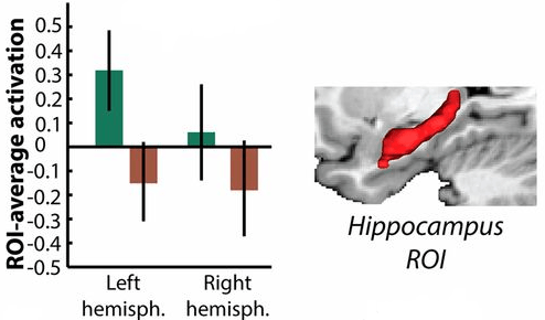
http://www.pnas.org/content/111/49/17654.full “Elevated hippocampal resting-state connectivity underlies deficient neurocognitive function in aging”


This post has somehow become a target for spammers, and I’ve disabled comments. Readers can comment on other posts and indicate that they want their comment to apply here, and I’ll re-enable comments.

Kids who have a larger and better-connected hippocampus learn math better when tutored

This 2013 Stanford study of 24 eight- and nine-year-old children found that measurements of limbic system areas predicted how well the 11 boys and 13 girls would respond to 8 weeks of one-on-one math tutoring!

“Pretutoring hippocampal volume predicted performance improvements. Furthermore, pretutoring intrinsic functional connectivity of the hippocampus with dorsolateral and ventrolateral prefrontal cortices and the basal ganglia also predicted performance improvements.

Brain regions associated with learning and memory, and not regions typically involved in arithmetic processing, are strong predictors of responsiveness to math tutoring in children. More generally, our study suggests that quantitative measures of brain structure and intrinsic brain organization can provide a more sensitive marker of skill acquisition than behavioral measures.”

None of the assessments, such as IQ and working memory tests, predicted how much benefit a child would receive from one-on-one math tutoring. The 16 children in the control group who didn’t receive one-on-one math tutoring didn’t improve their math performance over the 8-week period. Adults use different brain areas when solving math problems.


Much of the news coverage was from vested interests who dismissed the findings. A typical headline was “Your child’s brain on math: Don’t bother?”

The No Child Left Behind people were concerned that science could predict that some children were better suited to math tutoring than others. Psychiatrists and psychologists responded with general dismissals like small sample size, and the journalist let that stand without asking them how they disagreed with any of the specific P-, T- and other values found in the study’s supplementary material.

The researchers were careful to invoke a politically-correct meme of individual differences 19 times, including the study’s title!

“Individual differences” isn’t a causal explanation, however. The journalist whiffed and also gave a pass to the researchers on this uninformative-but-PC meme.

It certainly would have been within the scope of this study for the researchers to inquire further into causes for the findings. It possibly could have informed us of causal factors had the children’s test battery included emotional content, as did the subjects in the Early emotional experiences change our brains: Childhood maltreatment is associated with reduced volume in the hippocampus study.

http://www.pnas.org/content/110/20/8230.full “Neural predictors of individual differences in response to math tutoring in primary-grade school children”

Problematic research on suppressing unwanted memories

This 2014 French/UK human study found:

“Motivated forgetting mechanisms, known to disrupt conscious retention, also reduce unconscious expressions of memory, pointing to a neurobiological model of this process.”

There were multiple problems with this study.

1. The researchers excluded emotional content, although the study involved areas of the brain involved in processing emotions:

roi

How could the study’s findings apply to:

“The distressing intrusions that accompany posttraumatic stress disorder

when emotional memories were excluded? It was an unsupported assertion for one of the researchers to state:

“The better understanding of the neural mechanisms underlying this process arising from this study may help to better explain differences in how well people adapt to intrusive memories after a trauma.”

2. The term “unconscious” was used 27 times, including in the title, without defining it. The cited studies defined “unconscious” several meaningfully different ways. How could the findings achieve validity when they contained an undefined term?

3. The experiments involved short-term memories and visual perception, and the subjects took longer to visually perceive objects that they had been directed to suppress than those that they had been directed to think about. However, the researchers didn’t show that these experimental results could be extrapolated into findings about long-term unconscious memories.

4. Data manipulation:

  • The researchers noted:

    “We did not observe less hippocampal activation during no-think than think trials.”

  • This data didn’t fit what they wanted to find, so they:

    “Restricted the search volume to anatomically defined regions of interest.”

  • They still couldn’t make their predetermined finding, so they discarded:

    “An outlier which compromised the significance of this effect.”

The above process didn’t support the statement that immediately followed:

“Thus, suppression robustly engaged the brain regions associated with memory control, and this was accompanied by reduced activation in the hippocampus.”

Didn’t the reviewer have something to say about these four problem areas?

It was a letdown to read the details of the study when its title held out such promise for informing us about the unconscious influence of memories. Per the Scientific evidence page, it would really help a person as a first step to become somewhat aware of their unconscious memories and feelings, especially when these are expressed through behavior.

http://www.pnas.org/content/111/13/E1310.full “Suppressing unwanted memories reduces their unconscious influence via targeted cortical inhibition”

Translating PTSD research findings from animals to humans

This 2014 rodent study stressed the animals, measured their stress responses, then killed them and sampled genes in their amygdala, hippocampus, and blood. The researchers found that glucocorticoid receptor signaling genes were the primary pathway associated with “exposure-related individual differences in stress responses for the amygdala and blood. This pathway also placed first for the hippocampus in female rats (glucocorticoid receptor was second in male rats and prostate cancer signaling was first).

I’ll quote one press article’s coverage to show where the researchers wanted to go with the study’s findings:

“We found that most of the genes and pathways that are different in PTSD [post-traumatic stress disorder]-like animals compared to resilient animals are related to the glucocorticoid receptor, which suggests we might have identified a therapeutic target for treatment of PTSD.”

How about this lead sentence:

“There may some day be a blood test to determine whether someone suffers from Post-Traumatic Stress Disorder or is at risk for the psychiatric condition.”

Here’s another article’s paraphrase of a different researcher:

“Those are genes that become activated in the presence of stress. Like a key fitting into a lock, the hormone corticosterone, produced naturally by the body, connects to the receptor and has a calming effect.

In some rodents, and apparently in some people, the pathway appears to be defective, and this puts them at higher risk for PTSD.”

Also, from the study’s abstract:

“Corticosterone treatment 1 h[our] after PSS [predator-scent-stress]-exposure prevented anxiety and hyperarousal 7 d[ays] later in both sexes, confirming the GR [glucocorticoid receptor] involvement in the PSS behavioral response.”


Like other researchers continue to do, they stopped this study short of finding causes for the effects:

  1. What were the causes for genes in the glucocorticoid receptor signaling pathway being differentially expressed? “Exposure-related individual differences” isn’t a causal finding.
  2. If this pathway is “defective,” what exactly happened to make it that way?
  3. Did dampening the effects of stress with a shot of cortisol one hour after the stress treat the cause such that the rats were cured? Since the readers of the study and associated articles were led to infer that this treatment was a cure, why destroy the treated animals afterwards before the proofs of long-term efficacy were thoroughly documented and tested?

When studies like this are carried forward with humans, researchers should try to find the causes for these effects. It isn’t sufficient to pretend that there aren’t early-life causes for these effects. Such a pretense leads to the follow-on pretense that later-life consequences are mysteries such as “exposure-related individual differences” and not effects.

Researchers should act like the subjects are feeling human beings who can participate in treatments of both the causes and effects. They should remember that humans are not lab rats who need to be fixed.

http://www.pnas.org/content/111/37/13529.full “Expression profiling associates blood and brain glucocorticoid receptor signaling with trauma-related individual differences in both sexes”

Emotion inclusion in basic research drives discovery

What happens when a meme influences science? It would seem that researchers’ perceptions start to wheel around its axis.

A meme was the driver of this short 2015 commentary that stated:

“Most preclinical biomedical research, however, has been conducted with inadequate consideration of sex.”

If researchers discovered that gender hadn’t been adequately considered in research, maybe other “evolutionarily fundamental” factors had “inadequate consideration” as well.

Maybe one day soon, researchers will stop the current widespread exclusion of emotional content in studies, especially when the studies already included areas of the brain that are the biological bases of emotions.


To imagine a potentially beneficial future meme, I substituted the word “emotion” for “sex” in the title and part of two paragraphs of this commentary. How well does this fit?

[Emotion] inclusion in basic research drives discovery”

“Why is [emotion] so important?

Although many factors can influence an outcome, [emotion] is evolutionarily fundamental and affects the whole of the population

Across diverse disciplines, researchers risk drawing erroneous conclusions when they extrapolate outcome data from [emotional to non-emotional and vice versa].”

See A missed opportunity to study odor-evoked emotional memories for one deliberate exclusion of emotional content, although including emotions may have been appropriate, informative, and contributed to advancing science.

Click the hippocampus category to see a sample of human studies that may have included the biological seat of emotional memories, but only a scarce few of which included emotional content.

http://www.pnas.org/content/112/17/5257.full “Opinion: Sex inclusion in basic research drives discovery”

Epigenetic production of new brain neurons in the hippocampus

This 2015 La Jolla rodent study provided further details on the production of new neurons in the adult hippocampus:

“We propose that SOX2 sets a permissive epigenetic state in neural progenitor cells, thus enabling proper activation of the neuronal differentiation program under neurogenic cue.”

The researchers stated that the functions of these new brain neurons were:

“Newborn granule neurons generated from neural progenitor cells in the adult hippocampus play a key role in spatial learning and pattern separation.”

http://www.pnas.org/content/112/15/E1936.full “SOX2 primes the epigenetic landscape in neural precursors enabling proper gene activation during hippocampal neurogenesis

A missed opportunity to study odor-evoked emotional memories

The researchers of Can a study exclude the limbic system and adequately find how we process value? published another study. In this 2015 human study, subjects were monitored with fMRI scans while making choices on the identity and pleasantness of rewarding food odors.

I feel that the researchers missed quite a few good opportunities to advance science. Instead of making peripheral assessments of limbic system areas and citing numerous other studies, they could have included emotional content in their study and drawn their own conclusions.

Consider these opportunities:

  • Wouldn’t the odors used in the study such as chocolate cake and pizza and strawberry and potato chips – and other “comfort” foods – potentially be associated with emotional responses?
  • Don’t most humans have memories that include pleasant food odors?
  • Wouldn’t it have been informative to ask the subjects during fMRI scans to identify what emotions were evoked by the pleasant food odors?
  • Wouldn’t these resultant fMRI scans be expected to potentially show more strongly activated limbic system areas, given the hippocampus’ position as the seat of emotional memories?
  • Wouldn’t the additional emotional responses and memories and subsequent limbic system area activations potentially influence the subjects’ value judgments?

Instead, the researchers peripherally included limbic system areas in the study. The supplementary material included passages such as:

“Identity-specific value signals were found in not only the OFC, [orbitofrontal cortex] but also the ACC [anterior cingulate cortex] and hippocampus.”


Like the previous study, the current study’s focus was to provide evidence that areas of the cerebrum were in control when people made value judgments. The term “value” in the current study meant:

“the pleasantness of the odor.”

Like the previous study, areas of the limbic system weren’t addressed until the tail end of the supplementary material. The researchers cited other studies in an attempt to dismiss the role of the ACC in making value judgments, then said:

“Although we are unable to distinguish between these alternative explanations, our findings suggest that value-related signals in ACC—whether signed or unsigned—are specific to the identity of the expected outcome.”

Since the current study found that “identity” was encoded by cerebral areas, the above sentence was written to nudge the reader into inferring that the cerebrum dominated value judgments of “the pleasantness of the odor.”

The researchers similarly cited other studies in the last paragraph instead of specifically discussing how they studied the participation of the hippocampus part of the limbic system. They then speculated that the hippocampus’ contributions to value judgments in the current study were explained by the referenced studies:

“We speculate that the hippocampus is involved in retaining sensory-based information about specific rewards, which may be linked to value-based representations in OFC for later consolidation.”

Like the previous study, the researchers were begrudgingly diverted away from their focus on cerebral areas when they were forced to acknowledge the limbic system’s contributions to value judgments of “the pleasantness of the odor.”

http://www.pnas.org/content/112/16/5195.full “Identity-specific coding of future rewards in the human orbitofrontal cortex”