Chaos – not balance – and competition for resources are the natural order

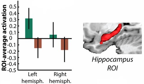
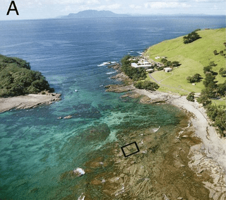
This 2015 Amsterdam/New Zealand/Cornell shore-life study found:

“Species abundances in natural ecosystems may never settle at a stable equilibrium.

Species in one of the world’s oldest marine reserves showed chaotic fluctuations for more than 20 years. The species replaced each other in cyclic order, yet the exact timing and abundances of the species were unpredictable.

Our findings provide a field demonstration of nonequilibrium coexistence of competing species through a cyclic succession at the edge of chaos.

Our findings show that natural ecosystems can sustain continued changes in species abundances.”

chaos

http://www.pnas.org/content/112/20/6389.full “Species fluctuations sustained by a cyclic succession at the edge of chaos”


The University of Amsterdam also participated in a 2013 study Evolution of microbial markets where evolutionary biologists studied microbes. Their related findings included:

“Cooperative interactions between individuals of different species.

Strategies important for microbes to optimize their success in potential biological markets:

  • (i) avoid bad trading partners;
  • (ii) build local business ties;
  • (iii) diversify or specialize;
  • (iv) become indispensable;
  • (v) save for a rainy day; and
  • (vi) eliminate the competition.”

A 2015 study How a well-adapted immune system is organized (the *.pdf file is linked because the html has errors) had a related finding that applied to our body’s immune system. The researchers found that the primary reason why each of our immune systems is unique is due to the effect of:

“Competition between receptor clones..NOT a biologically implausible centralized mechanism distributing resources system-wide.

The repertoire of lymphocyte receptors in the adaptive immune system protects organisms from diverse pathogens. A well-adapted repertoire should be tuned to the pathogenic environment to reduce the cost of infections.

Competitive dynamics can allow the immune repertoire to self-organize into a state that confers high protection against infections.”

Chaos and competition for resources are facts of life observed within ourselves and in nature from ocean life down to the microbe level.

Why are we often presented – as a fact of life – that what’s natural is for all aspects of our lives to be in balance? Emotional, economic, social, intellectual – you name it, we’re told that the natural model is one of “stable equilibrium.”


Two hypotheses of Dr. Arthur Janov’s Primal Therapy are relevant:

Trying for closure, though, becomes an act-out – a temporary fulfillment of a substitute need. But the underlying need remains unsatisfied, and soon drives further act-outs. Balance is never achieved.

With this viewpoint, can you see how behavior like the following shows the internal state of the actor as they attempt to thwart the natural reality of the situation?

  • A person in authority who demands that people cease their competition for a resource and instead, accept what the authority figure determines is fair and balanced. An example is limiting supplies with price controls after a disaster.
  • A person who disrupts cooperative behavior that provides a solution for the cooperators’ needs/wants and instead, interposes themselves in a directed solution. An example is requiring licenses for cooperative childcare.
  • A person who insists that peoples’ responses to chaos to form an optimal adaptation cease, and instead, conform to some other responses. An example is prohibiting free movement after a disaster.

It reveals even more about the internal states of people that the above examples become codified. Children are taught that the natural and solely acceptable way to behave is in accordance with these unnatural solutions.


There are some signs that unnatural solutions in society can be reversed. For example, here is a 2013 article about a UK village that benefited from removing all of its traffic signals and reverting to the natural order of human cooperation and competition.

At the individual level, though, it’s up to each one of us to recognize and reverse our unnatural states. We and the people around us will be pleased when we and they are no longer adversely affected by our unconscious act-outs that are driven by our internal states. There’s enough natural chaos without adding more with act-outs.

Our internal systems will suffer damage, for example, when our unconscious act-out is to be busy, always doing something, and we can’t relax. Stress adversely affects our internal systems until we understand and reverse the driving unnatural states.

Do popular science memes justify researchers’ cruelties to monkeys?

This 2015 Oxford study of 38 humans and 25 macaques drew correlations of brain activities between the two species. The study title included buzzwords such as “reward” and “decision making” and the study focused on the ever-popular “frontal cortex.”

Humans and macaques are separated by 25 million years of evolutionary adaptations and developments. Studies done with macaque subjects don’t automatically have human applicability.

Was a major reason for the study’s comparisons to provide justifications for keeping macaques as study subjects? Accepting these justifications and going along with the popular memes would ease the way for whatever cruelties researchers want to inflict on our primate relatives.

http://www.pnas.org/content/112/20/E2695.full “Connectivity reveals relationship of brain areas for reward-guided learning and decision making in human and monkey frontal cortex”

A mixed bag of findings about oxytocin, its receptor, and autism

This 2014 Stanford human study found:

“No empirical support for the OXT [oxytocin] deficit hypothesis of ASD [autism spectrum disorder], nor did plasma OXT concentrations differ by sex, OXTR [oxytocin receptor] SNPs [single nucleotide polymorphisms], or their interactions.”

Apparently, there was a:

“Prevalent but not well-interrogated OXT deficit hypothesis of ASD.”

The researchers followed up this worthwhile finding with three weak findings. The first, as stated by one of the study’s lead researchers, was:

“It didn’t matter if you were a typically developing child, a sibling or an individual with autism: Your social ability was related to a certain extent to your oxytocin levels.”

The second weak finding was that, regarding OXTR SNPs:

“The minor allele of rs2254298 predicted global social impairments on the SRS [Social Responsiveness Scale] and diagnostic severity on the ADI-R [Autism Diagnostic Interview-Revised]. In contrast, the major allele of rs53576 predicted impaired affect recognition performance on the NEPSY [A Developmental NEuroPSYchological Assessment].”

This was at odds with other relevant research, leading the researchers to state:

The functional significance of these two intronic variants remains unknown.”

The third weak finding irked me:

“Plasma OXT concentrations were highly heritable.”

because the researchers didn’t attempt to differentiate the contribution of the environment for the observed blood oxytocin levels, as did the similar How epigenetic DNA methylation of the oxytocin receptor gene affects the perception of anger and fear study.

I wonder what the reviewer’s feedback was about these weak findings. Did he make the researchers insert specific language into the lengthy paragraph about the study’s limitations, or did he give them a pass?

http://www.pnas.org/content/111/33/12258.full “Plasma oxytocin concentrations and OXTR polymorphisms predict social impairments in children with and without autism spectrum disorder”

Do our unique visual perceptions arise from brain structural differences?

This 2014 UK/German human study involved fMRI scans of the subjects inferior temporal cortex while viewing images:

“Brain representational idiosyncrasies accessible to fMRI are expressed in an individual’s perceptual judgments.

We found evidence for an individually unique representation predictive of perceptual idiosyncrasies in hIT [human inferior temporal cortex] (but not in early visual areas) and for personally meaningful (but not for unfamiliar) objects.”

Citing other studies, the researchers said:

“The size of primary visual cortex varies across individuals by a factor of about 2.5.

Although other areas might vary by smaller factors, many parts of the brain, including cortical and subcortical structures, show gross anatomical variation across individuals that is predictive of cognitive and behavioral differences.”

The researchers asserted:

“Functional differences as reported here ultimately must arise from differences in the physical structure of each individual brain.”

However, no evidence was provided for this assertion.

The researchers acknowledged this lack of evidence, but in a way that required further evidence:

“Our study demonstrates individual differences in high-level semantic representations but cannot address their structural basis. Our current interpretation is that the representational idiosyncrasies might arise from the microstructural plasticity of cortex, which is driven by individual experience.”


The researchers’ assertion beyond the study’s supporting data was at best a statement of their goal. Further, their bias to focus on the inferior temporal cortex area of the cerebrum led them to not investigate other areas of the brain that may have been involved with the “personally meaningful (but not for unfamiliar) objects” finding, such as the subjects’ limbic systems.

I hope that researchers won’t think that their research is complete when they reach their goal of finding “differences in the physical structure of each individual brain.” It would be far more informative to understand the causes for these effects.

http://www.pnas.org/content/111/40/14565.full “Unique semantic space in the brain of each beholder predicts perceived similarity”

Separating genetic from environmental factors when assessing educational achievement

This 2014 UK study of identical and fraternal twins found that an average of 62% of the differences among their scores on a significant test given at age 16 were due to genetic factors:

“Genetic influence is greater for achievement than for intelligence, and other behavioral traits are related to educational achievement largely for genetic reasons.”

However, the “genetic reasons” term didn’t mean that the researchers actually took genetic samples. From one news article:

“Identical twins share 100 percent of their genes while non-identical twins share just 50 percent of their genes. Because these sets of twins share the same environment, the scientists were able to compare identical and non-identical twins to estimate the relative contributions of genetic and environmental factors.”

This estimation method produced an artificial divide between genetic and environmental factors. Identical twins start out sharing 100% of their genes, but then their genes become expressed differently – often because of environmental factors – to produce unique individuals even before birth.

The sets of identical twins were definitely not the 100% same genetic makeup between themselves at age 16 as they were at conception, and that assumption was the foundation of the researchers’ model:

F2

“Bivariate estimates for additive genetic (A), shared environmental (C), and nonshared environmental (E) contributions to the correlations between GCSE and nine predictors. The total length of the bar indicates the phenotypic correlations.”

The researchers didn’t provide evidence that “genetic reasons” were causal factors to the stated extent. Although the model’s numbers may have indicated that the method’s results were valid, that didn’t necessarily mean that the reality of genetic and epigenetic influences on the subjects were represented to the stated precision by the results.

The weather analogy of Scientific evidence applies to this study’s methods:

“We can think about what we mean by evidence. For example, that when you see dark storm clouds overhead, that’s strong evidence that it’s about to rain. If you smell a certain scent, that’s maybe weak evidence that it’s about to rain. And if we see the dark storm clouds and then we smell the scent, the evidence doesn’t get weaker: if anything, it gets stronger.

But P-values in a circumstance like that, where you have a very small P-value in one dataset and a not-so-small P-value in a second dataset, you put the data together and the P-value will tend to sort of average.

So the P-value is not behaving like evidence.”

Better methods of estimating “the relative contributions of genetic and environmental factors” are available with actual genetic sampling. One way is to measure the degree of DNA methylation of genes as did:


The study and its news coverage were full of politically-correct buzzwords – for example, the researchers’ statement:

“The results also support the trend in education toward personalized learning.”

This “personalized learning” is a teacher not telling a student:

“You’re doing poorly at math. You need to pay attention in class and do the homework.”

but instead saying:

“You have a different learning style. We’ll tailor the math lessons to your style.”

The funniest thing I saw in the study’s news coverage was this one where someone argued that the researchers were wrong and that they needed educational psychologists on their staff to interpret the data. Guess the profession of the arguer!

http://www.pnas.org/content/111/42/15273.full “The high heritability of educational achievement reflects many genetically influenced traits, not just intelligence”

People who donated a kidney to a stranger have a larger amygdala

This 2014 Georgetown study was of people who had donated a kidney to a stranger. The study found that the subjects had a larger right amygdala part of their limbic systems:

“Our results support the possibility of a neural basis for extraordinary altruism.

In sum, our findings suggest that individuals who have performed an act of extraordinary altruism can be distinguished from healthy controls by increased right amygdala volume, as well as heightened responsiveness in right amygdala to fearful facial expressions, which may support enhanced recognition of these expressions.”

The researchers stopped short of causal explanations. They stated in the study’s abstract that:

“Individual variation in altruistic tendencies may be genetically mediated”

but didn’t develop any evidence to support this statement.

It would have been within the scope of the study had the researchers continued on to examine:

  • What may have happened in the subjects’ lives to possibly cause their neurobiological and psychological attributes?
  • What were the causes for the subjects’ extreme altruistic behavior?
  • Were these the same causes for their larger, more sensitive amygdala?

An accompanying PNAS commentary from a Harvard researcher made other points. However, the author showed his biases that the cerebrum rules human behavior with an out-of-left-field question at the end of a paragraph in which he developed specious reasoning.

The commentator was completely off base when he stated:

“Could it be that extraordinary altruists such as Maupin [a study participant] and the 19 individuals studied by Marsh et al. [the researchers] are special, not only because of how they feel when they see people in distress, but because of how they think?”

I don’t imagine that the brilliant commentator’s attempt to upstage the study’s subjects and put the spotlight on himself for some brilliant idea was much appreciated by anyone involved.

The amygdala is the central hub of a person’s limbic system. The study’s findings had very little to say about the subjects’ cerebral activity – thinking.

To postulate that the researchers missed that there was something different about the subjects’ thinking was out of touch with the realities of both the researchers’ scientific bases and the subjects. It’s another example of the current research mindset/social meme of cerebral dominance.

http://www.pnas.org/content/111/42/15036.full “Neural and cognitive characteristics of extraordinary altruists”

What causes disconnection between the limbic system and the cerebrum?

This 2014 Swedish human study with 339 subjects aged 25-80 years old found that as the subjects’ age increased, their hippocampus became less connected to their cerebrums:

“Age-related cortico–hippocampal functional connectivity disruption leads to a more functionally isolated hippocampus at rest, which translates into aberrant hippocampal decoupling and deficits in active mnemonic processing.”

The lead researcher said:

“What we can now show is that memory problems that come with increased age are most likely due to a process where the interaction among different regions of the hippocampus increases in response to less inhibitory cortical input. This in turn means that the hippocampus risks being more isolated from other important networks in the brain which impacts our ability to actively engage the hippocampus, for example to remember different events.”

Like other researchers commonly do, they excluded emotional content from the study. See another Swedish study Emotional memories and out-of-body–induced hippocampal amnesia as an example of why emotional memories are necessary in order to properly study the hippocampus.


1) As a result of excluding emotional content and other aspects of the study’ design such as using 25 as the beginning age of the subjects, all the researchers could muster as a explanatory factor was age. However, they had to couch their findings as “age-related” because age in and of itself wasn’t a causal explanation for the observed effects.

2) The findings weren’t even truly “age-related”  because, for example, the study didn’t necessarily apply to people below the age of 25. Had the study included 10-18 year old subjects, the researchers may have found that “less inhibitory cortical input” may also be present before puberty, as The prefrontal cortex develops more repressive function at puberty study indicated.

3) Had the study design included neurochemicals, the researchers may have found that “cortico–hippocampal functional connectivity disruption” was due to factors that influenced dopamine and glutamate levels, as A mechanistic study of neurotransmitters in the hippocampus indicated.

4) A finding that “cortico–hippocampal functional connectivity disruption” was influenced by other factors may also have been made had the study design included the subjects’ histories. Per my Welcome page, the findings of much of the recent research I’ve curated on this blog, and the references in those studies show that when basic needs aren’t met, especially early in people’s lives, and the painful conditions persist, enduring physiological changes may occur.

5) What the researchers noted in the study’s limitation paragraph were references to fMRI scans rather than limitations such as those mentioned above regarding the study design. The study provided unconvincing evidence for causes of “cortico–hippocampal functional connectivity disruption” and it wasn’t because of fMRI limitations.

http://www.pnas.org/content/111/49/17654.full “Elevated hippocampal resting-state connectivity underlies deficient neurocognitive function in aging”


This post has somehow become a target for spammers, and I’ve disabled comments. Readers can comment on other posts and indicate that they want their comment to apply here, and I’ll re-enable comments.

Kids who have a larger and better-connected hippocampus learn math better when tutored

This 2013 Stanford study of 24 eight- and nine-year-old children found that measurements of limbic system areas predicted how well the 11 boys and 13 girls would respond to 8 weeks of one-on-one math tutoring!

“Pretutoring hippocampal volume predicted performance improvements. Furthermore, pretutoring intrinsic functional connectivity of the hippocampus with dorsolateral and ventrolateral prefrontal cortices and the basal ganglia also predicted performance improvements.

Brain regions associated with learning and memory, and not regions typically involved in arithmetic processing, are strong predictors of responsiveness to math tutoring in children. More generally, our study suggests that quantitative measures of brain structure and intrinsic brain organization can provide a more sensitive marker of skill acquisition than behavioral measures.”

None of the assessments, such as IQ and working memory tests, predicted how much benefit a child would receive from one-on-one math tutoring. The 16 children in the control group who didn’t receive one-on-one math tutoring didn’t improve their math performance over the 8-week period. Adults use different brain areas when solving math problems.


Much of the news coverage was from vested interests who dismissed the findings. A typical headline was “Your child’s brain on math: Don’t bother?”

The No Child Left Behind people were concerned that science could predict that some children were better suited to math tutoring than others. Psychiatrists and psychologists responded with general dismissals like small sample size, and the journalist let that stand without asking them how they disagreed with any of the specific P-, T- and other values found in the study’s supplementary material.

The researchers were careful to invoke a politically-correct meme of individual differences 19 times, including the study’s title!

“Individual differences” isn’t a causal explanation, however. The journalist whiffed and also gave a pass to the researchers on this uninformative-but-PC meme.

It certainly would have been within the scope of this study for the researchers to inquire further into causes for the findings. It possibly could have informed us of causal factors had the children’s test battery included emotional content, as did the subjects in the Early emotional experiences change our brains: Childhood maltreatment is associated with reduced volume in the hippocampus study.

http://www.pnas.org/content/110/20/8230.full “Neural predictors of individual differences in response to math tutoring in primary-grade school children”

The critical period for some aspects of human sight can be extended past childhood

This 2013 human study provided further details of critical periods in human development. The study subjects were:

“11 children enrolled in a humanitarian and scientific effort in India that provides corrective surgery to children with treatable cataracts and subsequently studies their visual abilities.”

The researchers found:

“The human visual system can retain plasticity beyond critical periods, even after early and extended blindness.

We define “early-onset” blindness as occurring before 1 y of age. We define “extended” blindness as lasting at least until early childhood, when many visual abilities in normally developing children reach adult levels. Contrast sensitivity in particular develops until approximately age 7 in normally sighted humans.

Of the 11 children, five had no discernible improvement, whereas one child’s vision grew worse, probably because of post-surgical complications. Five of the patients showed remarkable enhancement, however, and of these, an 11-year-old and a 15-year-old showed 30-fold improvement in contrast sensitivity.

“The visual brain can be plastic for longer than we originally thought,” concludes Kalia. “Many of the kids dramatically improve their quality of life.”

http://www.pnas.org/content/111/5/2035.full “Development of pattern vision following early and extended blindness”

A missed opportunity to research the oxytocin receptor gene and autism

This 2013 study:

“Examined whether genetic variants of the OXTR [oxytocin receptor] affect face recognition memory in families with an autistic child.

We investigated whether common polymorphisms in the genes encoding the oxytocin and vasopressin 1a receptors influence social memory for faces.”

I feel that the researchers missed an opportunity to improve their assessment of the autism-related genetic contribution to the study’s findings by separating the degree of environmental influence on the oxytocin receptor gene expression, as did the How epigenetic DNA methylation of the oxytocin receptor gene affects the perception of anger and fear study.

An assessment of epigenetic DNA methylation of the oxytocin receptor gene may have been even more compelling because the researchers genetically sampled one non-autistic sibling in each of the autistic children’s families. I hope the study’s samples are still available, because they may offer the possibility of evaluating the contribution of the autistic children’s historical environment with potential confirmation from their siblings.

Both studies gave their subjects similar facial emotion recognition tests, with the current one deriving from findings about autism, and the second from findings about the amygdala. The studies also had common references, such as a 2010 study, A common allele in the oxytocin receptor gene (OXTR) impacts prosocial temperament and human hypothalamic-limbic structure and function.

http://www.pnas.org/content/111/5/1987.full “Common polymorphism in the oxytocin receptor gene (OXTR) is associated with human social recognition skills”

The prefrontal cortex develops more repressive function at puberty

This 2014 primate study found:

“The average magnitude of functional connections measured between neurons was lower overall in the prefrontal cortex of peripubertal [age when puberty starts] monkeys compared with adults. The difference resulted because negative functional connections (indicative of inhibitory interactions) were stronger and more prevalent in peripubertal compared with adult monkeys.”

The researchers found more inhibitory functional connections at the onset of puberty than during adulthood. This repressive functionality presumably develops at puberty because that’s when it’s relatively more needed:

“The bias toward increased inhibitory connectivity we report here for young monkeys might also be an intrinsic feature of human prefrontal cortex at a comparable stage of development.”

One hypothesis of Dr. Arthur Janov’s Primal Therapy is that repression is an important function that the prefrontal cortex evolved.

http://www.pnas.org/content/111/10/3853.full “Age-dependent changes in prefrontal intrinsic connectivity”

Wasting public science funds on voodoo dolls

This 2014 Ohio State human study found:

“Low glucose levels might be one factor that contributes to intimate partner violence.”

The study used pins stuck in voodoo dolls and noise blasts to measure aggression towards the subjects’ spouses. These measures had neither been correlated with “intimate partner violence” nor standardized, though.

The experimental data led to some fun-with-number associations:

“Lower evening glucose levels related to blasting one’s spouse with more intense and prolonged noise.

Lower levels of evening glucose were more aggressive in part because they also had greater aggressive impulses.”


Both the study’s design and implementation were such that they couldn’t provide evidence for causes of the effects. The expert opinion here expressed:

“Concerns to which extent this particular study is helpful in explaining the role of self-control (or lack thereof) in the etiology of intimate partner violence.”

and pointed out several methodological and statistical lacks.

National Science Foundation Grant BCS1104118 was a waste of public money while deserving areas of research go begging.

http://www.pnas.org/content/111/17/6254.full “Low glucose relates to greater aggression in married couples”

Agenda-driven research on emotional memories

I curated this 2013 study because one of the authors has made a career out of denying that people accurately remember and re-experience emotional memories. I’ll show how this viewpoint created problems with the study.

For background, one relevant hypothesis of Dr. Arthur Janov’s Primal Therapy is that there are differences in the levels of consciousness of: (1) an emotional memory; (2) the recall of an emotional memory; and (3) a verbal description of the recall of an emotional memory.

  1. The retrieval and re-experiencing of an emotional memory can engage our lower-level brain areas without our higher-level brain areas’ participation.
  2. The recall of 1 above is a product of our cerebrum in response to input from limbic system and lower brain areas.
  3. The verbal description of 2 above is a product of our brain’s language areas in response to input from the cerebral areas that recalled the emotional memory.

Clinical principles of Primal Therapy that follow are:

  • A patient won’t re-experience an emotional memory when they only just recall it.
  • It’s another level of consciousness even further removed from an emotional memory when someone describes their recall of the memory.

The researchers asserted that they studied emotional memories in one part of this study. Their method was to ask the subjects to recall and verbally describe the emotions they felt the week after 9/11/2001.

The researchers introduced factors to try to confuse the subjects about their recall of their emotions, and their verbal descriptions of their recall. The researchers were very sure that confusing the subjects’ cerebral recalls and descriptions produced evidence that the subjects’ emotional memories were changed and falsified.

Can you see how far removed the researchers were from studying emotional memories? They didn’t demonstrate that they understood how emotional memories were stored because they didn’t attempt to engage the subjects’ limbic system and lower brain areas.


Let’s illustrate the study’s inappropriate characterizations with an example. I burned my left index fingertip last week while toasting bread on an infrared oven grill. The pain is still stored with my emotional memory, and is probably why my memory is very clear.

I can recall the visual details of the grill, how my fingertip looked, the pain I initially felt, and the relief I felt when I held my finger under running cold water. I can retrieve and re-experience my emotional memory in a calm environment such as lying in bed with no aural or visual distractions.

Let’s imagine that the researchers analogously studied my burned fingertip accident. They would deny that I can accurately retrieve and re-experience my emotional memory of the accident if they could create problems with my verbal descriptions of my recall. For example, if I initially said that I pushed the kitchen faucet handle all the way in the cold direction, then after repeated questioning, I said that I wasn’t sure that the handle was pushed all the way over to Cold.


The researchers intentionally conflated the falsifiability of emotional memories with a strawman definition of false emotional memories.

They purposely misidentified both:

  • The subjects’ recalls of post-9/11 emotions; and
  • The subjects’ descriptions of their recalls

as emotional memories.

The study was designed to be lawyering, not science. The researchers DETRACTED from science.

Maybe their purposeful error could be overlooked if it was confined to this study. But it isn’t.

Imagine the damage this viewpoint creates when mental health professionals deny the reality of their patients’ feelings, experiences, and emotional memories!

http://www.pnas.org/content/110/52/20947.full “False memories in highly superior autobiographical memory individuals”

Who benefits when research promotes a meme of self-sacrifice?

The main purpose of this 2014 Illinois human study was to make findings directed toward high school students that:

“Well-being may depend on attending to higher values related to family, culture, and morality, rather than to immediate, selfish pleasure.”

The study’s messages to young people and to those who control young people were:

  • You have to give up trying to live your own life if you want to be happy.
  • For your own “well-being” just follow the “higher values” where other people tell you what to do and think.
  • Other people know how you should live your life better than you do. Science says so.

The researchers embedded many assertions into the study, most of which weren’t supported by the study’s data. The researchers’ main assertion was:

“Optimal well-being may be achieved through eudaimonic activities.”

The researchers repeated this assertion multiple times in multiple ways, such as citing philosophy and other research. The short version of the term “eudaimonic” was defined as: “Meaning and purpose, a life well-lived.”

The study’s ONLY measurement of “eudaimonic” activities was the subjects’

“Neural activation when making a donation to the family that involves self-sacrifice.”

The study’s main finding involving this SOLE measurement was:

“Eudaimonic decisions predicted longitudinal declines in depressive symptoms.”

Depressive symptoms were determined by “a self-report measure” where the subjects, 47 adolescents aged 15-17:

  • “Completed the internalizing symptoms subscale of the Youth Self-Report form of the Child Behavior Checklist
  • Underwent a brain scan during which they completed a family donation task and a risk-taking task.”

39 of the subjects returned one year later to reanswer the checklist.


I wonder what bases the reviewer used to approve the researchers’ methods.

1. In the study’s verbiage the researchers extrapolated the significance of the sole measurement of eudaimonic activities – the initial fMRI scan – many times past what it actually measured. One-time measurements of the blood flow in the ventral striatum of a few Los Angeles teenagers can’t validly be assigned as the bases for all of what the researchers went on and on about to glorify “prosocial eudaimonic decisions.”

2. No method checked the subjects’ personal impact of the experiments’ monetary rewards and donations. The subjects didn’t scale their personal relative importance of the monetary rewards and donations.

Consider the relative importance of a dime for a kid whose parents gave them a BMW to drive to high school. Compare that with a kid who searched the sidewalk for dropped coins as they walked to high school.

Absent subjective scaling, the monetary rewards and donations data couldn’t be used as the basis to produce informative results.

3. The balloon test used in this study to measure “risky hedonic decisions” was the same as in the Who benefits when research with no practical application becomes a politically correct meme? study. The same objection applies here: a video game task of popping balloons that engages the cerebrum was NOT informative to the cause-and-effect of the emotions and instincts and impulses from limbic system and lower brain areas that predominantly drive risky behavior.


Scientific justification for memes like the self-sacrifice promoted by this study helps rush people past what really happened in their lives. A popular cultural meme is to “live in the present” and purposefully overlook how we arrived at our present lives.

I wonder how we would evaluate the “higher values related to family, culture, and morality” if we felt and honestly understood our real history.

Do you feel that young people benefit when they sacrifice their lives in the name of “family, culture, and morality?” Who benefits when people don’t pause to reflect on how their history impacts what’s going on now with their lives?

http://www.pnas.org/content/111/18/6600.full “Neural sensitivity to eudaimonic and hedonic rewards differentially predict adolescent depressive symptoms over time”

Problematic research on suppressing unwanted memories

This 2014 French/UK human study found:

“Motivated forgetting mechanisms, known to disrupt conscious retention, also reduce unconscious expressions of memory, pointing to a neurobiological model of this process.”

There were multiple problems with this study.

1. The researchers excluded emotional content, although the study involved areas of the brain involved in processing emotions:

roi

How could the study’s findings apply to:

“The distressing intrusions that accompany posttraumatic stress disorder

when emotional memories were excluded? It was an unsupported assertion for one of the researchers to state:

“The better understanding of the neural mechanisms underlying this process arising from this study may help to better explain differences in how well people adapt to intrusive memories after a trauma.”

2. The term “unconscious” was used 27 times, including in the title, without defining it. The cited studies defined “unconscious” several meaningfully different ways. How could the findings achieve validity when they contained an undefined term?

3. The experiments involved short-term memories and visual perception, and the subjects took longer to visually perceive objects that they had been directed to suppress than those that they had been directed to think about. However, the researchers didn’t show that these experimental results could be extrapolated into findings about long-term unconscious memories.

4. Data manipulation:

  • The researchers noted:

    “We did not observe less hippocampal activation during no-think than think trials.”

  • This data didn’t fit what they wanted to find, so they:

    “Restricted the search volume to anatomically defined regions of interest.”

  • They still couldn’t make their predetermined finding, so they discarded:

    “An outlier which compromised the significance of this effect.”

The above process didn’t support the statement that immediately followed:

“Thus, suppression robustly engaged the brain regions associated with memory control, and this was accompanied by reduced activation in the hippocampus.”

Didn’t the reviewer have something to say about these four problem areas?

It was a letdown to read the details of the study when its title held out such promise for informing us about the unconscious influence of memories. Per the Scientific evidence page, it would really help a person as a first step to become somewhat aware of their unconscious memories and feelings, especially when these are expressed through behavior.

http://www.pnas.org/content/111/13/E1310.full “Suppressing unwanted memories reduces their unconscious influence via targeted cortical inhibition”